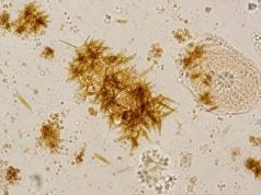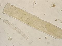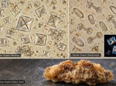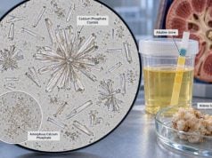
Amorphous crystals in urine are tiny mineral precipitates that look like fine, shapeless granules under the microscope. The two common types are amorphous urates, which usually appear in acidic urine, and amorphous phosphates, which are more typical of alkaline urine. In many people, these crystals are a temporary laboratory finding caused by urine pH, concentration, cooling, or a sample that sat before examination. They do not automatically mean kidney stones or kidney damage. The important questions are which type is present, what the urine pH was, whether the sample was fresh, and whether there are symptoms or other abnormal results. Persistent crystalluria, recurrent stones, very unusual urine pH, infection markers, or abnormal kidney function changes the interpretation. Because amorphous material can obscure cells and other sediment structures, laboratories may also need to distinguish it from bacteria, mucus, or more clinically important crystals before deciding what the finding means.
- Amorphous urates usually form in acidic urine: they are often yellow-brown or pinkish granules and may increase after cooling.
- Amorphous phosphates usually form in alkaline urine: they are often colorless and can make urine look cloudy.
- A single finding is often benign: crystal precipitation can happen after the urine leaves the body and does not by itself diagnose a stone.
- Urine pH is central to interpretation: the same granular-looking sediment has a different meaning in acidic versus alkaline urine.
- Follow-up matters when crystals persist with symptoms: recurrent flank pain, stones, UTI findings, or abnormal kidney tests warrants a broader evaluation.
Table of Contents
- What Amorphous Crystals Are
- Amorphous Urates vs Amorphous Phosphates
- How Urine pH and Sample Handling Affect Crystals
- Common Causes and Clinical Associations
- Do Amorphous Crystals Mean Kidney Stones?
- How to Interpret and Follow Up the Result
- When Crystals Need Prompt Medical Attention
What Amorphous Crystals Are
“Amorphous” describes appearance, not a single chemical compound. Instead of forming a distinctive geometric shape, amorphous crystals appear as numerous tiny granules with no obvious edges or repeated structure. They can clump together and may be difficult to distinguish from debris when viewed with ordinary bright-field microscopy.
In routine urine sediment, the two main amorphous categories are urates and phosphates. Their chemistry and clinical associations differ, but both commonly reflect temporary supersaturation: the urine contains more of a dissolved substance than can remain in solution under the current conditions. Temperature, pH, urine concentration, and the time between collection and analysis all influence whether precipitation occurs.
This is why crystalluria is not the same as kidney stone disease. Crystals can form in a specimen cup after voiding, especially as urine cools. They can also occur transiently in healthy people after dehydration, dietary changes, or shifts in urine pH. Stone formation requires additional processes such as sustained supersaturation, crystal growth, aggregation, and retention within the urinary tract.
The broader urine microscopy test is useful because amorphous granules should be interpreted alongside red and white blood cells, epithelial cells, bacteria, casts, and other crystal types. A report that says only “amorphous crystals: present” is less informative than one that also provides the urine pH and identifies whether the material is most consistent with urates or phosphates.
Cloudiness is another clue but is not specific. Phosphate precipitation can turn alkaline urine visibly cloudy, while abundant urates can produce a pink or brick-dust sediment, especially in a cooled specimen. Cloudy urine can also come from white blood cells, bacteria, mucus, semen, epithelial cells, or other crystals. A visual change therefore cannot identify the material without chemical testing or microscopy. This is particularly important when a person notices cloudy urine at home and assumes infection; phosphate precipitation can look dramatic even when there is no infection at all.
Laboratories sometimes report amorphous material semi-quantitatively as few, moderate, or many rather than providing a numeric crystal count. Those categories are method-dependent and are not equivalent to a disease-severity scale. “Many” crystals in a refrigerated specimen may be less clinically important than a smaller amount of a pathognomonic crystal, such as cystine, in a fresh sample. The identity, clinical setting, and reproducibility matter more than the descriptive quantity alone.
Amorphous Urates vs Amorphous Phosphates
The easiest practical distinction is the urine’s acid-base environment. Amorphous urates favor acidic urine, while amorphous phosphates favor alkaline urine. Their color and response to simple laboratory manipulations can also help.
| Feature | Amorphous urates | Amorphous phosphates |
|---|---|---|
| Usual urine pH | Acidic | Neutral to alkaline, especially alkaline |
| Typical appearance | Fine yellow-brown, reddish, or pink granules | Fine colorless to whitish granules |
| Effect of temperature | Often precipitate more after cooling; may dissolve with warming | Less defined by cooling than urates |
| Relation to pH | Favored by low pH | Favored by higher pH |
| Common meaning | Usually transient urate precipitation | Usually transient phosphate precipitation |
Amorphous urates are related to uric acid chemistry but should not be treated as identical to classic uric acid crystals. Uric acid crystals often form more recognizable rhomboids, rosettes, wedges, or barrels and are more directly associated with uric acid supersaturation and uric acid stone risk. A report of uric acid crystals in urine therefore carries different morphologic information than a report of amorphous urates.
Amorphous phosphates are also not the same as a confirmed calcium phosphate stone. Calcium phosphate can appear as different crystalline forms, including apatite-related material and brushite. These are influenced by alkaline urine, calcium concentration, phosphate concentration, citrate, and other factors. A separate finding of calcium phosphate crystals may deserve more targeted interpretation when stone disease is suspected.
How Urine pH and Sample Handling Affect Crystals
Urine pH changes mineral solubility. Acidic conditions make uric acid and urate-related precipitation more likely, while alkaline conditions favor precipitation of phosphate-containing salts. This relationship is useful, but the pH on one urine sample is only a snapshot. Meals, hydration, medications, vomiting, diarrhea, systemic acid-base status, infection, and time of day can all shift urine pH.
A urine pH test therefore provides context rather than a diagnosis. For example, an alkaline urine sample with abundant amorphous phosphates may simply reflect a post-meal alkaline tide. If the urine is persistently alkaline, however, clinicians may consider urinary infection with urease-producing bacteria, distal renal tubular acidosis, medication effects, or a metabolic pattern associated with calcium phosphate stones.
Specimen handling is equally important. Urine should ideally be examined while fresh. When a sample stands, carbon dioxide can escape and the pH can rise. Bacteria can multiply, cells can break down, and crystals can precipitate. Refrigeration preserves some components but strongly encourages precipitation of urates in susceptible samples. A sample that appears loaded with amorphous crystals after refrigeration may look quite different after it returns to room temperature.
This creates a useful rule: a crystal report should be interpreted as both a biological result and a specimen-handling result. If the finding conflicts with the rest of the urinalysis or the patient has no symptoms, repeat testing on a fresh sample may be more informative than immediately assuming disease.
Laboratories also use crystal solubility and optical features when morphology is unclear. Warming can help dissolve amorphous urates. Acetic acid can dissolve amorphous phosphates. Polarized light and phase-contrast microscopy may improve identification of other crystals when the sediment contains a mixture.
Common Causes and Clinical Associations
Most amorphous crystals arise from ordinary changes in concentration and pH. Several common situations can increase their appearance.
Low fluid intake or dehydration concentrates dissolved minerals. Concentrated urine is more likely to become supersaturated, particularly if the sample also cools before analysis. A first-morning specimen may therefore show more crystals than a well-hydrated daytime sample.
Diet and metabolic conditions can shift urine chemistry. High purine intake, increased uric acid production, fasting, or other causes of acidic urine can favor urate precipitation. Diets and medications that raise urine pH can favor phosphates. These effects are not perfectly predictable from diet alone because kidney handling and systemic acid-base balance also matter.
Urinary tract infection may produce alkaline urine when the organism splits urea and generates ammonia. In that setting, phosphate crystals may occur alongside bacteria, white blood cells, leukocyte esterase, nitrite, and urinary symptoms. The more specific infection-associated crystals are struvite, also called triple phosphate crystals. If microscopy reports characteristic struvite crystals, the connection to urease-producing infection is stronger than it is for amorphous phosphates alone.
Medication and alkali therapy can alter urine pH. Potassium citrate, sodium bicarbonate, carbonic anhydrase inhibitors, and other treatments may make urine more alkaline. Some medications can also crystallize directly, but drug crystals usually have their own morphology and should not automatically be labeled amorphous phosphates.
Sample delay or refrigeration may be the entire explanation. This is especially common with amorphous urates. A laboratory finding that appears only in an older, refrigerated sample but disappears in a prompt repeat specimen is less likely to reflect a persistent metabolic problem.
The key point is that amorphous crystals are nonspecific. They become clinically useful when they fit a larger pattern involving pH, stone history, symptoms, 24-hour urine chemistry, or repeated sediment findings.
Do Amorphous Crystals Mean Kidney Stones?
Not by themselves. Urinary crystal formation and stone formation are related, but they are not interchangeable. Many people form crystals transiently and never develop a stone. A stone requires crystals to persist, grow, aggregate, and remain in the kidney or urinary tract long enough to become a solid calculus.
Stone risk depends on the chemistry of the urine over time. Important factors include urine volume, calcium, oxalate, citrate, uric acid, sodium, pH, and the supersaturation of relevant minerals. A person with recurrent stones may therefore need a 24-hour urine stone risk profile rather than repeated spot microscopy alone.
Amorphous urates may occur in acidic, concentrated urine, a setting that can also favor uric acid crystallization. Yet a few amorphous urates do not prove hyperuricosuria or a uric acid stone. Similarly, amorphous phosphates can appear in alkaline urine without a calcium phosphate stone. The stone type should ideally be confirmed by analysis of a passed or removed stone when material is available.
Persistent crystal patterns deserve more attention when there is recurrent renal colic, a known stone on imaging, a strong family history, childhood stones, or metabolic disease. The presence of distinctive crystals such as cystine can be far more diagnostically specific than amorphous material. Common calcium oxalate crystals can also be seen in people without stones, so even recognizable crystals must be interpreted in context.
A practical way to think about the result is this: microscopy tells you what precipitated in that sample; metabolic testing and stone analysis tell you why a person repeatedly forms stones.
Crystal identification can also prevent unnecessary worry about toxicity. Calcium oxalate crystals, for example, may appear after ordinary dietary exposure and are not by themselves evidence of ethylene glycol poisoning; that diagnosis depends on the clinical emergency, acid-base findings, osmolar gap, kidney injury, and exposure history. Likewise, amorphous urates are not proof of gout, and amorphous phosphates are not proof of infection. Microscopy helps narrow possibilities, but the surrounding laboratory and clinical pattern determines significance. Repeated findings are more informative than an isolated specimen collected under unusual conditions.
How to Interpret and Follow Up the Result
If amorphous crystals are the only abnormal finding and the person has no urinary symptoms, follow-up is often simple. Hydration, a fresh repeat sample when needed, and review of urine pH may be enough. Treatment is usually not directed at the crystals themselves.
A more detailed evaluation becomes reasonable when the crystals are repeatedly abundant or occur with other abnormalities. Clinicians may review:
- Urine pH and specific gravity.
- Blood, protein, leukocyte esterase, and nitrite results.
- Microscopic red and white blood cell counts.
- Bacteria and epithelial cells, which help judge infection versus contamination.
- Serum creatinine and eGFR if kidney disease is a concern.
- Stone history and imaging when flank pain or recurrent stones are present.
- 24-hour urine chemistry for recurrent or high-risk stone formers.
An alkaline specimen with phosphates and bacteria should not automatically be treated as a UTI. Symptoms and a properly collected urine culture matter, because contamination and asymptomatic bacteriuria are common. Conversely, severe urinary symptoms with fever, flank pain, or systemic illness should not be dismissed because the crystal report looks benign.
A persistently high urine pH without infection may prompt evaluation for causes such as distal renal tubular acidosis, especially when calcium phosphate stones, low serum bicarbonate, or characteristic electrolyte patterns are present. Repeated acidic urine with uric acid stones may lead to urine alkalinization therapy, but that decision is based on stone type and metabolic assessment rather than amorphous urates alone.
Patients should also avoid trying to “correct” urine pH aggressively with supplements based only on one microscopy report. Raising urine pH can reduce uric acid crystallization but can increase calcium phosphate supersaturation if pushed too high. Stone prevention is safest when the target is matched to the actual stone and urine chemistry.
If a person has recurrent crystals but no documented stones, clinicians may first focus on low-risk measures such as adequate fluid intake and repeat testing under standardized conditions. For people who do form stones, prevention becomes more individualized. A stone analysis can distinguish calcium oxalate, calcium phosphate, uric acid, struvite, cystine, and mixed stones, while 24-hour urine testing can reveal low volume, hypercalciuria, hyperoxaluria, hypocitraturia, high sodium, high uric acid, or an unfavorable pH. This avoids treating the microscope image instead of the metabolic problem that drives stone formation.
Timing can also help interpret persistence. If crystals appear only in a first-morning sample but disappear after normal daytime hydration, concentration may be the dominant factor. If they remain abundant across fresh samples collected on different days, especially with consistent pH abnormalities or stone symptoms, the case for further metabolic evaluation is stronger.
When Crystals Need Prompt Medical Attention
Amorphous crystals rarely create an emergency by themselves. Urgent care is driven by symptoms and complications. Seek prompt medical assessment for severe flank or abdominal pain, fever or chills with urinary symptoms, repeated vomiting, inability to keep fluids down, visible blood in the urine, or markedly reduced urine output. These can signal an obstructing stone, kidney infection, or another problem that needs more than routine microscopy.
People with a single kidney, pregnancy, major immune suppression, known advanced kidney disease, or a history of complicated stones may need earlier assessment because obstruction or infection can carry higher risk. Persistent crystals accompanied by rising creatinine or significant proteinuria also deserve medical review even if there is little pain.
For most asymptomatic people, the useful response is not alarm but context: confirm the urine pH, consider whether the sample was fresh, look at the rest of the urinalysis, and repeat the test if the finding is unexpected. Amorphous urates and phosphates are often normal precipitation phenomena, but persistent or symptomatic crystalluria can be the first clue that a more targeted stone or metabolic evaluation is worthwhile.
References
- Urinary Crystals Identification and Analysis 2024 (Review)
- Differential identification of urine crystals with morphologic characteristics and solubility test 2022 (Review)
- The EFLM European Urinalysis Guideline 2023 2024 (Guideline)
- An updated approach to the evaluation of the urinary sediment 2025 (Review)
- Metabolic Evaluation and Recurrence Prevention for Urinary Stone Patients: An EAU Guidelines Update 2024 (Guideline)
Disclaimer
Amorphous crystals are a urine-microscopy finding and cannot diagnose a kidney stone, infection, or metabolic disorder by themselves. Discuss persistent crystalluria, recurrent stone symptoms, abnormal kidney function, or unusual urine pH with a qualified clinician who can interpret the entire urinalysis and decide whether additional testing is needed.