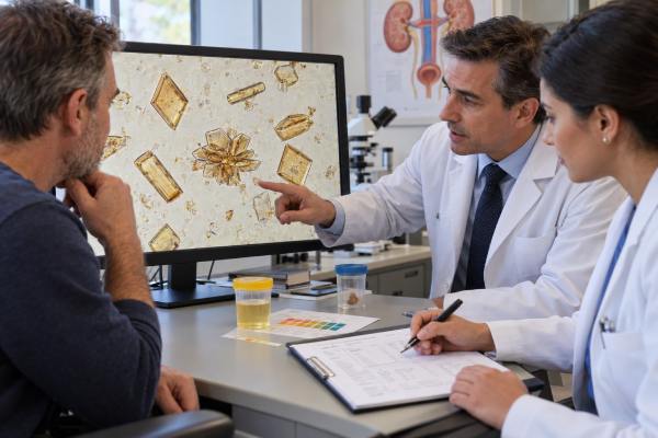
Uric acid crystals in urine are most often a sign that the urine is acidic enough for uric acid to come out of solution. A few crystals can appear temporarily in healthy people, especially after dehydration, fasting, a high-purine meal, or a period of concentrated urine. Persistent or abundant crystals matter more because they can reflect an environment that favors uric acid kidney stones. They are also more common in people with gout, metabolic syndrome, type 2 diabetes, obesity, chronic diarrhea, or other conditions that keep urine pH low.
The finding does not by itself diagnose gout, a kidney stone, or high blood uric acid. Urine microscopy is only one piece of the picture. The most useful next clues are urine pH, symptoms, kidney function, stone history, and—when stones are suspected—imaging and metabolic urine testing. Because uric acid is unusually soluble when urine becomes less acidic, identifying the problem can lead to practical prevention and, for confirmed uric acid stones, sometimes medical dissolution.
- Uric acid crystals usually form in acidic urine, commonly when urine pH is below about 5.5, but a single crystal finding can be temporary.
- Crystals are not the same as kidney stones. They show crystallization in the sample; stones require clinical assessment and often imaging for confirmation.
- Gout increases the chance of uric acid crystallization and stones, but urine crystals do not diagnose gout and can occur when the blood uric acid level is normal.
- Low urine volume and persistently low urine pH are major modifiable stone risks. Hydration and, when prescribed, urine alkalinization can reduce uric acid supersaturation.
- Seek prompt care for severe flank pain, fever, vomiting, inability to urinate, or worsening kidney function, because obstruction or infection can require urgent treatment.
Table of Contents
- What Uric Acid Crystals in Urine Mean
- Appearance, Urine pH, and Laboratory Reporting
- Common Causes and Risk Factors
- Gout, Uric Acid Kidney Stones, and the Connection
- How Uric Acid Crystal Results Are Evaluated
- Treatment, Prevention, and When to Get Care
What Uric Acid Crystals in Urine Mean
Uric acid crystals mean that dissolved uric acid has precipitated into solid microscopic particles in the urine. Uric acid is a normal end product of purine metabolism. Purines come from normal cell turnover and from foods such as organ meats, some red meats, certain seafoods, and other purine-rich foods. The kidneys filter uric acid from the blood and excrete part of it in urine.
Whether that uric acid stays dissolved depends heavily on urine pH. At a lower pH, a larger share is present as poorly soluble, non-ionized uric acid. When the urine is persistently acidic, the solution can become supersaturated and crystals can form. Concentrated urine makes this easier because the same amount of uric acid is dissolved in less water.
A microscopy report may therefore be abnormal without showing a specific disease. A person who was dehydrated for several hours can produce concentrated, acidic urine and have uric acid crystals on one sample. If the next well-hydrated sample is crystal-free and there are no symptoms or other abnormalities, the first result may have little clinical significance.
The finding becomes more important when it is repeated, abundant, accompanied by low urine pH, or associated with stone symptoms. Persistent crystalluria can indicate a urinary environment that favors uric acid stone formation. In a patient with acute kidney injury, very heavy crystalluria also raises concern for crystal-related tubular obstruction, although this is much less common than ordinary asymptomatic crystalluria.
Uric acid crystals are different from an elevated serum uric acid level. Blood and urine uric acid are related, but neither perfectly predicts the other. Some people form uric acid stones mainly because their urine stays too acidic even when total uric acid excretion is not unusually high.
Appearance, Urine pH, and Laboratory Reporting
Under the microscope, uric acid crystals are often yellow, amber, orange-brown, or reddish-brown. Their shape is variable. Classic forms include rhomboids, diamonds, wedges, barrels, and rosette-like clusters. Because the shapes can overlap with other crystals, trained microscopic identification and the urine pH help interpretation.
Most uric acid crystals form in acidic urine, often at a pH below about 5.5. A pH result is therefore one of the most useful companions to the microscopy report. However, pH changes over the day with meals, hydration, medications, and metabolism. A single pH of 5.0 does not prove a chronic problem, just as one pH of 6.5 does not rule one out.
Labs do not use one universal numerical “normal range” for crystals. Reports may say:
- none seen;
- rare or few;
- moderate;
- many or abundant; or
- a semiquantitative number per microscopic field.
That means a result should be interpreted using the laboratory’s own reporting system rather than comparing wording from another lab.
Sample handling also matters. Crystals can form after collection if urine sits for a long time or cools. Refrigeration is useful for preserving some urine components when testing is delayed, but cooling can promote precipitation of urates and phosphates. For accurate sediment interpretation, laboratories try to examine a fresh sample promptly and use standardized handling. If a crystal result seems inconsistent with the clinical picture, repeating a properly collected fresh specimen can be more informative than overinterpreting one sample.
A urinalysis usually adds other clues: specific gravity reflects urine concentration; blood may appear with a stone; protein can suggest kidney disease; and white blood cells, nitrite, or bacteria may point toward inflammation or infection. These findings do not prove the crystal caused the abnormality, but together they help define what the result means.
Common Causes and Risk Factors
The most common drivers of uric acid crystallization are low urine pH, low urine volume, and enough uric acid in the urine to reach supersaturation. Several situations can create that combination.
Dehydration and concentrated urine
Low fluid intake, heavy sweating, fever, vomiting, diarrhea, or prolonged exercise can reduce urine volume. Concentration raises the activity of uric acid and other stone-forming substances. Temporary crystals after a dehydrating day are therefore not unusual.
Metabolic syndrome and type 2 diabetes
Insulin resistance is strongly linked with persistently acidic urine. One important mechanism is reduced ammonium buffering in the kidney. The urine can remain too acidic even when the amount of uric acid excreted is not extreme. This helps explain why uric acid stones are more common in people with type 2 diabetes, obesity, hypertension, and metabolic syndrome.
Gout and high uric acid production
People with gout often have increased body urate burden and may excrete more uric acid in urine, especially under some dietary or treatment conditions. That can increase crystal and stone risk when urine pH is low. High cell turnover from certain blood disorders or cancer treatment can also sharply increase uric acid production and, in unusual circumstances, contribute to severe uric acid crystalluria and acute kidney injury.
Diet, fasting, and rapid weight change
A high intake of purine-rich animal foods can increase uric acid production. Very low-carbohydrate diets, prolonged fasting, or rapid weight loss can also lower urine pH and alter urinary chemistry. Diet is rarely the only cause, but it can amplify an existing tendency.
Chronic diarrhea or intestinal fluid loss
Chronic diarrhea can cause both volume depletion and bicarbonate loss, producing concentrated, acidic urine. People with inflammatory bowel disease, bowel surgery, ostomies, or chronic diarrheal disorders can therefore have a higher risk of uric acid stones.
Medications and medical conditions
Some drugs and diseases change urine pH, uric acid handling, or kidney function. The clinical meaning depends on the medication and the patient’s overall situation. A medication list, serum creatinine, electrolytes, and sometimes a 24-hour urine study are more useful than assuming the crystal finding has one cause.
| Pattern | Possible interpretation | Useful follow-up |
|---|---|---|
| Few crystals after dehydration | Temporary concentrated, acidic urine | Hydration and repeat urinalysis if clinically needed |
| Repeated crystals with urine pH below 5.5 | Persistent acidic urine and stone-forming environment | Stone history, diet review, metabolic testing |
| Crystals plus flank pain and blood in urine | Possible ureteral or kidney stone | Clinical evaluation and imaging when indicated |
| Heavy crystalluria with rising creatinine | Possible crystal-related kidney injury or severe metabolic disturbance | Prompt medical evaluation |
Gout, Uric Acid Kidney Stones, and the Connection
Gout and uric acid stones share urate biology, but they are not the same condition. Gout is an inflammatory arthritis caused by monosodium urate crystal deposition in joints and other tissues. Urine microscopy does not diagnose it. A person can have gout without urinary uric acid crystals, and a person can have urinary crystals without gout.
Uric acid kidney stones develop when urine remains supersaturated with uric acid long enough for crystals to nucleate, grow, and aggregate. Persistently low urine pH is often the dominant factor. Low urine volume and high uric acid excretion add to the risk.
Typical stone symptoms include sudden pain in the side or back that may radiate toward the lower abdomen or groin, nausea, vomiting, urinary urgency, and blood in the urine. Some kidney stones cause no symptoms until they move or obstruct urine flow.
Uric acid stones have an important treatment distinction: they can sometimes dissolve when the urine is alkalinized. Calcium-containing stones generally do not dissolve this way. Studies and stone guidelines support raising urine pH with prescribed alkali, most commonly potassium citrate when appropriate, while monitoring pH so it does not become excessively alkaline. In one contemporary series of patients treated for uric acid stones, potassium citrate–based therapy achieved complete chemolysis in most patients selected for dissolution treatment.
This is one reason stone composition matters. A stone that is passed or removed should be analyzed when possible. Noncontrast CT can detect uric acid stones even though they may be difficult or impossible to see on a standard plain abdominal X-ray. CT density, urine pH, and clinical history can suggest composition before a stone is retrieved, but stone analysis provides stronger confirmation.
People who form uric acid stones are generally considered at meaningful recurrence risk. A complete evaluation may include blood tests and one or two 24-hour urine collections measuring urine volume, pH, uric acid, citrate, sodium, calcium, oxalate, creatinine, and other components. The goal is not simply to find “high uric acid,” but to identify the combination of abnormalities that can actually be changed.
How Uric Acid Crystal Results Are Evaluated
A clinician usually starts with the context rather than the crystal count alone. Four questions are especially useful: Are there symptoms? Is the urine persistently acidic? Is kidney function normal? Is there a history of stones or gout?
For an incidental finding in a person who feels well, follow-up may be simple. Hydration status can be corrected and the urinalysis repeated if the result is unexpected or persistent. The repeat sample should be collected cleanly and processed promptly.
When symptoms or recurrent crystals are present, evaluation may include:
- urine pH and specific gravity;
- dipstick and microscopy for blood, protein, white blood cells, bacteria, and other crystals;
- serum creatinine and estimated glomerular filtration rate;
- serum uric acid, recognizing that it does not directly measure urine supersaturation;
- electrolytes and bicarbonate when an acid-base disorder is possible;
- urine culture when infection is suspected;
- noncontrast CT or ultrasound when stones or obstruction are suspected; and
- 24-hour urine testing for recurrent or high-risk stone formers.
A 24-hour urine uric acid result is different from a spot microscopy result. The 24-hour test quantifies how much uric acid is excreted over a day; microscopy shows whether crystals are physically present in that particular sample. A person can have high daily uric acid excretion without crystals if the urine is sufficiently dilute and less acidic. Conversely, concentrated, very acidic urine can crystallize uric acid even without extreme daily excretion.
Crystal identification can occasionally be uncertain. Other yellow-brown crystals and medication-related crystals may mimic uric acid. Polarized-light microscopy, shape, pH, clinical history, and sometimes specialized laboratory methods help resolve difficult cases.
Treatment, Prevention, and When to Get Care
There is no need to “treat the microscope” in every person with uric acid crystals. Management depends on why the crystals formed and whether they are causing or predicting a clinical problem.
For an isolated result related to concentrated urine, the practical first step is usually adequate fluid intake, unless a clinician has prescribed fluid restriction for heart failure, advanced kidney disease, or another condition. For people with recurrent kidney stones, guidelines commonly aim for enough fluid to produce more than about 2.5 L of urine per day, although the exact intake needed varies with climate, body size, activity, and medical conditions.
Dietary measures can include moderating excessive intake of purine-rich meats and seafood, avoiding dehydration, limiting very high sodium intake, and maintaining a sustainable weight rather than using crash diets. People with metabolic syndrome or diabetes may also benefit from improving glucose control and overall cardiometabolic health because these conditions are linked with low urine pH.
For confirmed uric acid stones or persistent high-risk urine chemistry, clinicians may prescribe alkali therapy. Potassium citrate is commonly used when safe because it raises urine pH and citrate. Treatment is individualized, particularly in people with reduced kidney function or high potassium levels. Home urine pH monitoring may be part of therapy. The target is deliberately controlled: too little alkalinization fails to dissolve uric acid, while excessive alkalinization can encourage other crystal types such as calcium phosphate.
Allopurinol or another urate-lowering drug may be useful when gout, marked hyperuricosuria, or another clear indication is present, but it is not a substitute for correcting persistently acidic urine in uric acid stone disease. Medication choice should be based on the full metabolic evaluation rather than on crystals alone.
Prompt medical assessment is important for severe or persistent flank pain, visible blood in urine, repeated vomiting, fever or chills, very low urine output, a known single kidney, pregnancy with stone symptoms, or worsening kidney function. Fever plus suspected obstruction is especially urgent because an infected, blocked urinary system may require drainage in addition to antimicrobial treatment.
The key point is that uric acid crystals are a clue, not a diagnosis. Their value comes from linking the microscopy finding with urine pH, hydration, metabolic risk, symptoms, and stone history. When that pattern shows persistent acidic urine, the problem is often modifiable.
How urine pH monitoring fits into stone prevention
For people with confirmed uric acid stones, urine pH is often more actionable than a single crystal count. A spot urinalysis gives one pH at one moment, while home monitoring can show whether urine stays acidic across the day and whether prescribed alkalinization is reaching its target. The exact target should come from the treating clinician because the goal may differ between dissolving an existing uric acid stone and preventing recurrence.
Timing matters. Urine pH naturally changes after meals and with sleep, hydration, and medication. A single morning value may be lower than values later in the day. When clinicians ask for home monitoring, they may recommend measurements at several consistent times rather than reacting to one isolated reading. A meter or high-quality pH paper can be used, but technique and storage affect accuracy.
More alkalinization is not always better. Raising pH enough to keep uric acid soluble can be useful, but persistently excessive alkalinity can increase calcium phosphate supersaturation in susceptible people. That is why stone prevention combines pH with urine volume, sodium, citrate, calcium, oxalate, and uric acid rather than chasing one number.
A person who only had incidental crystals and has never had a uric acid stone usually does not need to start alkali medication on their own. Repeated low pH, recurrent stones, kidney disease, or a complex metabolic history deserves clinician-guided evaluation before potassium-containing therapy is used.
References
- Medical treatment of uric acid kidney stones 2024 (Review)
- Metabolic Evaluation and Recurrence Prevention for Urinary Stone Patients: An EAU Guidelines Update 2024 (Guideline)
- Type 4 renal tubular acidosis and uric acid nephrolithiasis: two faces of the same coin? 2023 (Review)
- Pharmacologic treatment of kidney stones: Current medication and pH monitoring 2024 (Review)
- The EFLM European Urinalysis Guideline 2023 2024 (Guideline)
Disclaimer
Uric acid crystals should be interpreted with symptoms, urine pH, kidney function, medications, and stone history. This article provides general health information and is not a diagnosis or treatment plan; seek medical care for severe pain, fever, inability to urinate, persistent vomiting, or other concerning symptoms.