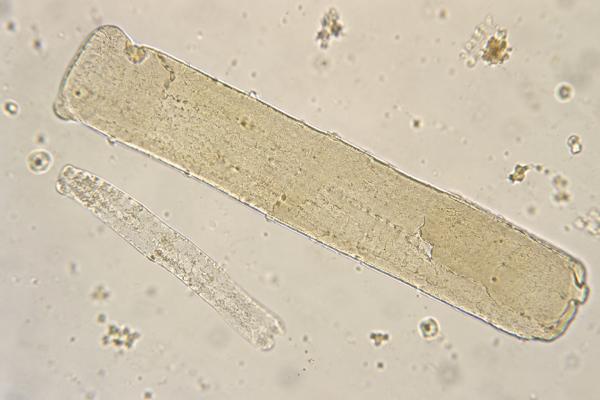
Broad casts in urine are unusually wide cylindrical structures formed inside enlarged or damaged kidney tubules and collecting ducts. They are an abnormal microscopy finding and are classically associated with advanced chronic kidney disease, severe tubular atrophy, and slow urine flow through dilated nephron segments. Broad casts are sometimes called “renal failure casts,” but that phrase can be misleading because one cast does not diagnose kidney failure or determine a CKD stage. The material inside a broad cast also matters. It may be waxy, granular, cellular, or mixed, and those features help indicate the type and chronicity of kidney injury. Broad waxy casts are especially associated with impaired renal function and prolonged tubular stasis. Interpretation should always include serum creatinine, estimated glomerular filtration rate, urine protein, blood pressure, symptoms, and the rest of the sediment. Because automated urinalysis may miss or misclassify pathological casts, a manual sediment review can be valuable when severe kidney disease is suspected.
- Broad casts are abnormal: their width suggests formation in dilated collecting ducts or severely altered tubules rather than a normal nephron.
- Broad waxy casts often point to advanced chronic kidney injury: they are especially associated with low eGFR and chronic tubular damage.
- Low urine flow favors cast formation: prolonged stasis allows uromodulin and trapped material to mold within the tubular lumen.
- One broad cast does not equal kidney failure: the result must be matched with creatinine, eGFR, proteinuria, and clinical findings.
- Manual microscopy may be needed: automated systems can have low sensitivity for pathological casts.
Table of Contents
- What Broad Casts Are
- How Broad Casts Form
- Broad, Waxy, and Granular Casts: Key Differences
- Conditions Associated With Broad Casts
- How to Interpret Broad Casts With Kidney Tests
- Repeat Testing and Follow-Up
- When Broad Casts Need Prompt Medical Attention
What Broad Casts Are
Urinary casts are microscopic molds of the kidney tubules. Their basic matrix is formed largely from uromodulin, a protein produced by cells in the thick ascending limb and distal nephron. When urine conditions favor precipitation, this protein can gel inside the tubular lumen and trap cells, cell fragments, lipids, pigments, or granular debris. The resulting cylinder is released into the urine and can be seen on sediment microscopy.
A broad cast is wider than an ordinary cast because it forms in a larger tubular space. Classically, this happens in dilated distal tubules or collecting ducts in kidneys with substantial chronic structural damage. As nephrons are lost, surviving tubules may become enlarged, and urine movement can slow. The cast takes on the shape of that wider lumen.
The term “broad cast” describes size, not composition. A broad cast may be waxy, coarsely or finely granular, or contain cells. That is why a report of “broad waxy casts” carries more information than “broad casts” alone. Waxy change suggests degeneration and prolonged residence time, whereas cellular material may point to a more active inflammatory or tubular process.
Older laboratory texts sometimes call broad waxy casts “renal failure casts.” The phrase reflects their historical association with markedly reduced renal function, but it can sound more definitive than the evidence supports. Modern interpretation is more careful: broad casts increase suspicion for severe chronic parenchymal disease, while kidney failure is diagnosed from filtration, symptoms, complications, and duration. A person with advanced CKD may have no broad casts in a particular specimen, and a reported broad cast still deserves confirmation if the rest of the evaluation is normal.
Cast quantity is also affected by how urine is concentrated and prepared. A centrifuged sediment from a small urine volume may appear to contain more casts than a differently standardized preparation. This is one reason laboratories often use semiquantitative reporting rather than a universal numeric threshold. The morphology and clinical setting usually matter more than a single count.
Broad casts are not expected in a healthy urine sediment. Small numbers of hyaline casts can occur after exercise, fever, or dehydration, but broad pathological casts generally indicate a more significant renal process. A full urine microscopy evaluation for kidney disease helps determine whether the broad cast is isolated or part of a larger pattern involving protein, blood cells, tubular cells, or other cast types.
How Broad Casts Form
Cast formation depends on both the kidney’s structure and the local urine environment. Uromodulin is more likely to form a gel when urine flow is slow and the tubular fluid is concentrated. Acidic conditions and high ionic strength can also favor protein precipitation. Once the matrix forms, it may trap whatever material is present in that nephron segment.
In chronic kidney disease, several changes can create ideal conditions for broad casts. Tubular atrophy and interstitial fibrosis distort the nephron. Some tubules become dilated, while fewer functioning nephrons carry the filtration workload. Reduced urine flow in damaged segments increases the time available for a cast to mature. A cast that remains in the tubule longer may lose clear cellular detail and become smoother, denser, and more waxy.
This is the basis for the traditional association between broad waxy casts and advanced renal failure. The cast reflects both a widened lumen and prolonged stasis. It is best understood as a morphological sign of severe nephron remodeling, not as a direct measurement of filtration.
Low urine flow can occur for several reasons besides advanced CKD, including volume depletion or severe acute kidney injury. Those conditions can promote cast formation, but a truly broad waxy cast still raises concern for significant underlying renal structural disease. Dehydration alone is much more commonly associated with hyaline casts than with classic broad waxy casts.
Specimen handling also matters. Casts can dissolve in very dilute or alkaline urine and may be missed if analysis is delayed. Conversely, artifacts such as mucus strands or fibers can be mistaken for casts. Experienced microscopy and prompt examination improve reliability.
A fresh first-morning sample can sometimes improve detection because it is more concentrated, but concentration alone does not create the structural changes responsible for a truly broad cast. If the urine is extremely dilute, casts may fragment or disappear before examination. If a specimen is very alkaline, the uromodulin matrix may dissolve more quickly. These preanalytic effects help explain why one sample can show casts and the next may not, even when the underlying kidney disease has not changed.
Broad, Waxy, and Granular Casts: Key Differences
Cast terminology can be confusing because width and composition are separate features. A cast can be broad and waxy at the same time, or broad and granular. Recognizing this prevents the common mistake of treating “broad,” “waxy,” and “granular” as three mutually exclusive categories.
| Cast description | Typical appearance | Common interpretation |
|---|---|---|
| Broad cast | Unusually wide cylinder | Suggests formation in dilated tubules or collecting ducts |
| Waxy cast | Dense, smooth, highly refractile, often with blunt or cracked edges | Associated with prolonged tubular stasis and impaired kidney function |
| Granular cast | Fine or coarse granules within the cast matrix | Often reflects cellular breakdown or tubular injury |
| Hyaline cast | Clear, nearly transparent matrix | Can be normal in small numbers or increase with low flow |
Waxy casts deserve particular attention because studies have linked them with reduced kidney function and higher proteinuria. A waxy cast finding can be quite specific for renal insufficiency, although it is not sensitive enough to rule out CKD when absent. Broad waxy casts strengthen the impression of advanced chronic tubular remodeling.
Granular casts are more strongly associated with tubular injury, especially acute tubular injury, although they can also appear in chronic disease. A granular cast pattern becomes more clinically significant when abundant “muddy brown” casts and renal tubular epithelial cells occur during acute kidney injury.
Hyaline casts are different because they contain mostly uromodulin without substantial cellular debris. They may increase with dehydration, strenuous exercise, diuretic use, or other low-flow states. The presence of ordinary hyaline casts should not be interpreted as equivalent to broad waxy casts.
Conditions Associated With Broad Casts
Advanced chronic kidney disease is the classic setting for broad casts. The underlying cause of CKD can vary widely. Long-standing diabetic kidney disease, hypertensive nephrosclerosis, chronic glomerular disease, hereditary kidney disorders, chronic tubulointerstitial disease, and obstructive nephropathy can all eventually produce nephron loss and tubular remodeling.
Broad casts do not reveal which of these diseases caused the damage. Instead, they provide structural information suggesting that some tubules are markedly dilated and urine transit is slow. The rest of the clinical record identifies the cause.
Severe acute kidney injury can also produce large casts, particularly when tubular injury is extensive and urine output is low. In that setting, granular or epithelial features may be more prominent than a purely waxy appearance. The distinction between acute, chronic, and acute-on-chronic disease depends on prior creatinine values, kidney size, anemia, mineral-bone abnormalities, proteinuria history, and imaging rather than cast morphology alone.
Obstruction can reduce urine flow and damage tubules. Bilateral obstruction or obstruction in a single functioning kidney can cause acute kidney injury, while long-standing obstruction can lead to chronic atrophy and dilation. Broad casts are not specific for obstruction, so imaging is needed when urinary blockage is suspected.
Heavy proteinuria can accompany many diseases that eventually produce broad casts. Protein in urine may also contribute to a more active sediment depending on the underlying glomerular disorder. A proteinuria evaluation helps quantify the degree of protein loss and determine whether glomerular disease is part of the picture.
Kidney size on ultrasound can sometimes add chronicity information. Small echogenic kidneys may support long-standing irreversible parenchymal disease, while normal or enlarged kidneys can occur in acute injury and in several chronic disorders such as diabetic kidney disease or infiltrative conditions. Imaging cannot identify cast composition, but it can reveal obstruction, cystic disease, asymmetric kidneys, or structural changes that help explain why broad casts are appearing. Trends over weeks or months then help distinguish a stable chronic abnormality from a new decline that needs urgent investigation.
How to Interpret Broad Casts With Kidney Tests
A broad cast result should trigger a review of kidney function rather than being read in isolation. The most important accompanying measures are serum creatinine, eGFR, urine protein or albumin, urine output, blood pressure, and the rest of the sediment.
Estimated glomerular filtration rate classifies filtration into G categories. CKD is defined by persistent kidney structural or functional abnormalities for at least three months, not by one cast report. A low eGFR result becomes more concerning when broad waxy casts, albuminuria, or other markers of kidney damage are also present.
A previous baseline is especially valuable. A creatinine that has been stable for years at an elevated level suggests chronic disease, while a sharp rise over days suggests acute kidney injury. Both can coexist. Someone with stage 4 CKD can develop dehydration, infection, medication-related injury, or obstruction and have an acute rise in creatinine on top of chronic damage.
Urine protein provides additional localization. Marked albuminuria suggests glomerular barrier injury, while lower-molecular-weight proteins or tubular cells may indicate tubulointerstitial disease. Red blood cell casts or acanthocytes point toward active glomerular bleeding and can signal a potentially treatable inflammatory process even in a person with chronic disease.
The numerical count of broad casts is less standardized than creatinine or albumin-to-creatinine ratio. Laboratories may report rare, few, moderate, or many casts per low-power field. The trend may be useful within the same laboratory, but clinicians generally do not stage CKD or measure treatment response by broad-cast count.
The same sediment can also contain evidence of acute injury superimposed on chronic disease. Broad waxy casts suggest chronic remodeling, while renal tubular epithelial cells and numerous granular casts can point to active tubular injury. Dysmorphic red cells or RBC casts can suggest active glomerular inflammation. Seeing both chronic and active features is clinically important because some acute contributors may be reversible even when baseline CKD is advanced.
The trend in kidney function adds important context. A broad cast in a person with stable mild chronic kidney disease is interpreted differently from the same finding during a rapid rise in creatinine, oliguria, or a new electrolyte disturbance. Urine sediment can show chronic tubular stasis, but blood tests and urine output show whether there is an active decline that needs urgent evaluation. Cast burden also varies from sample to sample, so severity should never be graded from one microscopic field alone.
Repeat Testing and Follow-Up
When broad casts are found unexpectedly, a repeat fresh urine sediment can confirm that the structure was a true cast rather than mucus, fiber, or another artifact. Confirmation is particularly useful when serum creatinine and urine protein are normal or when the result is inconsistent with the person’s history.
If kidney dysfunction is already known, follow-up focuses on severity, cause, and reversible contributors. Common tests include serum creatinine and eGFR, potassium, bicarbonate, calcium, phosphorus, complete blood count, urine albumin-to-creatinine ratio, and renal ultrasound when structural disease or obstruction is possible. The exact panel depends on CKD stage and suspected cause.
Medication review is important because nonsteroidal anti-inflammatory drugs, certain antibiotics, contrast exposure, diuretics, renin-angiotensin system blockers during severe volume depletion, and other agents can worsen kidney function in susceptible patients. Medication changes should be clinician-guided rather than made solely because a cast was reported.
Blood pressure, diabetes control, albuminuria, cardiovascular risk, and CKD complications often matter more for long-term management than whether broad casts remain visible. Modern CKD care uses eGFR and albuminuria to assess risk and guide therapy. A broad cast can alert clinicians to advanced structural damage, but it does not replace these standardized measures.
Manual microscopy may be worth requesting when an automated urinalysis reports “casts” without clear classification and the clinical picture suggests important kidney pathology. Studies comparing automated systems with expert manual review have found that pathological casts can be underdetected. This is one setting where an experienced sediment examination adds real diagnostic value.
When Broad Casts Need Prompt Medical Attention
Broad casts are not an emergency by themselves, but the kidney dysfunction associated with them can be. Prompt evaluation is needed when a person has markedly reduced urine output, rapidly worsening swelling, shortness of breath, confusion, persistent vomiting, severe weakness, or a known rapid rise in creatinine. These signs can accompany severe acute kidney injury or advanced renal failure.
Urgent care is also appropriate for high potassium, severe metabolic acidosis, pulmonary edema, or uremic symptoms when these are identified clinically or on blood testing. The need for dialysis is based on the overall clinical condition and biochemical complications, not on the presence or number of casts.
For a stable outpatient, broad casts should prompt organized kidney follow-up rather than panic. Confirm the result if necessary, compare current kidney function with prior values, quantify albuminuria or proteinuria, review medications and blood pressure, and determine whether the underlying disease is chronic, acute, or both. The cast is most useful as a clue to the architecture and flow conditions inside damaged nephrons.
References
- Diagnostic Utility of Urine Microscopy in Kidney Diseases 2024 (Review)
- An updated approach to the evaluation of the urinary sediment 2025 (Review)
- The clinical and pathological relevance of waxy casts in urine sediment 2022
- Executive summary of the KDIGO 2024 Clinical Practice Guideline for the Evaluation and Management of Chronic Kidney Disease: known knowns and known unknowns 2024 (Guideline)
- The Clinical Impact of Urinalysis Screened by Automated Microscopy Compared to Reference Manual Analysis 2024
Disclaimer
Broad casts are an abnormal urine-microscopy finding but do not diagnose kidney failure or determine CKD stage by themselves. Anyone with broad casts plus reduced urine output, rising creatinine, swelling, breathing difficulty, or other signs of worsening kidney function should receive timely medical evaluation.