
Urine microscopy can reveal kidney injury that a dipstick or blood test cannot fully explain. After a urine sample is concentrated and examined under a microscope, the sediment may show red blood cells, white blood cells, kidney tubular cells, casts, crystals, microorganisms, and other particles. The pattern matters more than any single finding. Red blood cell casts, for example, point toward bleeding and inflammation inside the kidney, while renal tubular epithelial cells and granular casts can support tubular injury. Waxy casts may accompany advanced kidney dysfunction, and certain crystals can suggest a stone-forming or drug-related process.
Microscopy does not replace serum creatinine, estimated glomerular filtration rate (eGFR), urine albumin testing, imaging, or kidney biopsy. Instead, it helps localize the problem and narrow the differential diagnosis. Results are most useful when the sample is fresh, collection is clean, and the findings are interpreted alongside symptoms, urine protein, kidney function, medications, and the clinical setting.
- Urine microscopy looks directly at formed particles in urine, including cells, casts, crystals, bacteria, and yeast.
- Casts are especially useful for kidney disease because they form inside renal tubules, so cellular casts usually indicate an intrarenal process rather than contamination from outside the kidney.
- Red blood cell casts suggest glomerular bleeding; white blood cell casts suggest renal inflammation; granular or tubular epithelial cell casts can support tubular injury.
- A normal or bland sediment does not rule out kidney disease. Many forms of chronic kidney disease have few distinctive microscopic findings.
- Fresh, properly collected urine improves accuracy. Delayed processing, very dilute urine, extreme pH, or contamination can alter cells, casts, and crystals.
Table of Contents
- What Urine Microscopy Shows in Kidney Disease
- Cells and What They Mean
- Casts and Kidney Localization
- Crystals and Other Sediment Findings
- Patterns in Common Kidney Problems
- Collection and Laboratory Accuracy
- How Results Guide Next Steps
What Urine Microscopy Shows in Kidney Disease
Urine microscopy examines the solid material suspended in urine. In a traditional sediment examination, the laboratory centrifuges a measured urine sample, removes much of the liquid, and inspects the concentrated sediment under low- and high-power magnification. Some laboratories use automated particle analyzers, digital imaging, or flow-based methods and confirm selected findings manually.
The test is valuable because kidney diseases often leave a microscopic “footprint.” Blood chemistry can show that filtration has changed, and albumin testing can show that the kidney is leaking protein, but microscopy can add clues about where the injury is occurring.
The main categories are:
- Red blood cells (RBCs): may come from anywhere in the urinary tract, but dysmorphic RBCs and RBC casts raise concern for a glomerular source.
- White blood cells (WBCs): reflect inflammation and may appear with infection, interstitial nephritis, glomerular disease, stones, or contamination.
- Renal tubular epithelial cells: increased numbers can indicate tubular epithelial injury.
- Casts: cylindrical structures molded within renal tubules; their contents can help localize and characterize disease.
- Crystals: may be benign, stone-related, metabolic, or drug-associated depending on type and context.
- Microorganisms: bacteria or yeast may represent infection, colonization, or contamination.
No isolated particle is perfectly diagnostic. A few RBCs can result from exercise, menstruation, stones, or sampling trauma. A few hyaline casts may be seen after exercise or dehydration. Even findings considered “active,” such as cellular casts, need clinical confirmation because identification can be difficult and sensitivity is limited.
A useful approach is to combine three layers of information: kidney function, urine chemistry, and urine sediment. For example, rising creatinine plus protein and blood on dipstick plus dysmorphic RBCs and RBC casts creates a much stronger case for glomerular injury than any one result alone.
Cells and What They Mean
Red blood cells
RBCs in urine are called hematuria. Microscopy can confirm true red cells when the dipstick is positive for blood and can sometimes help distinguish glomerular from nonglomerular bleeding. RBCs that have irregular shapes, blebs, or size variation are called dysmorphic. Acanthocytes—ring-shaped RBCs with membrane blebs—are particularly associated with glomerular bleeding when present in sufficient proportion and identified by experienced microscopy.
By contrast, uniformly shaped RBCs are more often described with bleeding from the ureter, bladder, prostate, or urethra, although morphology alone cannot locate every case. Kidney stones, tumors, infection, exercise, and sample contamination can all cause hematuria without a glomerular disease.
White blood cells
WBCs, especially neutrophils, increase with urinary inflammation. In bacterial urinary tract infection, WBCs may be accompanied by bacteria, leukocyte esterase, and symptoms. In kidney disease, pyuria can also occur with acute interstitial nephritis, glomerulonephritis, stones, tuberculosis, or other inflammatory conditions.
A WBC count by itself cannot distinguish cystitis from kidney inflammation. WBC casts are more localizing because casts form in the nephron; they suggest that leukocytes are coming from within the kidney rather than only the bladder or urethra.
Renal tubular epithelial cells
Renal tubular epithelial cells are normally shed in very small numbers. An increased number, especially with epithelial cell casts or coarse granular casts, supports tubular injury. This pattern can occur in acute tubular injury caused by ischemia, sepsis, toxins, severe illness, or nephrotoxic medications.
These cells can be challenging to distinguish from transitional epithelial cells, so laboratory expertise matters. Automated systems may flag them but may not classify them as reliably as an experienced observer in complex samples.
Squamous and transitional epithelial cells
Squamous epithelial cells usually come from the distal urethra or external genital tract. Many squamous cells often suggest contamination of a clean-catch sample. Transitional epithelial cells line the renal pelvis, ureters, bladder, and part of the urethra. Small numbers may be normal; increased numbers can occur after instrumentation or inflammation. Their presence is usually less specific for intrinsic kidney disease than renal tubular epithelial cells.
Casts and Kidney Localization
Casts are among the most informative urine microscopy findings because they form inside distal tubules and collecting ducts. Their basic matrix is largely uromodulin, a protein produced by tubular cells. Slow urine flow, concentration, and acidic conditions can favor cast formation. Cells, pigments, lipids, and debris can become trapped in the matrix.
| Cast type | Typical association | Key caution |
|---|---|---|
| Hyaline | May occur in healthy people, dehydration, exercise, fever, or kidney disease | Low specificity |
| Red blood cell | Glomerulonephritis or other glomerular bleeding | May be missed if sample is delayed or dilute |
| White blood cell | Pyelonephritis, acute interstitial nephritis, some inflammatory glomerular diseases | Not sensitive enough to exclude disease when absent |
| Renal tubular epithelial cell | Acute tubular injury | Requires accurate cell identification |
| Granular | Often tubular injury; coarse “muddy brown” casts are classic in acute tubular injury | Fine granular casts can be less specific |
| Fatty | Heavy proteinuria and nephrotic syndrome | Lipid droplets may show Maltese-cross appearance under polarized light |
| Waxy | Reduced nephron flow and advanced renal dysfunction | Specific for significant dysfunction in some studies but not sensitive |
| Broad | Advanced chronic kidney disease or severe nephron dilation/stasis | Interpret with eGFR and overall sediment |
RBC casts are an especially important finding. Because individual RBCs can enter urine anywhere along the urinary tract, free RBCs are not strongly localizing. RBCs enclosed in a tubular cast, however, indicate that bleeding occurred within the nephron. That raises concern for glomerulonephritis and usually deserves prompt evaluation if the finding fits the clinical picture.
“Muddy brown” granular casts and renal tubular epithelial cell casts are strongly associated with acute tubular injury. In a hospitalized patient with rising creatinine after shock, sepsis, surgery, or nephrotoxin exposure, this sediment pattern can support acute tubular injury over a purely prerenal process.
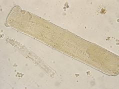
Waxy casts have a dense, smooth, often highly refractile appearance and can have blunt or cracked edges. They are associated with prolonged tubular stasis and impaired kidney function. Their presence can support advanced disease, but they do not by themselves establish a CKD stage or prove irreversible kidney failure.
Crystals and Other Sediment Findings
Crystals can be completely benign or clinically important. Their meaning depends on crystal type, urine pH, concentration, medications, symptoms, and kidney function.
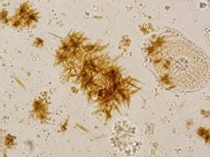
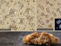
Common crystals include calcium oxalate, uric acid, and phosphate crystals. They may appear in healthy people, especially when urine is concentrated or has cooled after collection. Calcium oxalate crystals are often envelope- or dumbbell-shaped; uric acid crystals are usually yellow-brown and can be rhomboid, rosette, or barrel-shaped; phosphate crystals favor alkaline urine.
Some crystals are more concerning. Cystine crystals are classically hexagonal and can suggest cystinuria. Certain medication crystals can appear after exposure to drugs that precipitate in urine, particularly when the urine is concentrated or the dose is high. Crystal-associated acute kidney injury is uncommon but important because intratubular precipitation can obstruct flow and injure tubular cells.
Other useful sediment findings include:
- lipid droplets and oval fat bodies, often seen with nephrotic-range proteinuria;
- bacteria, which may support infection when collection quality, symptoms, and culture agree;
- yeast, which may be contamination, colonization, or candiduria;
- mucus and sperm, which are usually not evidence of kidney disease; and
- debris or artifacts, which can mimic cells or casts and require experienced review.
Crystals seen only after refrigeration or prolonged standing should be interpreted cautiously. A fresh repeat sample can help distinguish in-vivo crystalluria from precipitation that happened in the collection cup.
Patterns in Common Kidney Problems
The strongest use of urine microscopy is pattern recognition. Kidney diseases often produce combinations rather than one unique marker.
Glomerular disease
An “active” glomerular sediment may include dysmorphic RBCs, RBC casts, variable WBCs, and proteinuria. Examples include IgA nephropathy, lupus nephritis, postinfectious glomerulonephritis, and vasculitic glomerulonephritis. The urgency depends on kidney function, degree of proteinuria, blood pressure, systemic symptoms, and how rapidly abnormalities developed.
Microscopy can strengthen suspicion but does not identify the exact immune disease. Serologic tests and, in selected cases, kidney biopsy are needed for diagnosis and treatment planning.
Acute tubular injury
Acute tubular injury often produces renal tubular epithelial cells, epithelial cell casts, and granular casts. “Muddy brown” granular casts are a classic finding. The pattern can help explain an abrupt rise in creatinine after hypotension, severe infection, major surgery, pigment injury, or nephrotoxin exposure.
Acute interstitial nephritis
AIN can produce sterile pyuria, WBCs, WBC casts, mild-to-moderate proteinuria, and sometimes hematuria. Drugs are common triggers, but infections and systemic diseases are also possible. The classic combination of fever, rash, and eosinophilia is not present in many patients, so the medication timeline and kidney function trend often matter more.
Pyelonephritis
Kidney infection may show pyuria, bacteria, and sometimes WBC casts. Fever, flank pain, urinary symptoms, and urine culture are central to diagnosis. Absence of a WBC cast does not exclude pyelonephritis.
Chronic kidney disease
CKD may have a bland sediment, especially in hypertensive or vascular disease. Other forms of CKD remain active microscopically. Persistent proteinuria, hematuria, cellular casts, broad casts, or waxy casts can provide clues about the underlying disease and severity. CKD diagnosis and staging, however, depend on abnormalities lasting at least three months and use eGFR and markers of kidney damage—not sediment alone.
Collection and Laboratory Accuracy
Good microscopy begins before the slide reaches the microscope. A clean-catch, midstream specimen reduces contamination from skin and genital surfaces. The sample should be labeled accurately and delivered promptly.
Cells and casts can deteriorate in very dilute or alkaline urine and during long delays. Contemporary urinalysis guidance emphasizes timely particle examination, with refrigeration when a delay cannot be avoided. Refrigerated urine should be handled according to laboratory protocol because cooling can create or increase crystal precipitation.
First-morning urine is concentrated and can be useful for some measurements, but prolonged bladder storage can also damage fragile cells and casts. For sediment analysis in some clinical settings, a fresh second-void specimen can provide better-preserved formed elements.
Manual and automated methods each have strengths. Automation improves speed, standardization, and throughput. Skilled manual microscopy remains valuable when clinicians are looking for pathological casts, dysmorphic RBCs, unusual crystals, or complex sediment that automated classifiers may not characterize correctly.
A report that says “no casts seen” also reflects sampling. Casts may be sparse and unevenly distributed. One aliquot may not contain a structure that is present elsewhere in the urine. This is why microscopy is best used as a probability-changing test rather than an absolute rule-out test.
How Results Guide Next Steps
The next step depends on the pattern and clinical setting. For mild, isolated findings in a person with normal kidney function, repeating a properly collected urinalysis may be enough. Persistent or high-risk findings need broader evaluation.
Common follow-up includes serum creatinine and eGFR, urine albumin-to-creatinine or protein-to-creatinine ratio, blood pressure, electrolytes, medication review, and repeat urine microscopy. A urine culture is appropriate when infection is suspected. Ultrasound or other imaging may be used for stones, obstruction, structural disease, or unexplained kidney dysfunction.
Prompt nephrology evaluation is particularly important when microscopy shows RBC casts, marked dysmorphic hematuria with proteinuria, rapidly rising creatinine, nephrotic-range protein loss, or persistent cellular casts. These findings can accompany diseases where early diagnosis changes treatment.
Symptoms can also make the result urgent. Seek timely care for reduced urine output, new swelling with shortness of breath, severe hypertension symptoms, fever with flank pain, visible blood with clots, severe stone-like pain, or rapidly worsening illness.
A useful microscopy report is not a final diagnosis. Its value is in converting a broad problem—“kidney function is abnormal”—into a more focused question: Is the pattern glomerular, tubular, interstitial, infectious, obstructive, or metabolic? When paired with blood and urine chemistry, that localization can shorten the path to the right next test.
Why microscopy can disagree with other kidney tests
It is common for urine microscopy, eGFR, and albumin measurements to tell different parts of the story. A person can have a low eGFR with a nearly bland sediment if the damage is chronic and no longer shedding many inflammatory cells. Another patient can have striking RBC casts early in glomerulonephritis while the serum creatinine is still near baseline. Neither result cancels the other.
Albuminuria is similar. Heavy albumin loss points toward glomerular barrier damage, but the sediment can range from bland to highly active depending on the disease. Conversely, acute tubular injury may cause a large creatinine rise with only modest albuminuria because the main lesion is tubular rather than glomerular.
Timing explains some disagreement. Serum creatinine changes after filtration has fallen and can lag behind the initial injury. Urinary particles may appear earlier, disappear as the process evolves, or be missed because casts are shed intermittently. Hydration also changes how concentrated the sediment appears.
These differences are clinically useful rather than frustrating. When creatinine, protein, and microscopy are considered together, they help separate filtration loss from active inflammation, glomerular leakage, or tubular injury. A discordant result sometimes prompts repeat microscopy, quantitative protein testing, imaging, serologic studies, or biopsy instead of assuming one test is “wrong.”
Questions worth asking about an abnormal sediment report
If a report contains an unfamiliar cast or cell type, useful questions include whether the result was confirmed manually, whether the sample was fresh, and whether the abnormality has appeared before. Ask how the finding fits the creatinine trend and urine protein level. For persistent hematuria, ask whether RBC morphology was assessed and whether a glomerular or urologic source is more likely. For pyuria, ask whether culture or medication review is needed. These questions turn a descriptive laboratory report into a more focused diagnostic plan.
References
- Diagnostic Utility of Urine Microscopy in Kidney Diseases 2024 (Review)
- An updated approach to the evaluation of the urinary sediment 2025 (Review)
- The EFLM European Urinalysis Guideline 2023 2024 (Guideline)
- Urinary Vacuolar Casts Are a Unique Type of Casts in Advanced Proteinuric Glomerulopathies 2024
- KDIGO 2024 CKD Guideline 2024 (Guideline)
Disclaimer
Urine sediment findings must be interpreted with kidney function, urine protein, symptoms, medications, and sample quality. This article is for general education and does not replace medical evaluation, especially when creatinine is rising, urine output falls, or cellular casts are reported.