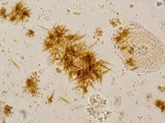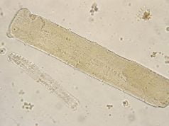
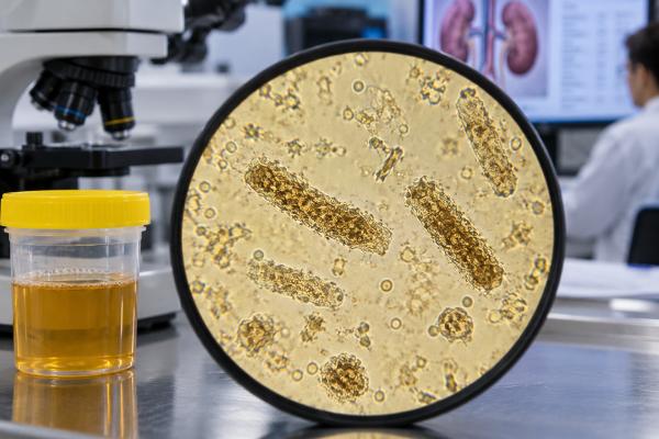
Granular casts are tube-shaped structures in urine that contain fine or coarse cellular debris. A small number of nonspecific granular casts can appear after exercise or dehydration, but abundant coarse or “muddy brown” granular casts are strongly associated with acute tubular injury, a common form of intrinsic acute kidney injury. They form when cells and proteins break down inside kidney tubules and become trapped in a uromodulin cast matrix. Their meaning depends on appearance, quantity, kidney-function trend, urine output, medications, blood pressure, and the rest of the sediment. In a patient whose creatinine is rising, muddy-brown granular casts can be more informative than urinary sodium indices alone and can support tubular injury even when fractional excretion of sodium is below 1%. Granular casts do not identify the original cause, so clinicians still look for ischemia, sepsis, nephrotoxins, pigment injury, obstruction, and other kidney disorders.
- Granular casts are not all equally abnormal: rare fine granules can be nonspecific, while numerous coarse muddy-brown casts strongly support acute tubular injury.
- They form from tubular debris: injured renal tubular cells and proteins break down and become incorporated into casts inside the nephron.
- Kidney function determines significance: rising creatinine, reduced urine output, high potassium, or fluid overload makes a granular-cast finding more urgent.
- Muddy-brown casts can outweigh a low FENa: acute tubular injury can still be present even when fractional excretion of sodium is below 1%.
- The cause still has to be found: shock, severe infection, medications, toxins, rhabdomyolysis, and other insults can all produce a tubular-injury sediment.
Table of Contents
- What Granular Casts Are
- Fine, Coarse, and Muddy-Brown Granular Casts
- Granular Casts and Acute Tubular Injury
- Common Causes of Granular Casts
- Microscopy, Cast Counts, and Urine Sediment Scores
- Granular Casts vs Other Urinary Casts
- Clinical Patterns That Change the Meaning
- Evaluation, Follow-Up, and Recovery
- When the Result Is Urgent
What Granular Casts Are
Casts are cylindrical molds formed inside kidney tubules. Their basic scaffold is usually uromodulin, a protein produced in the thick ascending limb and distal nephron. As urine flows through the tubule, the matrix can trap cells, cell fragments, pigments, crystals, and proteins.
Granular casts contain particulate debris rather than clearly intact cells. Their granules may come from degenerated renal tubular epithelial cells, lysosomal material, filtered proteins, or other cellular breakdown products. The result can range from a lightly granular transparent cast to a dense brown cylinder packed with coarse material.
This explains why granular casts overlap with epithelial cell casts. A cast that initially contains recognizable tubular cells can become more granular as those cells degenerate. The sediment therefore reflects a continuum rather than perfectly separate categories.
There is no single universal normal cutoff. Laboratories may report casts per low-power field or use qualitative terms such as rare, few, moderate, or many. A rare granular cast in concentrated urine does not carry the same meaning as abundant muddy-brown casts in a patient with acute kidney injury.
Fine, Coarse, and Muddy-Brown Granular Casts
Granular casts are commonly described as fine or coarse. Fine granular casts contain smaller particles and can be relatively pale. Coarse granular casts contain larger, denser granules and are generally more concerning for active tubular damage when numerous.
The best-known pattern is the muddy-brown granular cast. These casts have a dark tan, brown, or burnt-amber appearance and are associated with acute tubular injury. They may be broad or irregular and can contain dense debris from sloughed tubular cells.
Modern biopsy-correlated studies have shown very high specificity of muddy-brown granular casts for acute tubular injury when confidently identified in patients with AKI. That does not mean every brown structure in urine is diagnostic. Pigments, crystals, and artifacts can mimic casts, and expertise in sediment microscopy matters.
A 2024 study measuring muddy-brown casts found wide variation in their length and width, which reinforces that size alone is not the defining feature. Their granular composition and characteristic appearance are more important than a strict dimension.
Color can also be affected by hemoglobin, myoglobin, bilirubin, or medication-related pigments. When rhabdomyolysis or hemolysis is suspected, laboratory testing is needed to determine whether pigmented granular material is contributing to the cast appearance.
Granular Casts and Acute Tubular Injury
Acute tubular injury, or ATI, is a structural injury to renal tubular epithelial cells. It commonly occurs after sustained ischemia, severe infection, major surgery, toxins, or medication exposure. The older term “acute tubular necrosis” is still widely used, but not every patient has widespread necrosis on pathology.
As injured cells lose attachment, they shed into the tubular lumen. Some appear intact as renal tubular epithelial cells. Others degenerate and contribute to epithelial and granular casts. A sediment containing numerous RTECs, epithelial casts, and muddy-brown granular casts is therefore a classic ATI pattern.
This can help distinguish intrinsic tubular injury from a purely prerenal state. In uncomplicated volume depletion before structural damage develops, the sediment is often bland or shows only hyaline casts. If low blood flow persists long enough to injure cells, the sediment can evolve from bland to granular.
Urine microscopy is particularly useful because common chemical indices have limitations. Fractional excretion of sodium, or FENa, can be low in early ATI, in people with chronic kidney disease, and in other mixed states. A 2022 prospective study found muddy-brown granular casts in many patients whose FENa was still below 1%; biopsy-proven ATI remained strongly associated with the casts.
For this reason, a low FENa should not be used to overrule convincing sediment evidence of ATI. The reverse is also true: an elevated FENa without supportive clinical context does not prove tubular injury.
Common Causes of Granular Casts
Granular casts describe the tubular response, not the original insult. Common causes include:
- Ischemia and shock: severe bleeding, prolonged hypotension, cardiac events, major surgery, or other states that reduce kidney perfusion.
- Sepsis and critical illness: inflammation, microcirculatory changes, medications, and hemodynamic instability can combine to injure tubules.
- Nephrotoxic medications: certain antibiotics, chemotherapy drugs, and other agents can directly damage tubular cells.
- Pigment injury: myoglobin from rhabdomyolysis and hemoglobin from severe intravascular hemolysis can injure tubules and produce pigmented granular casts.
- Crystal-related injury: drug crystals, uric acid, oxalate, or other precipitates can obstruct tubules and cause secondary epithelial damage.
- Tubulointerstitial disease: inflammatory disorders can produce granular casts in addition to white cells or white blood cell casts.
- Severe glomerular disease: downstream tubular injury can accompany heavy proteinuria or inflammatory glomerulonephritis.
The patient’s history narrows the list. A person with septic shock after surgery has a different pretest probability from someone who develops AKI after starting a high-risk medication or after a prolonged seizure with severe muscle breakdown.
A related drug crystals in urine result can be especially useful when crystals and granular casts appear together during medication-associated AKI.
Microscopy, Cast Counts, and Urine Sediment Scores
Urine sediment is usually examined at low power to locate casts and at higher power to identify their contents. A fresh sample improves preservation. Casts can dissolve or degrade in dilute, alkaline, or old urine, so a negative result does not completely exclude tubular injury.
Some nephrology practices use urine microscopy scoring systems based on the number of RTECs and granular casts. Higher scores have been associated with a greater likelihood of ATI and worse AKI severity in several cohorts. A 2025 Kidney360 study also evaluated microscopy scores for early detection and prediction of AKI in at-risk patients.
These scores are useful because they convert a descriptive sediment into a semiquantitative pattern, but they are not universal laboratory standards. The same specimen may be reported differently by an automated analyzer, routine laboratory microscopy, and nephrologist-performed manual microscopy.
A 2023 study comparing laboratory and nephrologist microscopy found that nephrologists were more likely to recognize pathologic casts and that sediment-based diagnoses of ATI or glomerulonephritis could align well with biopsy. This supports manual review when the automated report is vague but clinical suspicion is high.
Serial microscopy can also matter. The first sample early in AKI may be bland; later samples can show more granular debris. If kidney function is not following the expected course, repeating sediment examination can reveal a pattern that was not visible initially.
Granular Casts vs Other Urinary Casts
| Cast type | Main contents | Typical meaning |
|---|---|---|
| Hyaline | Mostly uromodulin protein | Can be normal with dehydration or exercise; also seen in prerenal states |
| Granular | Degenerated cellular and protein debris | ATI when abundant, coarse, or muddy-brown |
| Epithelial | Renal tubular epithelial cells | Active tubular epithelial injury |
| RBC | Red blood cells | Strong clue to glomerular bleeding |
| WBC | White blood cells | Interstitial nephritis or pyelonephritis in the right setting |
| Waxy | Highly degenerated cast material | Prolonged tubular stasis, often advanced kidney disease |
The distinction is not always clean. Mixed cellular and granular casts occur, and aging casts can change appearance. This is why the overall urine microscopy pattern in kidney disease is more useful than one label.
Clinical Patterns That Change the Meaning
Granular casts are most useful when the cast pattern matches the clinical course. Several common combinations show why context matters.
Muddy-brown casts + a creatinine rise after hypotension or shock is a strong ATI pattern. The history provides the likely cause, while microscopy provides evidence that the injury has become structural rather than remaining purely prerenal.
Muddy-brown casts + FENa below 1% should not be dismissed. A low FENa can persist in ATI and can be affected by chronic kidney disease, heart failure, sepsis, or timing. When microscopy shows convincing muddy-brown casts, clinicians do not usually use FENa alone to rule out tubular injury.
Granular casts + dark urine after muscle injury raises concern for pigment nephropathy from myoglobin. Creatine kinase, potassium, phosphorus, calcium, and kidney function become important. Urine dipstick may be strongly positive for blood even when few RBCs are visible because the dipstick detects myoglobin as heme activity.
Granular casts + pyuria or WBC casts broadens the differential to interstitial inflammation or kidney infection with secondary tubular injury. Medication timing, fever, flank pain, bacteriuria, and culture help separate sterile drug-related inflammation from pyelonephritis.
Granular casts + dysmorphic RBCs or RBC casts suggests that a glomerular process may be causing downstream tubular stress. Glomerulonephritis can produce both an active nephritic sediment and secondary ATI, particularly in severe disease.
Rare fine granular casts with normal creatinine are much less specific. They may follow exercise, fever, or transient concentrated urine. A repeat sample after recovery from a temporary stressor can clarify whether the finding persists.
The trajectory is also important. Early ATI can have a relatively bland sediment, while later urine shows more RTECs and granular debris. During recovery, casts may remain visible even as urine output improves. One sample is therefore a snapshot, not a complete timeline of injury.
Evaluation, Follow-Up, and Recovery
When granular casts appear with AKI, clinicians first assess severity: creatinine trend, urine output, potassium, bicarbonate, volume status, and symptoms. A broader acute kidney injury blood test panel helps identify complications that may require urgent treatment.
The next goal is to find and reverse the trigger. That may mean treating infection and shock, restoring appropriate circulation, stopping or dose-adjusting a nephrotoxic drug, managing rhabdomyolysis, or relieving urinary obstruction. Fluids help in some patients but can worsen pulmonary edema in others, so treatment must match the volume state.
Granular casts do not need a treatment of their own. As tubular cells recover and the underlying injury resolves, the sediment often becomes less active. Creatinine can improve more slowly than urine output, and urinary debris may persist temporarily during recovery.
After significant AKI, follow-up matters even if kidney function appears to return to baseline. AKI increases future risk for chronic kidney disease, especially after severe or repeated episodes. Repeat creatinine, urine protein assessment, blood pressure, and medication review may be appropriate after discharge.
A kidney biopsy is usually unnecessary when the clinical story and muddy-brown sediment clearly support ATI and recovery proceeds as expected. Biopsy becomes more relevant when kidney function continues to worsen, the sediment shows glomerular features, or the diagnosis remains uncertain enough to change treatment.
Granular casts can also help with risk assessment, but they are not a stand-alone prognostic test. Studies of urine sediment scores have found that greater numbers of granular casts and renal tubular epithelial cells tend to occur in more established tubular injury and can be associated with worse short-term kidney outcomes in selected AKI populations. That relationship does not mean a cast count can predict an individual patient’s need for dialysis or chance of full recovery. Baseline kidney function, hemodynamics, infection, toxin exposure, urine output, and response to treatment remain more important clinical variables.
Serial urine microscopy can be useful when the diagnosis is uncertain. A patient who initially has a bland sediment may later develop obvious muddy-brown casts as tubular injury declares itself. Conversely, a recovering patient may still shed granular material for a time even while creatinine is stabilizing. The sediment and serum creatinine therefore do not always change on the same schedule.
This is especially relevant in mixed AKI, where prerenal physiology and tubular injury coexist. A person with heart failure, sepsis, or prolonged dehydration may have concentrated urine and a low fractional excretion of sodium while also having structural tubular damage. In that setting, granular casts provide direct morphologic evidence that complements—not replaces—hemodynamic indices.
A useful follow-up question is whether the sediment is becoming more or less active together with the clinical course. Fewer pathologic casts, improving urine output, falling creatinine, and correction of the trigger all support recovery. Persistent or increasing casts with worsening kidney function should prompt reassessment for ongoing ischemia, an unrecognized nephrotoxin, obstruction, inflammation, or another kidney process.
Importantly, an improving creatinine does not require the urine sediment to become completely normal on the same day. Tubular debris can continue to pass during repair. Decisions about discharge, dialysis, medication dosing, and follow-up are therefore based on the whole clinical course rather than on a requirement that granular casts disappear.
When the Result Is Urgent
A report of rare granular casts in someone with normal kidney function can usually be discussed non-urgently. The situation changes when casts accompany signs of acute kidney injury.
Seek prompt medical evaluation for markedly reduced urine output, rapidly rising creatinine, persistent vomiting or dehydration, severe weakness, confusion, significant swelling, shortness of breath, or known high potassium. Severe infection, low blood pressure, major trauma, or dark urine after muscle injury also warrants urgent assessment.
Granular casts are most useful as evidence of tubular stress or injury. Abundant muddy-brown casts are a strong clue to ATI, but the clinical task is still to identify why the tubules were injured and whether complications need immediate treatment.
References
- Dimensions of muddy brown granular casts in patients with acute tubular injury 2024
- Concomitant Identification of Muddy Brown Granular Casts and Low Fractional Excretion of Urinary Sodium in AKI. 2022
- Diagnostic Utility of Urine Microscopy in Kidney Diseases 2024 (Review)
- The Utility of Urine Microscopy Score for Early Detection and Prediction of Acute Kidney Injury in At-Risk Patients. 2025
- Urine Microscopy: The Enduring Value of a Classic Test. 2025
- Urine Sediment Examination: Comparison Between Laboratory-Performed Versus Nephrologist-Performed Microscopy and Accuracy in Predicting Pathologic Diagnosis in Patients with Acute Kidney Injury 2023
Disclaimer
Granular casts can support a diagnosis of kidney tubular injury, but they do not identify the cause or severity by themselves. Interpretation should include creatinine, urine output, electrolytes, medications, blood pressure, systemic illness, and other urine findings. Markedly reduced urine, rapidly worsening kidney function, severe swelling, breathlessness, confusion, or other signs of acute illness require prompt medical assessment.