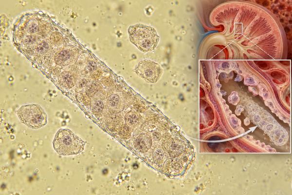
Epithelial cell casts in urine are tube-shaped structures containing renal tubular epithelial cells. Because urinary casts form inside kidney tubules, an epithelial cell cast is more specific for kidney-tubule involvement than simply finding free epithelial cells in a urine sample. These casts are most often associated with acute tubular injury, including ischemic injury from low blood flow and toxic injury from medications, pigments, or other exposures. They can also appear in other tubulointerstitial kidney disorders. The finding is not interpreted by itself: clinicians look at the number and type of casts, free renal tubular epithelial cells, granular or muddy-brown casts, serum creatinine, urine output, medication exposure, blood pressure, and the overall illness. A few nonspecific particles can be misclassified by automated systems, so manual sediment review may be important. Epithelial cell casts plus a rising creatinine or falling urine output should prompt timely assessment for acute kidney injury and its cause.
- Epithelial cell casts come from the kidney tubules: they contain renal tubular epithelial cells embedded in a protein matrix formed within the nephron.
- They often support acute tubular injury: especially when seen with free tubular cells and granular or muddy-brown casts.
- There is no single normal numeric range: pathologic casts are usually reported qualitatively or per low-power field, and laboratory methods differ.
- The result does not identify the cause by itself: ischemia, medications, toxins, severe illness, and other tubulointerstitial processes must be considered.
- Kidney-function changes determine urgency: rising creatinine, oliguria, high potassium, fluid overload, or severe illness needs prompt medical review.
Table of Contents
- What Epithelial Cell Casts Are
- How Epithelial Cell Casts Form
- What Epithelial Cell Casts Usually Mean
- Causes of Tubular Cell Shedding and Casts
- Microscopy Patterns and Related Findings
- How Doctors Evaluate the Result
- Clinical Patterns That Change the Interpretation
- Prognosis, Follow-Up, and Repeat Testing
- When the Finding Is Urgent
What Epithelial Cell Casts Are
A urinary cast is a cylindrical mold of a kidney tubule. Casts form when the protein uromodulin, historically called Tamm-Horsfall protein, gels inside the distal nephron and traps cells, debris, pigments, lipids, or crystals. Their tube-like shape reflects the lumen in which they formed.
An epithelial cell cast specifically contains renal tubular epithelial cells. These cells line the nephron and normally remain attached to the tubular basement membrane. When tubules are stressed or injured, cells can detach and enter the lumen. If they become embedded in a cast matrix, the sediment may show a recognizable epithelial cast.
This makes the finding different from squamous epithelial cells that commonly enter a urine sample from skin or the external genital tract. It is also more localizing than finding free renal tubular epithelial cells in urine. Free tubular cells can indicate kidney injury, but an epithelial cast demonstrates that cells were incorporated into material formed within the nephron.
Epithelial cell casts are generally considered pathologic rather than a normal incidental finding. However, there is no universal cutoff such as “more than X casts equals acute tubular injury.” Laboratories may report rare, few, moderate, or many casts, or count them per low-power field. The type, abundance, and clinical context determine significance.
How Epithelial Cell Casts Form
Cast formation is favored by concentrated urine, low tubular flow, and conditions that promote uromodulin aggregation. Uromodulin is produced by cells in the thick ascending limb and early distal tubule. It can create a transparent hyaline matrix that becomes the scaffold for other sediment elements.
In acute tubular injury, damaged epithelial cells lose polarity and attachment, swell, die, or slough into the lumen. Some remain intact enough to be recognized microscopically. Others break down into coarse cellular debris. When intact cells are trapped early, an epithelial cell cast forms. As those cells degenerate, the cast may become granular and eventually resemble a dense muddy-brown granular cast.
This progression helps explain why a single urine sample may contain a mixture of free tubular epithelial cells, epithelial cell casts, granular casts, and cellular debris. It also explains why serial microscopy can change over time as the injury evolves or recovery begins.
A fresh sample improves recognition. Prolonged standing, alkaline urine, and low osmolality can damage cells and casts, making them harder to identify. Modern guidance therefore emphasizes timely examination and standardized preparation when manual microscopy is used.
What Epithelial Cell Casts Usually Mean
The most common interpretation is active tubular epithelial injury. In a patient with acute kidney injury, epithelial cell casts increase concern for intrinsic renal injury rather than a purely prerenal reduction in blood flow that has not yet damaged tubules.
Acute tubular injury is the preferred pathophysiologic term for many cases historically labeled acute tubular necrosis. Not every patient has widespread tissue necrosis. The process may include reversible cell stress, focal cell death, sloughing, inflammation, and abnormal repair.
Urine microscopy can help distinguish patterns. Prerenal states often have a bland sediment or only hyaline casts. Acute tubular injury is more likely to produce renal tubular epithelial cells, epithelial casts, and granular casts. The combination is more informative than any one element.
This is why nephrologists sometimes use semiquantitative urine microscopy scores based on numbers of tubular epithelial cells and granular casts. Higher scores have been associated with a greater likelihood or severity of tubular injury in studied AKI populations. The score supports clinical judgment; it does not replace serum creatinine, urine output, hemodynamic assessment, or imaging when obstruction is possible.
An epithelial cast should not automatically be labeled “ATN” without considering the rest of the case. Tubular cells can be shed in several kidney disorders, and mixed sediments are common in critically ill people.
Causes of Tubular Cell Shedding and Casts
Acute tubular injury has many triggers. The same microscopy pattern can result from very different upstream problems.
Ischemic injury occurs when kidney tissue receives inadequate oxygen and blood flow. Major surgery, shock, severe bleeding, sepsis, prolonged hypotension, or cardiac failure can contribute. A short prerenal episode may resolve without tubular damage, but severe or sustained hypoperfusion can progress to structural injury and cellular shedding.
Medication and toxin exposure can directly injure tubular cells. Examples include some antimicrobial agents, chemotherapy drugs, radiographic contrast in susceptible settings, and other nephrotoxins. Some drugs cause crystals that obstruct and injure tubules rather than direct cytotoxicity. Reviewing recent prescriptions, infusions, supplements, and exposures is therefore central to the work-up.
Pigment-related injury can follow severe muscle breakdown, which releases myoglobin, or marked intravascular hemolysis, which releases hemoglobin. Pigments can injure tubules and contribute to granular or pigmented casts.
Inflammatory tubulointerstitial disease can also produce tubular epithelial cells. Acute interstitial nephritis more often shows pyuria and may show white blood cell casts, while the sediment can overlap with acute tubular injury. White blood cell casts shift the differential toward interstitial inflammation or kidney infection when the clinical setting fits.
Glomerular diseases may secondarily injure tubules when heavy protein, blood, or inflammatory mediators reach downstream segments. In these situations, epithelial cell casts can coexist with dysmorphic RBCs or RBC casts rather than defining a purely tubular disorder.
Microscopy Patterns and Related Findings
Correct identification matters because several casts can look similar after degeneration. Experienced microscopy interprets the entire sediment rather than naming one isolated structure.
| Finding | Typical appearance | Common interpretation |
|---|---|---|
| Epithelial cell cast | Tubular cells embedded in a cylindrical matrix | Active renal tubular epithelial injury |
| Granular cast | Fine or coarse granules; may be brown | Tubular injury when abundant or muddy-brown |
| Hyaline cast | Nearly transparent protein matrix | Can occur with dehydration, exercise, or prerenal states |
| RBC cast | Red cells within a cast | Strongly supports glomerular bleeding |
| WBC cast | White cells within a cast | Interstitial nephritis or pyelonephritis in the right context |
The relationship between epithelial and granular casts in acute kidney injury is especially important. Tubular cells may degenerate within a cast, making a cellular cast progressively more granular. Dense muddy-brown casts are a classic marker of acute tubular injury.
Automated analyzers can count many cells accurately but may be less reliable for unusual casts. A 2023 comparison found nephrologist-performed sediment examination more likely than routine laboratory analysis to identify pathologic casts and dysmorphic RBCs in patients with AKI. That does not mean automated testing is poor; it means clinically important abnormal particles may deserve manual review.
How Doctors Evaluate the Result
The evaluation starts with a simple question: Is kidney function changing? Serum creatinine trends and urine output determine whether the microscopy finding is occurring with acute kidney injury. A single creatinine value can be misleading without a prior baseline.
Clinicians then look for the mechanism. Blood pressure, volume status, recent surgery, infection, medication exposure, contrast studies, muscle injury, hemolysis, and urinary symptoms all matter. Blood tests commonly include electrolytes, bicarbonate, BUN, creatinine, and sometimes creatine kinase or hemolysis markers.
A broader acute kidney injury blood test panel helps identify complications such as high potassium or metabolic acidosis. Urinalysis adds protein, blood, glucose, pH, and specific gravity. Microscopy can reveal whether the sediment is bland, tubular, nephritic, inflammatory, or crystalline.
Kidney and bladder ultrasound is useful when urinary obstruction is possible, especially with oliguria, prostate disease, pelvic malignancy, stones, or hydronephrosis risk. Serologic testing is added when glomerulonephritis or systemic inflammatory disease is suspected.
Kidney biopsy is not required for typical acute tubular injury that fits the clinical course and improves as expected. It becomes more relevant when the diagnosis is uncertain, kidney function continues to worsen without explanation, or the urine pattern suggests a treatable glomerular or interstitial disease.
Clinical Patterns That Change the Interpretation
The same epithelial cell cast can carry different weight depending on the surrounding findings. Looking at common patterns helps prevent both overcalling and undercalling tubular injury.
Epithelial cell casts plus muddy-brown granular casts and rising creatinine is a classic tubular-injury pattern. In a patient who recently had shock, major surgery, severe infection, or a nephrotoxic exposure, this combination makes acute tubular injury much more likely. The urine findings may be especially useful when other indices, such as fractional excretion of sodium, are difficult to interpret because of diuretics, chronic kidney disease, or mixed physiology.
Epithelial cell casts plus pyuria or white blood cell casts broadens the differential. Acute interstitial nephritis, pyelonephritis, or another inflammatory tubulointerstitial process may be present. Fever, rash, eosinophilia, drug timing, flank pain, urinary symptoms, and urine culture help distinguish these causes. Urine eosinophils are not reliable enough to confirm or exclude acute interstitial nephritis by themselves.
Epithelial cell casts plus heavy proteinuria or dysmorphic RBCs suggests that a glomerular process may be damaging tubules secondarily. Severe protein leakage can stress proximal tubular cells, while hematuria and inflammatory debris can add downstream injury. In that setting, a purely “tubular” label would miss the broader kidney disease.
Epithelial cell casts with crystals can occur when crystal deposition injures tubular epithelium. Medication crystals, uric acid, oxalate, or other pathologic crystals can obstruct tubules and provoke cell shedding. The crystal type, urine pH, exposure history, and kidney-function trend determine what the finding means.
A few questionable casts with normal creatinine and normal urine output may need confirmation rather than an immediate diagnosis of AKI. Cast identification can be difficult, particularly in dilute urine or automated images. A fresh repeat sample and manual review can clarify whether the reported structures are true cellular casts, free epithelial-cell clusters, mucus, or other sediment material.
The timing of sampling also matters. Early ischemic kidney stress can produce a relatively bland sediment before cells and granular material become abundant. Later in the course, cellular elements may degenerate into coarse granules. During recovery, the sediment can continue to show residual cells even as creatinine improves. This is why the urine sediment is best read as a snapshot within a changing clinical course, not a fixed score detached from time.
Prognosis, Follow-Up, and Repeat Testing
Epithelial cell casts show tubular injury, but they do not by themselves predict whether kidney function will fully recover. Outcome depends on the severity and duration of the insult, baseline kidney function, age, comorbid disease, and whether the cause can be reversed.
Many patients with acute tubular injury recover at least partially as surviving tubular cells repair and repopulate the epithelium. Severe or repeated injury can lead to incomplete recovery and a higher future risk of chronic kidney disease. This is one reason follow-up creatinine and urine testing after an AKI episode is important even when a person feels better.
Repeat microscopy can be useful when the initial sample is nondiagnostic but clinical suspicion remains high. Sediment findings can evolve; granular casts or tubular cells may become more obvious later. Conversely, declining numbers of pathologic casts during clinical recovery can be reassuring, although there is no standardized “cast clearance” target used for every patient.
Treatment targets the cause rather than the cast. That may mean restoring circulation, treating sepsis, stopping or adjusting a nephrotoxic medication, relieving obstruction, managing rhabdomyolysis, or treating an immune-mediated kidney disease. Fluids are helpful in some settings but can be harmful in fluid-overloaded patients, so they should be tailored to the clinical state.
When the Finding Is Urgent
An incidental report of a rare epithelial cast with normal kidney function should be discussed with a clinician, but urgency rises when the sediment accompanies objective AKI.
Prompt or urgent assessment is appropriate for rapidly rising creatinine, markedly reduced urine output, inability to urinate, severe dehydration, persistent vomiting, significant swelling, shortness of breath, confusion, chest symptoms, or known high potassium. People who are septic, hypotensive, or receiving high-risk nephrotoxic therapy need especially close monitoring.
The practical message is that epithelial cell casts are evidence from inside the kidney tubules. Their presence should trigger a search for tubular injury and its cause, with the speed of evaluation determined by kidney-function change and the patient’s overall condition.
For an outpatient with no symptoms, a useful next step is often to confirm the kidney-function trend and repeat a properly collected urinalysis if the original specimen was old, dilute, or otherwise questionable. Reviewing recent medication changes is also important because dose changes, new antibiotics, contrast exposure, or over-the-counter products may provide the missing context.
In hospitalized patients, the same finding can be far more consequential. Creatinine may lag behind the actual injury, so falling urine output, hemodynamic instability, or new electrolyte abnormalities can make an epithelial-cast pattern clinically important before a large creatinine rise appears. Recovery is followed mainly with urine output, creatinine, electrolytes, volume status, and treatment of the underlying cause—not by waiting for every abnormal cast to disappear.
After a significant AKI episode, follow-up may also include blood pressure and urine protein or albumin testing because even apparently recovered patients can have residual kidney risk. The timing depends on the severity of the episode and baseline kidney health. Persistent creatinine elevation, proteinuria, recurrent abnormal sediment, or reduced estimated GFR may justify nephrology follow-up.
References
- Diagnostic Utility of Urine Microscopy in Kidney Diseases 2024 (Review)
- Renal tubular epithelial cells response to injury in acute kidney injury. 2024 (Review)
- Urinary renal tubular epithelial cells and casts as predictors of renal outcomes in patients with biopsy-proven diabetic nephropathy 2024
- Urine Microscopy: The Enduring Value of a Classic Test. 2025
- The Utility of Urine Microscopy Score for Early Detection and Prediction of Acute Kidney Injury in At-Risk Patients. 2025
- The EFLM European Urinalysis Guideline 2023 2024 (Guideline)
Disclaimer
Epithelial cell casts can indicate renal tubular injury but do not identify the cause or severity by themselves. Results should be interpreted with serum creatinine, urine output, other sediment findings, medications, and the clinical setting. Rapidly worsening kidney function, markedly reduced urine, breathing difficulty, severe swelling, confusion, or other signs of acute illness require prompt medical evaluation.