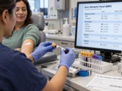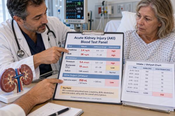
Acute kidney injury (AKI) is a sudden decline in kidney function that develops over hours to days. No single blood test proves AKI by itself. Clinicians usually recognize it by a change in serum creatinine, reduced urine output, or both, then use other blood and urine results to judge severity and search for the cause. A typical AKI evaluation includes creatinine, blood urea nitrogen (BUN), electrolytes such as potassium and sodium, bicarbonate, glucose, and often calcium or phosphorus. Urinalysis, urine microscopy, medication review, fluid status, and sometimes imaging add important context.
The key is the trend. A creatinine value that looks only mildly abnormal can represent major kidney dysfunction if it rose quickly from a lower baseline. Likewise, a person can have important AKI before creatinine rises much, especially when urine output falls. AKI can be reversible, but it can also cause dangerous potassium, acid-base, and fluid problems, so timely interpretation matters.
- AKI is usually identified by a rapid creatinine rise, low urine output, or both, not by one isolated kidney blood-test value.
- A creatinine increase of at least 0.3 mg/dL within 48 hours or to at least 1.5 times baseline within 7 days meets standard AKI criteria.
- Urine output below 0.5 mL/kg/hour for 6 hours can meet AKI criteria even if creatinine has not risen yet.
- Potassium, bicarbonate, and fluid balance help show urgency because severe hyperkalemia, acidosis, or fluid overload may need immediate treatment.
- BUN can rise with AKI but is less specific than creatinine because dehydration, bleeding, protein intake, steroids, and other factors also affect it.
Table of Contents
- What an AKI Panel Measures
- Creatinine and Urine Output Criteria
- BUN, Electrolytes, and Bicarbonate
- What Different Result Patterns Can Mean
- How Doctors Confirm the Cause
- When AKI Results Are Urgent
- Repeat Testing, Recovery, and Follow-Up
What an AKI Panel Measures
An “AKI blood test panel” is not one universally fixed laboratory order. It is a practical group of tests used to detect a sudden loss of kidney function, identify complications, and help separate possible causes. In a hospital, many of these values come from a basic or comprehensive metabolic panel, while kidney-specific testing and urine studies are added as needed.
The core measurements usually include:
- Serum creatinine: the main blood marker used to define and stage AKI. It reflects filtration indirectly and is most useful when compared with a recent baseline.
- BUN: measures the nitrogen portion of urea in blood. It often rises when filtration falls, but it is strongly affected by hydration, protein breakdown, gastrointestinal bleeding, and medications.
- Potassium: can rise when impaired kidneys cannot excrete enough potassium. Severe elevation can disturb the heart rhythm.
- Sodium: may be low, normal, or high depending on fluid balance, losses, intake, and hormonal responses.
- Bicarbonate or total CO2: helps assess metabolic acidosis, which can develop when the kidneys cannot remove acid effectively.
- Glucose: important because marked hyperglycemia can alter fluid balance and contribute to dehydration.
- Calcium and phosphorus: often checked in more severe illness or when kidney dysfunction is prolonged.
A basic metabolic panel provides creatinine, BUN, sodium, potassium, chloride, bicarbonate, and glucose in one common test set. Clinicians may also order a broader kidney function blood test panel when the clinical question extends beyond a rapidly evolving AKI episode.
These results answer different questions. Creatinine and urine output help establish that kidney function has changed. Potassium and bicarbonate help show whether the change is creating dangerous complications. BUN, urinalysis, blood pressure, symptoms, and the medical history help point toward why it happened.
Creatinine and Urine Output Criteria
The standard AKI definition focuses on change over time. AKI is present when at least one of the following occurs: serum creatinine rises by 0.3 mg/dL or more within 48 hours, serum creatinine rises to 1.5 times or more than baseline within the previous 7 days, or urine volume falls below 0.5 mL/kg/hour for at least 6 hours.
This is why a “normal-range” creatinine does not always rule out AKI. For example, a rise from 0.6 to 0.9 mg/dL is a 50% increase and can meet AKI criteria even though 0.9 mg/dL may still fall inside many laboratory reference ranges. The creatinine blood test is therefore interpreted against a personal baseline whenever one is available.
AKI is also staged by severity:
| AKI stage | Serum creatinine criterion | Urine output criterion |
|---|---|---|
| Stage 1 | 1.5–1.9 times baseline, or increase of at least 0.3 mg/dL | Below 0.5 mL/kg/hour for 6–12 hours |
| Stage 2 | 2.0–2.9 times baseline | Below 0.5 mL/kg/hour for at least 12 hours |
| Stage 3 | At least 3 times baseline, creatinine at least 4.0 mg/dL, or kidney replacement therapy started | Below 0.3 mL/kg/hour for at least 24 hours, or no urine for at least 12 hours |
Urine output deserves special attention because it can change before creatinine. A systematic review published in 2024 found that urine-output criteria often identified AKI more frequently and earlier than creatinine criteria. But urine volume is not perfectly specific. Dehydration, low blood pressure, obstruction, diuretics, heart failure, and normal physiologic variation can all affect it.
Creatinine also has delays and limitations. After filtration suddenly falls, several hours to a day or longer may pass before blood creatinine reaches a clearly abnormal value. Large fluid shifts can dilute it, and people with low muscle mass may generate less creatinine. That is why clinicians often repeat creatinine and track urine rather than waiting for one dramatic abnormal result.
BUN, Electrolytes, and Bicarbonate
BUN, potassium, sodium, chloride, and bicarbonate do not define AKI on their own, but they often show how the illness is affecting the body.
BUN commonly rises when filtration falls because urea is retained. A typical adult BUN reference range is roughly 7–20 mg/dL, although laboratories differ. BUN may rise disproportionately during dehydration because the kidneys reabsorb more urea along with water. It can also increase with gastrointestinal bleeding, high protein intake, fever, catabolic illness, corticosteroids, and tissue breakdown. The BUN test therefore works best as part of a pattern rather than as a stand-alone measure of kidney injury.
Potassium is one of the most important safety values. Many laboratories use an adult reference range around 3.5–5.0 mmol/L. AKI can cause potassium to accumulate, especially with severe filtration loss, tissue breakdown, acidosis, or medications that raise potassium. A level above about 6.0 mmol/L, rapidly rising potassium, symptoms, or ECG changes can represent an emergency; exact thresholds depend on the patient and clinical setting. A dedicated potassium blood test in kidney disease is often repeated when the first result is concerning.
Bicarbonate is a useful acid-base marker. A common reference interval is approximately 22–29 mmol/L, though laboratories vary. Low bicarbonate may suggest metabolic acidosis from reduced kidney acid excretion, lactic acidosis, diarrhea, diabetic ketoacidosis, or other causes. Severe acidosis in AKI can become an indication for urgent kidney replacement therapy when it does not respond to treatment.
Sodium is more about water balance than kidney filtration alone. A low sodium level may appear with excess body water, heart failure, cirrhosis, certain medications, or inappropriate antidiuretic hormone activity. High sodium often points to water loss that exceeds sodium loss. In AKI, the direction of sodium change depends heavily on the underlying illness and treatment.
What Different Result Patterns Can Mean
Clinicians commonly group AKI causes into three broad categories: reduced kidney perfusion, damage within the kidney, and blockage of urine flow. Blood tests cannot always distinguish them, but patterns can provide clues.
Reduced blood flow to the kidneys
Vomiting, diarrhea, bleeding, fever, severe infection, overuse of diuretics, heart failure, and low blood pressure can reduce effective blood flow to the kidneys. Creatinine and BUN may both rise. BUN may rise more than creatinine, producing a higher BUN/creatinine ratio, but that ratio is not reliable enough to diagnose dehydration or “prerenal” AKI by itself.
A person who has lost fluid may also have low blood pressure, fast heart rate, dry mucous membranes, concentrated urine, and a history of poor intake or fluid loss. Kidney function can improve quickly when the cause is corrected, but not every low-flow state responds simply to more fluid. Someone with heart failure, for example, may be fluid overloaded while kidney perfusion is still poor.
Injury within the kidney
Kidney tissue can be damaged by prolonged low blood flow, severe infection, medications, toxins, inflammation, or immune disease. Acute tubular injury is a common hospital cause. In these cases, creatinine may keep rising despite correction of blood pressure or hydration.
Urinalysis and urine microscopy become particularly useful. Blood, protein, white cells, casts, or other sediment findings may point toward glomerular disease, interstitial nephritis, or tubular injury. Creatine kinase may be checked when rhabdomyolysis is suspected because muscle breakdown releases myoglobin and potassium and can trigger severe AKI.
Urinary tract obstruction
A blockage can occur from an enlarged prostate, stone, tumor, blood clot, or another structural problem. Creatinine may rise rapidly, but urine output can be variable. Complete blockage affecting both kidneys or the only functioning kidney may cause very low or absent urine output. Kidney and bladder ultrasound is often used because blood tests alone cannot show where the obstruction is.
How Doctors Confirm the Cause
An AKI panel starts the investigation rather than finishing it. Doctors combine the lab pattern with timing, symptoms, medications, vital signs, urine findings, and imaging.
A practical evaluation often includes these steps:
- Confirm the trend. Repeat creatinine and review older results to establish the true baseline and speed of change.
- Measure urine output. In hospitalized patients, intake and output may be recorded hourly or over defined intervals.
- Review medications and recent exposures. NSAIDs, ACE inhibitors, ARBs, diuretics, some antibiotics, chemotherapy drugs, contrast exposure, supplements, and other agents can be relevant depending on the situation. Some drugs are appropriate to continue; others may need temporary adjustment under medical supervision.
- Assess fluid and circulation status. Blood pressure, heart rate, swelling, lung findings, weight change, and recent fluid losses help guide treatment.
- Check urinalysis and urine sediment. Protein, blood, leukocytes, casts, and crystals may point toward a specific kidney process.
- Look for obstruction when appropriate. Ultrasound is common when urinary retention, stones, pelvic disease, a solitary kidney, or unexplained AKI raises concern.
- Order targeted tests. Depending on the suspected cause, clinicians may check creatine kinase, blood cultures, autoimmune markers, complement levels, hemolysis tests, or other studies.
Estimated GFR is often automatically reported with creatinine, but it has an important limitation in rapidly changing AKI: standard eGFR equations assume creatinine is relatively stable. During an acute rise or fall, the reported eGFR can lag behind the true change in filtration and should not be read like a steady-state chronic kidney measurement.
Biomarkers such as NGAL, KIM-1, and cell-cycle arrest markers have been studied for earlier detection or risk assessment. They can add information in selected clinical settings, but serum creatinine and urine output remain the main routine criteria used to diagnose AKI.
A single creatinine value is much easier to interpret when a recent baseline is available. For example, a result that looks only mildly elevated compared with the laboratory reference interval may represent a major change for someone whose usual creatinine is much lower. The opposite can also occur: a person with stable chronic kidney disease may have a chronically elevated creatinine without meeting criteria for a new acute kidney injury. Clinicians therefore compare the current value with prior results, the timing of the change, urine output, medications, blood pressure, fluid losses, and the clinical setting. When no reliable baseline exists, the diagnosis may remain uncertain until repeat measurements show a trend.
The direction of change matters as much as the first abnormal number. Creatinine often lags behind the kidney insult, so an early value can underestimate the severity of a rapidly evolving problem. Falling urine output may appear sooner, but urine output can also change for reasons that are not kidney failure, including diuretic use and urinary obstruction. For that reason, doctors usually interpret both signals together rather than treating either one as a perfect standalone test. Repeat creatinine and electrolyte testing over hours or days can show whether kidney function is worsening, stabilizing, or recovering.
When AKI Results Are Urgent
The urgency of AKI depends less on the creatinine number alone and more on the whole clinical picture. A modest creatinine rise can be serious if it occurs quickly or alongside dangerous potassium, acidosis, low blood pressure, infection, or reduced urine output.
Seek urgent medical assessment for symptoms or results that suggest severe AKI complications, such as:
- very little or no urine for many hours;
- new shortness of breath, rapidly increasing swelling, or signs of fluid in the lungs;
- severe weakness, palpitations, fainting, or an abnormal heart rhythm, which may occur with major potassium disturbances;
- confusion, extreme drowsiness, seizures, or marked illness;
- persistent vomiting or inability to keep fluids down;
- severe dehydration, bleeding, or very low blood pressure;
- suspected sepsis, including fever or hypothermia with rapid breathing, confusion, or circulatory instability.
Kidney replacement therapy, often called dialysis in this setting, is considered when complications cannot be controlled medically. Common reasons include refractory hyperkalemia, severe metabolic acidosis, pulmonary edema or fluid overload that is not responding to treatment, certain toxin exposures, or severe uremic complications. There is no single creatinine or BUN cutoff that automatically means dialysis is required.
A person with a new creatinine rise should not try to “flush the kidneys” by forcing large amounts of water without knowing the cause. Extra fluid can help some people who are volume depleted, but it can worsen swelling and breathing in heart failure or advanced kidney dysfunction.
Repeat Testing, Recovery, and Follow-Up
AKI is dynamic, so repeat testing is central to care. In severe or rapidly changing cases, creatinine, potassium, bicarbonate, and other electrolytes may be checked several times in a day. In milder stable cases, the interval may be longer. The goal is to see whether kidney function is still worsening, has stabilized, or is recovering.
Recovery may be rapid after reversible dehydration or obstruction, or it may take days to weeks after more substantial kidney injury. Urine output can improve before creatinine falls because blood creatinine needs time to clear. During recovery, some patients enter a high-urine-output phase and can lose water and electrolytes quickly, so monitoring may still be necessary even when creatinine is improving.
After an AKI episode, follow-up matters because AKI increases the risk of later chronic kidney disease. A clinician may repeat creatinine and eGFR after discharge, review blood pressure and medications, and check urine albumin or protein. The timing depends on severity and recovery, but people with incomplete recovery, recurrent AKI, substantial protein or blood in the urine, or advanced preexisting kidney disease often need closer nephrology follow-up. A CKD blood test panel may be useful once kidney function has stabilized enough for chronic assessment.
The most informative question after any AKI panel is not “Is this number high?” but “How has it changed, what complication is it causing, and what explains the change?” That approach keeps creatinine, BUN, electrolytes, and urine output in their proper clinical context.
References
- Urine output is an early and strong predictor of acute kidney injury and associated mortality: a systematic literature review of 50 clinical studies 2024 (Systematic Review)
- Biomarkers of acute kidney injury: From discovery to the future of clinical practice 2024 (Review)
- Acute Kidney Injury: Biomarker-Guided Diagnosis and Management 2022 (Review)
- Overview of Diagnostic Criteria and Epidemiology of Acute Kidney Injury and Acute Kidney Disease in the Critically Ill Patient 2022 (Review)
- Diagnosis, evaluation, and management of acute kidney injury: a KDIGO summary (Part 1) 2013 (Guideline Summary)
Disclaimer
This article is for general education and does not diagnose acute kidney injury or replace medical care. AKI can change quickly and may cause life-threatening potassium, acid-base, or fluid problems; seek prompt medical evaluation for a new rapid creatinine rise, very low urine output, severe illness, breathing difficulty, or other urgent symptoms. Laboratory ranges and treatment decisions vary by patient and facility.