Drug crystals in urine are particles formed when a medication or one of its metabolites becomes concentrated enough to precipitate in the urinary tract. The finding can be harmless crystalluria, but it can also be an early clue to crystal-related kidney injury, especially when kidney function is worsening. Risk depends on the drug, dose, urine pH, urine flow, hydration, kidney function, and how quickly the medicine is given. Medications linked to crystalluria include high-dose amoxicillin, acyclovir, methotrexate, sulfonamides, some HIV protease inhibitors, triamterene, ciprofloxacin, and others. Microscopy can sometimes show a recognizable crystal pattern, but appearance alone may not identify a drug with certainty. The result is most useful when interpreted with the medication list, serum creatinine, urine output, urine pH, symptoms, and timing. New oliguria, flank pain, gross hematuria, or a rising creatinine while taking a crystal-forming drug deserves prompt clinical review.
- Drug crystals do not automatically mean kidney damage: some people have transient crystalluria without acute kidney injury.
- Risk rises when urine is highly concentrated: dehydration, low urine output, high drug exposure, and impaired kidney clearance can favor precipitation.
- Urine pH matters: some drugs precipitate more readily in acidic urine, while others become less soluble at different pH ranges.
- Microscopy can provide a rapid clue: free crystals, crystal-containing casts, renal tubular cells, and granular casts may support crystal-related tubular injury.
- Seek prompt care for red flags: sharply reduced urine, visible blood, severe flank pain, vomiting with dehydration, or a rising creatinine during treatment needs timely assessment.
Table of Contents
- What Drug Crystals in Urine Mean
- Medications That Can Form Urinary Crystals
- What Drug Crystals Look Like Under the Microscope
- When Drug Crystals Can Injure the Kidneys
- How a Drug-Crystal Result Is Interpreted
- Follow-Up, Treatment, and Prevention
- Drug Crystals vs Kidney Stones and Other Crystals
- When to Get Urgent Medical Care
What Drug Crystals in Urine Mean
Drug crystalluria means that a medication, a medication metabolite, or occasionally a substance whose excretion was altered by a medication has formed solid crystals in urine. It is a microscopy finding, not a diagnosis by itself. The kidneys continuously filter blood and concentrate many substances into urine. If the concentration of a poorly soluble compound exceeds what the urine can keep dissolved, crystals may form.
The key distinction is between crystalluria and crystal nephropathy. Crystalluria simply describes crystals in the urine. Crystal nephropathy means crystals have contributed to kidney injury, usually by obstructing tubules, directly damaging tubular cells, or triggering local inflammation. A person can therefore have visible drug crystals while serum creatinine remains normal, and another person can develop acute kidney injury with clinically important intrarenal crystal deposition.
A routine urine microscopy test can detect many formed elements, including cells, casts, and crystals. For suspected medication-related crystalluria, the context matters as much as the shape: the medication name, dose, route, treatment duration, urine pH, hydration status, and recent kidney-function trend help determine whether the crystals are meaningful.
Crystals can also appear after a specimen has cooled or stood for too long. That is why laboratories try to examine fresh urine promptly. Refrigeration is useful for preserving many urine components, but it can also increase precipitation of some crystals, so crystal interpretation may require a separate fresh, nonrefrigerated aliquot.
Medications That Can Form Urinary Crystals
Several drugs are well known to precipitate in urine under the right conditions. The list is not exhaustive, and not every person taking one of these medicines develops crystalluria.
| Medication or class | Typical clinical setting | Important risk factors or clues |
|---|---|---|
| Amoxicillin | High-dose therapy, especially intravenous treatment | High exposure, low urine pH, hypovolemia, abrupt hematuria or cloudy urine |
| Acyclovir | Often intravenous antiviral therapy | Rapid infusion, dehydration, high dose, reduced kidney clearance |
| Methotrexate | High-dose oncology regimens | Acidic urine, delayed drug clearance, volume depletion |
| Sulfadiazine or sulfamethoxazole | Antimicrobial therapy | Acidic, concentrated urine; high exposure |
| Atazanavir or indinavir | HIV treatment | Crystalluria, stones, flank symptoms, obstruction |
| Triamterene | Diuretic therapy | Drug or metabolite precipitation; may contribute to stones |
| Ciprofloxacin | Antibiotic therapy | High exposure, dehydration, urine conditions that favor precipitation |
Amoxicillin is an important modern example because crystalluria is increasingly recognized during high-dose intravenous therapy. Reported rates vary by population and dose. The crystals are usually composed of amoxicillin trihydrate. Crystalluria may be asymptomatic, but gross hematuria, cloudy urine, and an abrupt creatinine rise can point toward amoxicillin-induced crystal nephropathy.
Acyclovir is excreted through the kidneys and can precipitate in the distal nephron, particularly when intravenous doses are high, infusion is rapid, or hydration is poor. High-dose methotrexate can also precipitate, especially in acidic urine; oncology protocols therefore use careful hydration, urine alkalinization, drug-level monitoring, and rescue strategies when indicated.
Some medicines increase stone risk through metabolic effects rather than by forming the stone or crystal themselves. For example, carbonic anhydrase inhibitors can change urine pH and mineral handling. That mechanism is different from seeing crystals made directly from the medication.
What Drug Crystals Look Like Under the Microscope
Drug crystals can be distinctive, but overlap is common. Bright-field, phase-contrast, and polarized-light microscopy may all help. Polarized light is especially useful because many drug crystals are birefringent, meaning they interact with polarized light in a characteristic way.
Examples include:
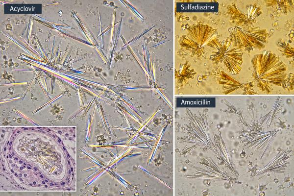
- Amoxicillin: often described as needle-like structures that aggregate into broom-brush, sheaf, or wheat-like forms.
- Acyclovir: commonly appears as long, thin needles, sometimes with sharp or blunt ends and strong birefringence.
- Sulfonamides: may form sheaves, rosettes, shells, or fan-like structures with radial striations.
- Methotrexate: may appear as yellow-brown or golden needle-like material and can be seen in free crystals or crystal-containing casts.
- Protease inhibitors: indinavir and atazanavir can produce needle, plate, rectangular, fan, starburst, or rosette-like forms depending on the compound and conditions.
Microscopy also looks for evidence of accompanying tubular injury. Renal tubular epithelial cells in urine indicate shedding from the nephron and become more concerning when numerous. Granular casts, particularly muddy-brown casts in the appropriate setting, support acute tubular injury. Crystal-containing casts are especially suggestive that precipitation is occurring within the tubules rather than only after urine left the kidney.
Automated urine analyzers can flag abnormal particles, but unusual crystals often need manual review. If identification would change treatment, a laboratory may use additional methods such as infrared spectroscopy, specialized microscopy, or direct analysis of a passed stone. The report may therefore say “crystals, unidentified” or “drug-like crystals” rather than naming a medication with certainty.
When Drug Crystals Can Injure the Kidneys
The kidney risk comes from a combination of supersaturation, tubular obstruction, cell injury, and inflammation. A crystal-forming drug can reach a high concentration in tubular fluid because the kidneys excrete it. If urine volume falls, the same drug amount is dissolved in less water, making precipitation more likely. Crystals can then block tubular flow, scrape or injure epithelial surfaces, and trigger an inflammatory response.
Several factors increase vulnerability:
- dehydration from fever, vomiting, diarrhea, poor intake, or diuretics;
- high medication doses or rapid intravenous administration;
- reduced kidney function that slows drug clearance;
- urine pH that reduces the drug’s solubility;
- prolonged exposure or high circulating drug levels;
- concurrent nephrotoxic illness, sepsis, or other kidney stress;
- older age or pre-existing chronic kidney disease in some settings.
An abrupt rise in creatinine after starting a known crystal-forming medication is more important than crystalluria alone. Clinicians often review the timing carefully: did kidney function change hours to days after drug exposure, and is there an alternative explanation such as low blood pressure, sepsis, urinary obstruction, or acute interstitial nephritis?
Medication crystals can cause different patterns of injury. Some people develop transient microscopic hematuria. Others develop oliguria, acute tubular injury, obstructive nephropathy, or, rarely, a need for dialysis. A formal acute kidney injury blood test panel helps track creatinine, electrolytes, and other consequences, while urine output remains a critical bedside marker.
Drug crystals should not be confused with common metabolic crystals such as uric acid or calcium oxalate. The latter can occur without medications and may relate more directly to metabolic factors or kidney stone risk.
How a Drug-Crystal Result Is Interpreted
A useful interpretation answers three questions: Are the crystals real? Could the current medication produce them? Is there evidence that the kidneys are being harmed?
First, specimen quality matters. A clean-catch sample collected into a proper container and examined promptly reduces artifacts. A delayed or refrigerated sample may develop crystals after collection. Urine pH and specific gravity help explain whether the sample is acidic, alkaline, or highly concentrated.
Second, the complete medication list should include prescriptions, over-the-counter products, supplements, recent intravenous medications, and drugs that may already have been stopped. The timing of exposure is essential because a recently administered medication may still be present in urine.
Third, clinicians compare microscopy with kidney-function data. Serum creatinine, estimated GFR, urine output, and electrolytes show whether filtration is stable. Protein, blood, white blood cells, and casts add localization clues. A broader urine sediment test can help distinguish a predominantly crystalline pattern from inflammatory or glomerular disease.
There is no universal “normal range” for drug crystals. Ideally, no medication-derived crystals are seen, but the clinical importance depends on amount and context. A small number in an otherwise well person with stable kidney function can have a very different meaning from abundant crystals plus rising creatinine and falling urine output.
The crystal count itself is also not perfectly standardized across laboratories. Reports may use terms such as rare, few, moderate, many, crystals per high-power field, or analyzer-based particle counts. Trends are most useful when the same laboratory method is used.
Follow-Up, Treatment, and Prevention
Management focuses on the suspected drug and the factors that allowed crystals to form. Treatment must be individualized because a strategy that improves solubility for one drug may be inappropriate for another.
Common clinical steps include reviewing the medication dose for current kidney function, correcting dehydration, and stopping or reducing the suspected drug when medically appropriate. If an antimicrobial or cancer treatment is essential, clinicians may substitute another agent, adjust dosing, alter infusion rate, or use a protocol designed to reduce precipitation risk.
Hydration increases tubular flow and lowers urinary drug concentration, but aggressive fluids are not safe for everyone. People with heart failure, advanced kidney disease, or severe liver disease may need closely supervised fluid management. Urine pH manipulation is also drug-specific. For high-dose methotrexate, alkalinization is standard because methotrexate and its metabolites are less soluble in acidic urine. This is not a reason to self-treat urinary pH with baking soda or supplements.
Follow-up may include repeat creatinine and electrolytes, urine output measurement, repeat microscopy, and sometimes drug concentrations. If obstruction is suspected, kidney and bladder ultrasound or other imaging may be needed. Persistent or severe acute kidney injury can require nephrology consultation and, in uncommon cases, dialysis.
Prevention is often straightforward: maintain appropriate hydration when instructed, use doses adjusted to kidney function, avoid unnecessary overlapping nephrotoxins, and follow medication-specific monitoring. Patients receiving high-risk therapies such as high-dose methotrexate generally have protocolized prevention rather than relying on routine outpatient hydration advice.
Drug Crystals vs Kidney Stones and Other Crystals
Drug crystalluria, crystal nephropathy, and kidney stones overlap, but they are not interchangeable terms. Crystalluria means microscopic crystals are present in urine. Crystal nephropathy means crystals are contributing to kidney tissue or tubular injury. A kidney stone is a larger solid structure that forms within the urinary collecting system and may cause renal colic or obstruction.
Some medications can cause more than one of these problems. A poorly soluble drug can first appear as microscopic crystals, then aggregate or deposit within tubules, and in some circumstances contribute to a macroscopic stone. Protease inhibitors such as indinavir or atazanavir are classic examples of medicines that may be associated with crystalluria, obstruction, and drug-containing stones. Other drugs may change urine chemistry and promote ordinary mineral stones without the medication itself making up the stone.
The distinction matters because the tests are different. Urine microscopy is best for seeing free crystals and casts. Imaging such as ultrasound or CT is used when a stone or urinary obstruction is suspected. A passed or removed stone can be analyzed to determine its chemical composition. Standard stone analysis is especially useful when clinicians need to know whether the stone contains drug material or a common mineral such as calcium oxalate or uric acid.
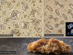
Common urinary crystals can also be mistaken for drug crystals. Uric acid, calcium oxalate, calcium phosphate, and amorphous urate or phosphate crystals may occur in people who are not taking a crystal-forming medication. Their shape and preferred urine pH can provide clues, but atypical forms can overlap. A patient taking amoxicillin who has calcium oxalate crystals does not automatically have amoxicillin crystalluria.
This is why medication timing and clinical context are essential. If a person has stable creatinine, normal urine output, no blood or pain, and only a few common crystals, medication-induced kidney injury may be unlikely. In contrast, a new abundance of unusual crystals appearing soon after high-risk drug exposure, especially with tubular cells or casts and a rising creatinine, is much more concerning.
Several interpretation mistakes are worth avoiding:
- Assuming every crystal is pathologic. Some crystal types can appear transiently in concentrated urine or after specimen cooling.
- Assuming every drug crystal means AKI. Crystalluria may precede injury or occur without a measurable creatinine change.
- Ignoring a rising creatinine because crystals are absent. Drug-related AKI can occur through mechanisms other than crystals, including direct tubular toxicity or acute interstitial nephritis.
- Treating the urine pH without identifying the drug. Solubility behavior differs among medications, so unsupervised acidification or alkalinization can be ineffective or harmful.
- Using one microscopy result in isolation. Repeat testing can be useful when the initial sediment is nondiagnostic and kidney function continues to worsen.
A negative microscopy result therefore does not completely exclude drug-related kidney injury, and a positive crystal result does not prove causation. The most reliable interpretation joins the sediment findings to the medication exposure, kidney-function trend, urine flow, and overall clinical picture.
When to Get Urgent Medical Care
Crystalluria itself is often found on a laboratory report rather than felt as a symptom. The urgency comes from signs of obstruction, acute kidney injury, severe dehydration, or a serious medication reaction.
Contact the prescribing clinician promptly if a urine report mentions abundant or unusual crystals while you are taking a medication known to crystallize, particularly if a recent creatinine level has increased. Do not stop an essential prescription on your own unless a clinician has instructed you to do so.
Seek urgent medical assessment for markedly reduced or absent urine, new visible blood in the urine, severe flank or abdominal pain, repeated vomiting, inability to keep fluids down, swelling with shortness of breath, severe weakness, confusion, or symptoms occurring during high-dose intravenous medication treatment. These can reflect clinically significant kidney injury or another urgent problem.
The most important point is that drug crystals are a signal to integrate with kidney function and medication exposure, not a stand-alone verdict. When recognized early, many medication-related crystalline kidney injuries improve after the offending exposure and contributing factors are corrected.
References
- Drug-Induced Acute Kidney Injury 2022 (Review)
- Amoxicillin crystalluria and amoxicillin-induced crystal nephropathy: a narrative review. 2025 (Review)
- Biology of drug-induced acute nephrotoxicity: focus on the tubulointerstitium 2026 (Review)
- Diagnostic Utility of Urine Microscopy in Kidney Diseases 2024 (Review)
- The EFLM European Urinalysis Guideline 2023 2024 (Guideline)
- Urinary Crystals Identification and Analysis 2024 (Review)
Disclaimer
Drug crystals in urine can range from an incidental laboratory finding to a sign of medication-related acute kidney injury. Interpretation and treatment depend on the specific medication, dose, kidney function, symptoms, and other laboratory findings. Do not stop or change a prescribed medicine solely because crystals were reported; contact the prescribing clinician for individualized guidance, and seek urgent care for markedly reduced urine, severe pain, visible blood, or symptoms of acute illness.