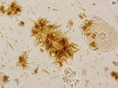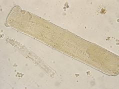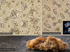

Struvite crystals in urine are magnesium ammonium phosphate crystals that often form in alkaline urine. Their most important association is with urinary infections caused by bacteria that produce the enzyme urease. Urease breaks down urea, raises urine pH, and creates conditions that favor struvite formation. On microscopy, struvite commonly appears as rectangular, colorless “coffin-lid” crystals, although shapes can vary. Finding a few crystals does not prove that a person has a kidney stone or an active infection. The result becomes more significant when struvite appears with persistently alkaline urine, urinary symptoms, bacteria, white blood cells, recurrent infections with urease-producing organisms, or imaging evidence of stones. Infection-related struvite stones can grow quickly and may form branching staghorn calculi that fill part of the kidney collecting system. Because bacteria can remain within a stone, treatment may require both infection control and complete stone management rather than antibiotics alone.
- Struvite is magnesium ammonium phosphate, a crystal that commonly forms in alkaline urine.
- Urease-producing urinary bacteria are the classic trigger; Proteus species are well known, but other organisms can also produce urease.
- Struvite crystals do not automatically mean a struvite stone: symptoms, urine culture, urine pH, and imaging determine the clinical significance.
- Recurrent UTI plus high urine pH and struvite crystals raises concern for an infection stone, especially with flank pain or a history of staghorn calculi.
- Fever or chills with flank pain and suspected urinary obstruction is an emergency because an infected obstructed kidney can progress to sepsis.
Table of Contents
- What Struvite Crystals Are
- Why Struvite Forms in Alkaline, Infected Urine
- Microscopy Appearance and Result Meaning
- Struvite Stones, Staghorn Calculi, and UTI Risk
- Testing and Diagnosis After Struvite Crystals Are Found
- Treatment, Prevention, and When to Seek Care
What Struvite Crystals Are
Struvite is a mineral made of magnesium ammonium phosphate hexahydrate. In urine microscopy, the term may also overlap with “triple phosphate,” an older name that refers to the same classic crystal type. A separate article on triple phosphate crystals covers that terminology in more detail.
Crystals form when dissolved substances in urine become supersaturated and begin to precipitate. For struvite, the chemical environment usually includes abundant magnesium, ammonium, and phosphate together with an alkaline pH. The ammonium and high pH are especially linked to urease activity from certain bacteria.
Struvite crystalluria is not the same thing as a struvite stone. Crystals can appear transiently in a urine specimen, while a stone is a solid structure that has grown within the urinary tract. Conversely, a person with a struvite stone may not shed obvious crystals in every urine sample. Microscopy therefore contributes evidence but cannot replace imaging. The same principle applies in reverse: an imaging-confirmed stone requires composition testing or clinical context before it is labeled struvite or an infection stone.
The finding also has different significance depending on the sample. Urine that sits too long before analysis can become more alkaline as bacteria multiply and urea breaks down. Crystals may then form after collection rather than inside the urinary tract. Proper specimen handling helps prevent this artifact.
The words struvite, triple phosphate, and magnesium ammonium phosphate are often used as if they are interchangeable in urine microscopy. In everyday laboratory reporting, that is usually reasonable. In stone analysis, however, composition can be more complex. Infection stones frequently contain struvite together with carbonate apatite or other minerals. A microscopy label describes the crystals seen in that specimen; it does not provide a full chemical analysis of a stone.
Unlike calcium oxalate or uric acid crystals, struvite has a particularly strong relationship to infection. That association is why clinicians pay attention when struvite appears together with bacteria, pyuria, urinary symptoms, or recurrent infections.
Why Struvite Forms in Alkaline, Infected Urine
The key chemical step is urease. Urease-producing microorganisms split urea into ammonia and carbon dioxide. Ammonia accepts hydrogen ions, making the urine more alkaline, while ammonium becomes available to combine with magnesium and phosphate. This promotes precipitation of struvite and often carbonate apatite.
Struvite formation generally favors urine above neutral pH, and infection stones often develop when urine is persistently strongly alkaline. A single alkaline result, however, is not proof of infection. Diet, medications, sample delay, and other metabolic factors can change urine pH. A urine pH test for kidney stones is useful only when interpreted with the rest of the clinical picture.
Proteus species are classic urease-producing urinary pathogens. Other organisms that may produce urease include some species of Klebsiella, Morganella, Providencia, Staphylococcus, and Corynebacterium. The exact organism matters because urease production varies by species and even by strain. Escherichia coli, the most common cause of routine UTI, is usually urease-negative, although it can coexist with other bacteria or be cultured in a person whose stone formed under different conditions.
This is one reason microscopy cannot identify the causative organism. Seeing struvite crystals may suggest a urease-driven process, but a urine culture is needed to determine which bacteria are growing and whether they are clinically significant.
The process can become self-reinforcing. An infection raises pH and promotes mineral precipitation. Once an infection stone develops, bacteria can persist within pores or biofilm on the stone. The stone then acts as a reservoir that makes infection harder to eradicate and increases the chance of recurrence.
Not every person with a urease-positive UTI forms a stone. Stone formation also depends on urinary anatomy, drainage, infection duration, mineral concentrations, prior stones, and other host factors. Conditions that impair urine flow, such as obstruction, urinary tract reconstruction, neurogenic bladder, or long-term catheter use, can increase the chance that bacteria persist and mineral deposits enlarge. This does not mean every person with these conditions develops struvite stones, but recurrent infection deserves closer attention. The presence of crystals shows that the urine environment can support precipitation at that moment; it does not predict with certainty that a stone will develop.
Microscopy Appearance and Result Meaning
Classic struvite crystals are colorless and rectangular with slanted ends, producing the familiar “coffin-lid” appearance. They may also form prism-like, feathered, or less typical shapes. Crystal appearance changes with concentration, pH, temperature, and how the sample is prepared.
Laboratories may report struvite qualitatively as rare, few, moderate, or many, or simply note that the crystals are present. There is no single universal crystal count that defines infection or stone disease. A high crystal burden can support concern when the rest of the findings point in the same direction, but quantity alone does not establish the diagnosis.
A useful interpretation looks at related results:
| Finding | How it changes interpretation |
|---|---|
| High urine pH | Supports the alkaline environment needed for struvite but is not specific for infection |
| White blood cells or leukocyte esterase | Supports urinary inflammation and can occur with UTI |
| Bacteria | Raises suspicion for infection but may also reflect contamination |
| Positive urine culture with a urease-producing organism | Strongly supports an infection-related mechanism |
| Blood in urine | Can occur with stones, infection, or another urinary lesion |
| Flank pain or imaging evidence of a stone | Makes clinically important stone disease much more likely |
The combination of crystals, pH, cells, and bacteria is part of urine microscopy, but culture and imaging answer different questions. Microscopy shows what is present in the urine sample. Culture identifies viable organisms. Imaging shows whether a stone, obstruction, or structural problem is present.
Sample age is a major source of error. Bacterial overgrowth after collection can increase urine pH, and cooling can cause additional crystals to precipitate. Laboratories therefore prefer fresh specimens or validated storage conditions. If crystals appear in a delayed or questionable sample and do not fit the patient’s symptoms, repeating a properly collected specimen can be more informative than assuming the finding is pathologic.
Struvite Stones, Staghorn Calculi, and UTI Risk
Struvite stones are often called infection stones because they are closely linked to urease-producing urinary infections. Many contain a mixture of struvite and carbonate apatite rather than pure struvite. They can enlarge faster than many metabolic stones because ongoing infection continually creates favorable chemistry.
A large infection stone can branch into several calyces of the kidney and form a staghorn calculus. Staghorn describes the shape rather than the exact chemical composition; not all staghorn stones are struvite, and not all struvite stones are staghorn. Still, infection stones are an important cause.
Symptoms vary. Some people have recurrent UTIs, flank discomfort, fever, blood in urine, or urinary odor. Others have surprisingly few symptoms despite a large stone. Chronic obstruction and recurrent infection can gradually damage kidney tissue. In severe cases, infection can spread into the bloodstream.
One clinically important situation is an infected obstructed urinary system. A stone that blocks urine flow while infection is present can raise pressure in the kidney and prevent adequate drainage. Antibiotics alone may not control the problem because infected urine remains trapped. Urgent drainage with a ureteral stent or nephrostomy may be needed before definitive stone removal.
Recurrent infection with the same urease-producing organism can be a clue that a stone or structural reservoir remains. Persistent alkaline urine and struvite crystals add supporting evidence but do not replace imaging. Likewise, recurrent infections should not automatically be blamed on a stone without evaluating bladder emptying, anatomy, catheters, sexual and hormonal factors, and other causes.
A bacteria finding on urine microscopy cannot distinguish infection from contamination by itself. Symptoms, collection quality, pyuria, and culture are needed, especially before deciding on antibiotics.
Testing and Diagnosis After Struvite Crystals Are Found
If struvite crystals are an isolated finding in a person without symptoms, clinicians may simply review urine pH, sample quality, and history. Persistent or abundant crystals, recurrent UTI, flank pain, fever, hematuria, or a known stone history warrants a more focused evaluation.
Urinalysis can show pH, leukocyte esterase, nitrite, blood, protein, white blood cells, red blood cells, bacteria, and crystals. Nitrite can support bacterial infection when positive, but it is not positive with every organism or every infection. A negative result cannot exclude UTI.
A urine culture is especially important when an infection stone is possible. Culture identifies the organism and can provide susceptibility results for antibiotic selection. A urine culture and sensitivity test is more informative than trying to infer the bacterium from crystal shape.
Imaging is used to determine whether a stone is actually present. Noncontrast CT is highly sensitive for urinary stones and can show size, location, branching, and obstruction. Ultrasound avoids radiation and can detect hydronephrosis and many renal stones, though it can miss smaller ureteral stones. The best test depends on symptoms, pregnancy status, age, prior imaging, and clinical urgency.
The degree of obstruction matters as much as the stone’s composition. A small ureteral stone can create an acute emergency if it blocks an infected kidney, while a large nonobstructing renal stone may present more gradually. Imaging can also show whether both kidneys are involved and whether there is thinning or damage from long-standing obstruction. In recurrent infection-stone disease, comparison with older scans helps determine whether a stone is new, growing, or residual after a previous procedure. These details guide decisions about drainage, timing of definitive removal, and follow-up more directly than the number of crystals seen under the microscope.
Blood tests may include creatinine and estimated GFR to assess kidney function, complete blood count when infection is suspected, and metabolic measurements when broader stone risk is being evaluated. In an acutely ill person, blood cultures and sepsis assessment may be necessary.
After a stone is removed, stone analysis can establish its composition. This is more definitive than assuming composition from urine crystals. A patient can form mixed stones, and crystal findings can change over time.
When cultures are repeatedly negative despite suspected infection stones, clinicians may review whether antibiotics were taken before collection, whether the sample was obtained correctly, and whether the organism is difficult to grow using routine methods. Stone cultures or cultures from the renal pelvis can sometimes differ from a standard bladder urine culture. This is especially relevant around surgery, where infection risk depends on bacteria that may be harbored within the stone itself.
A second common mistake is treating urine pH as a diagnosis. Strongly alkaline urine supports struvite chemistry, but pH can rise after a specimen stands, and diets or medications can also make urine more alkaline. Likewise, a normal pH on one day does not completely exclude an infection stone because pH fluctuates and treatment may have changed the urine environment. Repeated results and culture data are more useful than one isolated number.
People with recurrent stones may also undergo a metabolic stone evaluation, often including one or more 24-hour urine collections. Infection stones have a different primary mechanism from calcium or uric acid stones, but a patient can have overlapping metabolic risk factors. Identifying them can guide prevention after infection and stone burden are addressed.
Treatment, Prevention, and When to Seek Care
Treatment depends on whether the finding represents crystals only, an active UTI, or an infection stone. Crystals alone are not treated with antibiotics. If there is a symptomatic bacterial infection, antibiotics are selected according to clinical severity, local resistance patterns, and culture results when available.
Established struvite stones often require urologic treatment because bacteria can persist inside the stone. Complete stone clearance is an important goal when feasible. Large or staghorn stones are commonly managed with procedures such as percutaneous nephrolithotomy, sometimes combined with staged or additional techniques. The exact plan depends on stone size, anatomy, kidney function, infection, and surgical risk.
Antibiotics may be used before, around, or after stone procedures depending on culture results and infection risk. Long-term suppressive antibiotics are not appropriate for everyone. In selected patients who cannot achieve complete stone removal, specialists may consider additional medical strategies, including urease inhibition, but these treatments can have significant adverse effects and require close supervision.
Prevention focuses on removing infection reservoirs, treating recurrent infections appropriately, maintaining adequate hydration when medically safe, and correcting anatomic or emptying problems when possible. People who use long-term urinary catheters or have neurogenic bladder may have additional infection and drainage risks, so prevention plans often include catheter and bladder-management strategies rather than diet alone. Follow-up imaging may also be used after treatment because residual fragments can serve as a starting point for recurrent infection stone growth. Simply trying to acidify urine at home is not a reliable treatment for an infection stone and can create other risks. The underlying infection and stone burden need direct assessment.
Seek prompt medical care for recurrent UTI symptoms, persistent high urine pH with struvite crystals, repeated growth of urease-producing organisms, flank pain, visible blood, or a known stone with new urinary symptoms. A UTI urinalysis can help identify inflammatory and bacterial clues, but culture is often needed in recurrent or complicated cases.
Seek emergency care for fever or shaking chills with severe flank pain, vomiting with inability to keep fluids down, confusion, fainting, very low urine output, or signs of sepsis, particularly when a stone or obstruction is possible. Infected obstruction is a urologic emergency because rapid drainage can be lifesaving.
The practical message is to separate three findings: struvite crystals, urinary infection, and struvite stones. They are related but not interchangeable. When all three align—alkaline urine, a urease-producing infection, and imaging-confirmed stone disease—the case needs coordinated infection and stone management rather than treatment of the microscopy result alone.
References
- Struvite and Triple Phosphate Renal Calculi 2023 (Review)
- International Alliance of Urolithiasis (IAU) guideline on staghorn calculi management 2024 (Guideline)
- Unraveling the association of bacteria and urinary stones in patients with urolithiasis: an update review article 2024 (Review)
- Spectrum of Bacterial Pathogens from Urinary Infections Associated with Struvite and Metabolic Stones 2023
- Urinary Crystals Identification and Analysis 2024 (Review)
- The EFLM European Urinalysis Guideline 2023 2024 (Guideline)
Disclaimer
This article provides general information about struvite crystals, infection stones, and urinary infection and does not diagnose or treat an individual condition. Crystal findings should be interpreted with symptoms, urine pH, culture results, kidney function, and imaging when indicated. Fever with severe flank pain or suspected urinary obstruction requires urgent medical evaluation.