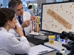
Triple phosphate crystals are usually another name for struvite, a magnesium ammonium phosphate crystal that forms most readily in alkaline urine. On urine microscopy, they classically look like colorless rectangular prisms with angled ends, often described as “coffin-lid” crystals. Their main clinical importance is the connection with urinary infections caused by urease-producing bacteria. These organisms split urea, generate ammonia, and raise urine pH, creating the chemical conditions in which triple phosphate can precipitate. A crystal report alone does not prove a urinary tract infection or an infection stone, because urine can become alkaline for other reasons and crystals may form after a specimen sits too long. Concern rises when triple phosphate crystals occur with urinary symptoms, pyuria, bacteria, persistently high pH, recurrent cultures growing urease-producing organisms, or imaging evidence of a stone. Infection-related stones may enlarge into staghorn calculi and can be difficult to eradicate unless both the infection and the stone burden are addressed.
- Triple phosphate crystals are generally struvite crystals, made of magnesium ammonium phosphate.
- Their classic shape is a colorless “coffin-lid” prism, although less typical forms can occur.
- They are strongly associated with alkaline urine and urease-producing bacteria, especially in recurrent or complicated urinary infections.
- Crystals alone do not diagnose a UTI or kidney stone; culture, symptoms, urine pH, and imaging determine the significance.
- Fever with flank pain, vomiting, low urine output, or suspected obstruction needs urgent care because an infected blocked urinary system can become life-threatening.
Table of Contents
- What Triple Phosphate Crystals Are
- Microscopy Appearance and the Role of Urine pH
- Triple Phosphate, UTI, and Urease-Producing Bacteria
- From Crystals to Struvite and Staghorn Stones
- Testing and Interpreting an Abnormal Result
- Treatment, Prevention, and When to Seek Care
What Triple Phosphate Crystals Are
The term triple phosphate is a traditional laboratory name for crystals composed mainly of magnesium ammonium phosphate. It remains common on urinalysis reports even though “struvite” is the mineral term more often used in stone literature and urology. The mineral name is struvite. In routine urine microscopy, “triple phosphate crystals” and “struvite crystals” generally refer to the same classic crystal type.
The older name reflects the combination of magnesium, ammonium, and phosphate found within the microscopic crystal structure itself. The full mineral formula includes water molecules as part of the crystal structure. These chemicals are normally present in urine in dissolved form, but they can precipitate when concentrations and pH favor crystallization.
The strongest driver is an alkaline environment rich in ammonium. This is why triple phosphate has a much closer relationship to infection than many other common urinary crystals. Urease-producing bacteria create ammonium and raise pH, making precipitation much easier.
Finding crystals does not prove a stone exists, and the absence of crystals on one sample does not rule out an existing struvite stone. Crystalluria means that crystals are present in the urine sample. A kidney stone is a larger solid structure that has developed within the urinary tract. Some people shed crystals without having an imaging-visible stone, while people with established stones may have no crystals in a particular specimen.
A related page on struvite crystals and infection stones discusses the mineral and stone relationship from the struvite perspective. In practice, the two terms often appear on different laboratory reports for the same basic crystal.
Triple phosphate also should not be confused with every crystal that occurs in alkaline urine. Calcium phosphate and amorphous phosphate can also precipitate at higher pH, but they have different chemistry and clinical associations. Correct identification matters because classic triple phosphate raises a more specific question about urease-producing infection.
Microscopy Appearance and the Role of Urine pH
Triple phosphate crystals are classically colorless rectangular prisms with oblique ends, producing the familiar “coffin-lid” appearance. The shape can be striking when well formed, but not every crystal looks textbook-perfect. Variants can appear as elongated prisms, feathered forms, or irregular aggregates.
Microscopic appearance is influenced by urine concentration, temperature, pH, and how long the specimen stands before analysis. A crystal that forms after collection may look identical to one that formed in the urinary tract, which is why pre-analytic handling matters.
Triple phosphate favors alkaline urine. A urine pH above 7 supports the chemical environment, and infection stones often develop in more strongly alkaline conditions. However, one alkaline pH result is not diagnostic of a urease-positive infection. Meals, medications, renal acid-base handling, vegetarian dietary patterns, and delayed specimen processing can all raise urine pH.
A delayed specimen is particularly important. Bacteria can multiply while urine sits at room temperature, convert urea, and make the sample more alkaline. The sample can also cool, changing the solubility of several salts. Together, these pre-analytic changes can produce a crystal pattern that was less prominent when the urine was freshly voided. This is why an unexpected result should be checked against specimen collection and processing times rather than interpreted in isolation. That shift can trigger phosphate crystallization in the cup. A fresh sample or properly preserved specimen gives a more reliable picture of what was happening in the body.
A urine pH test is most useful when interpreted with microscopy, symptoms, and culture. Repeated strongly alkaline results are more meaningful than one isolated value, especially when the same urease-producing organism keeps appearing.
Laboratories may report crystals as rare, few, moderate, many, or present without a standardized count. There is no universal “normal range” for triple phosphate that predicts stone formation. A few crystals in an old alkaline sample can be less important than a persistent pattern of crystals, pyuria, and recurrent culture-proven infection.
Other phosphate crystals create common microscopy pitfalls. Amorphous phosphates look granular rather than coffin-shaped. Calcium phosphate can form wedges, rosettes, or plates. If morphology is uncertain, fresh manual microscopy and the urine chemistry can help. Rare or atypical particles may also be reviewed by an experienced laboratory professional rather than relying only on automated classification.
This distinction has practical consequences. Calcium phosphate crystalluria can accompany metabolic stone disease without any bacterial infection, while classic triple phosphate prompts a closer look for urease-producing organisms. Treating every alkaline phosphate crystal as struvite can therefore lead to unnecessary antibiotics, while dismissing repeated coffin-lid crystals in a patient with recurrent Proteus infections can miss an infection-stone pattern. The laboratory shape, urine pH, culture history, and imaging should point in the same direction before a stone is labeled infection-related. Stone analysis remains the best way to establish the composition of material that has actually formed a calculus.
Triple Phosphate, UTI, and Urease-Producing Bacteria
The link between triple phosphate and UTI is based on bacterial urease. This enzyme breaks urea into ammonia and carbon dioxide. Ammonia then shifts urine toward alkalinity and supplies ammonium ions. Magnesium, ammonium, and phosphate can combine and precipitate as struvite.
Proteus species are classic urease-producing urinary pathogens. Other organisms can also produce urease, including some strains or species of Klebsiella, Morganella, Providencia, Staphylococcus, and Corynebacterium. The exact pattern varies, so crystal type cannot tell which organism is present.
This is why urine culture and susceptibility testing is important in recurrent or complicated cases. Culture identifies viable bacteria and can help select an effective antibiotic. Microscopy may show bacteria, but it cannot reliably determine the species or antibiotic resistance profile.
Escherichia coli causes many ordinary UTIs but is usually urease-negative. A patient can still have E. coli in the urine and triple phosphate crystals for unrelated reasons, or E. coli can coexist with another organism. Laboratory findings should therefore be integrated rather than forcing every result into one explanation.
Symptoms that make infection more likely include burning with urination, urgency, frequency, lower abdominal discomfort, foul-smelling urine, fever, chills, and flank pain. White blood cells, leukocyte esterase, and bacteria provide additional evidence. Nitrite can be positive with some urinary pathogens but may remain negative despite true infection.
| Finding | Interpretation |
|---|---|
| Triple phosphate crystals alone | Shows alkaline phosphate crystallization but does not prove infection |
| Crystals plus high pH and pyuria | Raises concern for urinary inflammation and possible urease-positive UTI |
| Crystals plus recurrent Proteus culture | Strongly supports an infection-related environment |
| Crystals plus flank pain and stone on imaging | Raises concern for a struvite or mixed infection stone |
| Crystals in an old specimen without symptoms | May reflect post-collection precipitation or bacterial overgrowth |
A UTI should not be diagnosed from crystal morphology alone. Likewise, antibiotics should not be started solely because “triple phosphate” appears on a microscopy report. Symptoms, culture, pregnancy status, urinary anatomy, catheter use, and overall illness determine whether treatment is indicated.
From Crystals to Struvite and Staghorn Stones
Persistent urease-positive infection can create the conditions for infection stones. Struvite often forms with carbonate apatite, so an infection stone may be chemically mixed rather than pure magnesium ammonium phosphate.
These stones can grow rapidly because the bacteria continue altering urine chemistry. A large stone may extend through the renal pelvis and several calyces, creating a branching staghorn calculus. Staghorn describes shape, not composition: some staghorn stones are metabolic rather than infection-based, and some struvite stones never become staghorn.
The presence of a stone changes the infection problem. Bacteria can remain protected within the stone matrix or biofilm, making eradication difficult even when a bladder urine culture temporarily becomes negative. Recurrent infection with the same urease-producing organism can therefore be a clue that a residual stone or structural reservoir remains.
Possible symptoms include recurrent UTI, flank discomfort, blood in urine, fever, or urinary odor, but large stones can sometimes produce few symptoms. Over time, obstruction, recurrent inflammation, and infection can damage kidney tissue.
One of the most dangerous situations is infection behind an obstruction. A stone can block urine flow while bacteria multiply in the collecting system. The kidney becomes pressurized and infected urine cannot drain. This can progress quickly to sepsis. Emergency management may require drainage with a ureteral stent or percutaneous nephrostomy before definitive stone removal.
Crystals are therefore best viewed as an early clue to a urine environment, not proof that this dangerous scenario exists. Imaging is what determines whether a stone or obstruction is present. Noncontrast CT is highly sensitive for urinary stones, while ultrasound can identify many kidney stones and hydronephrosis without radiation.
Stone analysis after removal provides the most direct information about composition. It can confirm struvite and identify other mineral components. This is more reliable than assuming the composition of an imaging-visible stone from one urine crystal result.
Testing and Interpreting an Abnormal Result
When triple phosphate crystals are reported, the first question is whether the person has symptoms or a history that makes infection stones plausible. An asymptomatic person with a single crystal finding may need only review of sample quality and urine pH. A person with recurrent febrile UTI, persistently alkaline urine, or flank pain needs a more thorough assessment.
A repeat urinalysis can be useful if the specimen was old, contaminated, or unexpected. The repeat should be collected according to laboratory instructions and analyzed promptly. The rest of the urinalysis helps determine whether crystals are isolated or accompanied by blood, pyuria, leukocyte esterase, nitrite, bacteria, protein, or other abnormalities.
Urine culture is central when infection is suspected. In complicated stone disease, clinicians may compare bladder cultures with cultures obtained closer to the kidney or from the stone during a procedure because bacteria inside a stone can differ from organisms recovered in a routine voided specimen.
Imaging is appropriate when symptoms or recurrent infection suggest a stone. CT provides detailed information about stone size, location, branching, and obstruction. Ultrasound may be favored in pregnancy, in some younger patients, or when avoiding radiation is a priority. The choice depends on the clinical question and urgency.
Blood tests can include creatinine and estimated GFR, complete blood count, and markers of systemic infection. If a person is acutely ill, blood cultures and lactate or other sepsis-related tests may be required. These tests assess consequences that urine crystals cannot measure.
Patients with recurrent stones may undergo metabolic testing after acute infection and obstruction are controlled. A 24-hour urine collection can assess volume, calcium, citrate, oxalate, uric acid, sodium, pH, and other variables. Even when infection is the main driver, a patient can have additional metabolic risks that make future stones more likely.
Several interpretation mistakes are common:
- assuming that all alkaline urine means infection;
- assuming that every triple phosphate crystal means a stone;
- treating crystals with antibiotics without evidence of symptomatic infection;
- dismissing recurrent urease-positive cultures after a temporary response to antibiotics;
- relying on a single voided culture when a large infection stone is already known;
- trying to identify stone composition from urine microscopy alone.
A structured approach avoids both unnecessary treatment and missed infection stones.
Treatment, Prevention, and When to Seek Care
Treatment depends on the underlying problem. An isolated crystal finding does not need a medication. A symptomatic UTI is treated with an appropriate antibiotic, often guided by culture in complicated or recurrent disease. A confirmed infection stone may require urologic removal in addition to antibiotics.
Complete stone clearance is important because residual fragments can harbor bacteria and serve as a starting point for regrowth. Large or staghorn stones are often treated with percutaneous nephrolithotomy, sometimes in more than one stage or with additional procedures. The treatment plan depends on anatomy, stone burden, kidney function, infection status, and procedural risk.
Antibiotics are used around stone treatment when infection is present or the procedure carries infection risk. Long-term suppressive therapy is reserved for selected cases rather than used automatically. A urease inhibitor may be considered in unusual situations when complete stone clearance is not possible, but adverse effects limit its use and specialist supervision is required.
Prevention focuses on eliminating persistent infection when possible, correcting urinary obstruction or drainage problems, maintaining adequate urine volume when medically safe, and following up after stone treatment. Follow-up can include repeat cultures and imaging, especially after treatment of a large infection stone, because small residual fragments may enlarge again. The schedule is individualized to stone burden, procedure, kidney function, infection history, and recurrence risk rather than based on the crystal count. People with long-term catheters, neurogenic bladder, urinary reconstruction, or recurrent obstruction may need individualized strategies because bacterial colonization and impaired drainage increase complexity.
Trying to make urine acidic with supplements or extreme diets is not a substitute for infection and stone treatment. Urine chemistry is influenced by many factors, and aggressive self-treatment can cause electrolyte problems or interact with medical conditions. The main modifiable target in true infection stones is the urease-producing infection and any retained stone material.
Contact a healthcare professional for recurrent burning or urgency, repeated high-pH urine with triple phosphate crystals, recurrent cultures growing urease-producing organisms, visible blood, or flank pain. A UTI urinalysis can help characterize the inflammatory pattern, but culture is often needed in recurrent or complicated infection.
Seek emergency care for fever or shaking chills with severe flank pain, persistent vomiting, confusion, fainting, very low urine output, or known stone disease with signs of infection. Infection plus obstruction is a time-sensitive urologic emergency.
The key is to interpret triple phosphate crystals as one part of a chain: alkaline urine can permit crystallization, urease-producing bacteria can drive the chemistry, and persistent infection can promote struvite stone growth. Identifying which links are actually present determines whether the right next step is repeat testing, culture, imaging, antibiotic treatment, or urologic stone management.
References
- Struvite and Triple Phosphate Renal Calculi 2023 (Review)
- International Alliance of Urolithiasis (IAU) guideline on staghorn calculi management 2024 (Guideline)
- Unraveling the association of bacteria and urinary stones in patients with urolithiasis: an update review article 2024 (Review)
- Spectrum of Bacterial Pathogens from Urinary Infections Associated with Struvite and Metabolic Stones 2023
- Urinary Crystals Identification and Analysis 2024 (Review)
- The EFLM European Urinalysis Guideline 2023 2024 (Guideline)
Disclaimer
This article provides general information about triple phosphate crystals, struvite, urinary infection, and infection stones and is not a diagnosis. Crystal results should be interpreted with urine pH, symptoms, culture results, kidney function, and imaging when appropriate. Fever with severe flank pain or suspected urinary obstruction requires urgent medical evaluation.