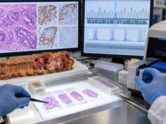
An FGFR2 fusion test looks for rearrangements that join the fibroblast growth factor receptor 2 gene to another gene and create an abnormally active growth signal. These alterations are concentrated in intrahepatic cholangiocarcinoma (iCCA), where they occur in roughly 10%–15% of cases in many series. A clinically relevant positive result can identify a patient with previously treated advanced disease who may benefit from an FGFR inhibitor. The test is technically more complex than a simple point-mutation assay because FGFR2 has many possible fusion partners and breakpoints. Partner-agnostic RNA-based next-generation sequencing is especially useful because it detects the expressed fusion transcript directly, while some DNA-only panels can miss rearrangements in large intronic regions. FISH can detect a rearrangement but does not always show the partner or whether the resulting transcript is in-frame and functional. A positive report should therefore be interpreted using the exact alteration, assay method, tumor location, treatment setting, and current drug indication.
- FGFR2 fusions/rearrangements are most common in intrahepatic cholangiocarcinoma and are uncommon in extrahepatic disease.
- A positive clinically relevant FGFR2 fusion can support treatment with an approved FGFR inhibitor in defined advanced-disease settings.
- RNA-based NGS is often preferred for fusion detection because it directly identifies expressed fusion transcripts and can be partner-agnostic.
- A negative DNA-only test does not always exclude an FGFR2 fusion if intronic coverage is limited or tissue quality is poor.
- Acquired FGFR2 kinase-domain mutations can cause resistance during FGFR inhibitor treatment and may be detectable by repeat tissue or ctDNA testing.
Table of Contents
- What an FGFR2 Fusion Is
- Who Should Be Tested
- FGFR2 Testing Methods
- What a Positive Result Means
- Targeted Treatment Implications
- Resistance and Repeat Testing
- Negative, Indeterminate, and Uncertain Results
What an FGFR2 Fusion Is
FGFR2 is a receptor tyrosine kinase. Under normal conditions, binding of fibroblast growth factors causes receptors to dimerize and activate downstream pathways such as RAS–MAPK and PI3K–AKT. These signals help regulate cell growth, survival, and differentiation.
In some intrahepatic cholangiocarcinomas, a chromosomal rearrangement joins the 5′ portion of FGFR2 to the 3′ portion of another gene. The partner often supplies a dimerization domain or otherwise removes normal regulatory control, allowing the FGFR2 kinase to signal continuously.
Many different partner genes have been reported. BICC1 is one of the most common, but dozens of others exist. This diversity is why a test that searches only for a short list of known partners can miss clinically relevant fusions.
The clinically important rearrangements generally preserve the FGFR2 kinase domain. A report should therefore distinguish a clearly oncogenic in-frame fusion from a rearrangement of uncertain functional significance.
FGFR2 fusions are not the same as FGFR2 amplification or a single-nucleotide mutation. Those alteration classes can have different biological and therapeutic meaning.
They are also not inherited in the usual hereditary-cancer sense. In cholangiocarcinoma, FGFR2 fusions are typically somatic changes confined to the tumor.
Who Should Be Tested
Patients with advanced or metastatic intrahepatic cholangiocarcinoma are the highest-priority group because FGFR2 fusions are enriched in this anatomic subtype and can change systemic treatment options.
Current biliary-tract guidance supports broad molecular profiling early in advanced disease rather than waiting until chemotherapy has failed. The reason is practical: tissue sequencing and RNA testing can take time, and a new biopsy may be difficult to arrange when the patient needs a second-line decision.
Testing can also be reasonable in earlier-stage iCCA when adequate tissue is available, especially if recurrence risk is high and the molecular information may become useful later. Institutional practice varies.
Extrahepatic cholangiocarcinomas rarely harbor FGFR2 fusions, but broad NGS panels may still include FGFR2 because an unusual actionable alteration can occasionally occur. The entire profile matters more than a single prevalence estimate.
A cholangiocarcinoma biomarker panel typically tests FGFR2 together with IDH1, BRAF, HER2, MSI/MMR, NTRK, RET, and other targets so tissue is used efficiently.
FGFR2 Testing Methods
The method matters more for FGFR2 than for many point mutations because fusion breakpoints are structurally diverse.
RNA-based next-generation sequencing
RNA-based NGS directly reads the fusion transcript produced by the tumor. Partner-agnostic hybrid-capture or single-primer approaches can identify novel partners because they do not require the laboratory to guess the fusion in advance.
RNA has a biological advantage: if an in-frame fusion is expressed, the assay sees the chimeric message that can produce the abnormal protein. The disadvantage is that RNA degrades more readily than DNA in formalin-fixed tissue, so old or poorly preserved specimens may fail quality control.
DNA-based next-generation sequencing
DNA panels can detect point mutations, insertions/deletions, copy-number changes, and many rearrangements in one assay. The challenge is that FGFR2 breakpoints often occur in introns that are large, repetitive, or incompletely covered. A DNA-only negative result may therefore be falsely reassuring if the panel has weak intronic coverage.
When DNA shows a suspicious FGFR2 rearrangement with an unknown partner, reflex RNA testing can confirm whether an oncogenic transcript is actually expressed.
FISH
Break-apart fluorescence in situ hybridization can show that the FGFR2 locus is rearranged. It does not require knowledge of the partner and can work on limited tissue. However, FISH usually cannot identify the partner gene, determine the precise breakpoint, or prove that the rearrangement creates an expressed in-frame fusion.
Because each method has strengths and weaknesses, combined DNA/RNA profiling is often the most complete strategy when tissue allows.
What a Positive Result Means
A clearly positive result indicates an FGFR2 fusion or qualifying rearrangement that is considered an oncogenic driver. The report may list the fusion as FGFR2::BICC1, FGFR2-BICC1, or another partner combination.
A useful report should include:
- the exact fusion partner or rearrangement description;
- the exons involved when known;
- whether the fusion is predicted or confirmed to be in-frame;
- the testing platform and specimen type;
- supporting read counts or quality metrics when reported; and
- an interpretation of clinical actionability.
Not every structural change near FGFR2 is automatically actionable. Some rearrangements may not preserve the kinase domain or may not produce a functional transcript. A molecular pathologist may need to interpret unusual breakpoints.
FGFR2 positivity is primarily a predictive biomarker. It identifies a tumor dependency that can be targeted pharmacologically. It is not a serum marker and does not quantify tumor burden.
The presence of an FGFR2 fusion can also correlate with a distinctive clinical phenotype, including occurrence in younger patients and certain histologic or molecular features, but these associations do not replace the actual molecular test.
Targeted Treatment Implications
FGFR inhibitors have established the clinical value of testing. Pemigatinib and futibatinib are examples of FGFR-targeted drugs used for previously treated advanced cholangiocarcinoma with qualifying FGFR2 fusions or rearrangements, depending on regulatory region and label.
These drugs inhibit FGFR signaling and can produce objective tumor responses in a substantial subset of molecularly selected patients. The benefit is not universal, and responses can eventually be limited by acquired resistance.
FGFR inhibitors also have class-specific adverse effects. Common issues include hyperphosphatemia, nail and skin changes, dry mouth, diarrhea, fatigue, and ocular toxicity. Phosphate levels and ophthalmologic symptoms require monitoring. Futibatinib is an irreversible FGFR inhibitor, while pemigatinib binds reversibly; their resistance profiles can differ.
A positive FGFR2 result does not mean treatment should begin before first-line therapy when the approval applies to previously treated disease. The exact line of therapy, prior regimen, performance status, and current guideline should be checked.
FGFR2 is one target among several in iCCA. An IDH1/IDH2 result, for example, can define a different targeted-treatment pathway.
Resistance and Repeat Testing
Cancer cells can evolve under the selective pressure of FGFR inhibition. One of the best-described resistance mechanisms is acquisition of secondary FGFR2 kinase-domain mutations that reduce drug binding or reactivate signaling.
Multiple resistance mutations can arise at the same time in different tumor clones. This creates a “polyclonal” resistance pattern that may be difficult to capture with a single tissue biopsy.
Circulating tumor DNA can be useful in this setting because plasma may sample DNA from several metastatic sites at once. Emerging kinase-domain mutations can sometimes be detected before or at radiographic progression.
Repeat molecular testing after progression can therefore answer a different question from the original diagnostic test. The first test asks, “Is FGFR2 driving this cancer?” A resistance test asks, “How has the FGFR2-driven cancer changed under treatment?”
Resistance information may support enrollment in clinical trials of next-generation FGFR inhibitors designed to overcome specific mutations. It may also explain why a tumor that initially responded has begun growing again.
Not every progression requires repeat testing, and no resistance finding automatically dictates an approved next therapy. The value depends on available trials, tissue access, ctDNA shedding, and the patient’s treatment goals.
Negative, Indeterminate, and Uncertain Results
A negative result is only as strong as the assay used. If a high-quality partner-agnostic RNA panel is negative with adequate tumor content, a clinically relevant expressed FGFR2 fusion becomes unlikely. If a small DNA-only panel with limited intronic coverage is negative, residual uncertainty may be higher.
An indeterminate result means the test could not answer the question. Reasons include low tumor percentage, degraded RNA, insufficient nucleic acid, failed sequencing quality, or ambiguous FISH signal. Indeterminate should not be translated into “FGFR2 wild type.”
An uncertain rearrangement may identify a structural event without enough evidence that it creates an oncogenic in-frame product. Reflex RNA sequencing or expert molecular-pathology review can clarify such findings.
When a test is negative but clinical need remains high, ask:
- Was RNA fusion testing included?
- Was the assay partner-agnostic?
- Was tumor content adequate?
- Did the DNA panel cover FGFR2 introns well?
- Is another tumor block available?
- Could plasma ctDNA or a repeat biopsy provide additional information?
The goal is not to repeat testing indefinitely. It is to make sure that a potentially actionable fusion was not missed because the first method was technically unsuited to the alteration type.
A final practical point is that FGFR2 fusion status usually remains stable as the founding driver, while resistance mutations can change over time. That distinction explains why initial tumor profiling and later ctDNA testing can both be useful without asking the same question twice.
Why partner-agnostic testing matters
FGFR2 can fuse with many different genes. A closed assay that contains primers only for a few known partners may detect common fusions but miss a novel or rare partner that activates the receptor in the same way. Partner-agnostic methods are designed to find the rearrangement even when the laboratory did not predict the partner in advance.
Hybrid-capture RNA sequencing and certain single-primer extension methods are especially useful for this purpose. They can identify the exact fusion junction and often show whether the transcript is in-frame. This level of detail helps a molecular pathologist determine whether the event is likely to be a true oncogenic driver.
A report that says “FGFR2 rearrangement detected by FISH” can still be clinically important, but additional RNA characterization may clarify an unusual case, especially if the structural event does not fit a typical pattern.
How fusion structure determines whether FGFR2 remains oncogenic
The transforming activity of an FGFR2 fusion depends on preserving the receptor’s kinase domain while disrupting normal regulatory control. In intrahepatic cholangiocarcinoma, clinically important rearrangements commonly involve the region around exon 17/intron 17 and retain the kinase-coding portion of the gene.
The partner gene often contributes a domain that promotes dimerization, allowing two fusion proteins to pair without the normal fibroblast growth factor signal. This creates constitutive downstream signaling.
A rearrangement that removes the kinase domain or creates an out-of-frame transcript may not have the same meaning. This is why the exact breakpoint and transcript structure can matter when a report identifies a novel fusion.
What an FGFR2-positive result does not tell you
The result does not show tumor burden, stage, or how quickly the cancer is growing. A small localized iCCA and a widely metastatic iCCA can both carry the same fusion. Imaging and pathology determine extent of disease.
The result also does not guarantee response to an FGFR inhibitor. Some tumors have primary resistance, and others respond initially but later acquire secondary mutations or activate alternative pathways. Treatment benefit is a probability derived from trials, not a certainty created by the biomarker.
Finally, the result is usually not inherited. Family members do not need germline FGFR2 testing solely because a cholangiocarcinoma fusion was found. If the broader tumor profile suggests a possible hereditary alteration in another gene, that separate finding may justify germline evaluation.
How to read a fusion report line by line
Start with the specimen: tissue, cytology, or plasma. Next, identify whether the assay was DNA, RNA, FISH, or a combined method. Then find the exact alteration and ask whether the report calls it pathogenic, oncogenic, or clinically actionable.
If a partner gene is listed, check whether the fusion is described as in-frame. If the partner is unknown, determine whether the report still establishes a qualifying FGFR2 rearrangement for the intended therapy. Look for comments about assay limitations, low tumor fraction, or failed RNA quality.
Finally, separate the diagnostic finding from the treatment recommendation. Molecular laboratories may list potential drugs based on the gene, but the oncologist must verify the cancer type, line of therapy, regulatory approval, prior treatment, organ function, and current guideline before prescribing.
This stepwise reading prevents a common precision-oncology error: assuming that any alteration in an actionable gene is equivalent to the exact alteration studied in the pivotal trials.
Why archival tissue can still be useful
An FGFR2 fusion is usually a founding driver, so an archival specimen from the original intrahepatic tumor can remain informative even if the patient later develops metastatic disease. If the block contains adequate viable tumor and RNA quality is acceptable, testing does not always require a fresh biopsy.
A new specimen becomes more valuable when the old tissue is exhausted, RNA has degraded, or the clinical question has shifted from finding the original fusion to studying acquired resistance. The age of the sample alone is therefore less important than its quality and the question being asked.
Bottom-line interpretation
An FGFR2 result is only as useful as the method that produced it. A clearly expressed, in-frame fusion detected by a partner-agnostic RNA assay provides strong evidence of an actionable driver in iCCA. A negative low-coverage DNA assay provides less certainty, while an indeterminate assay provides no true answer at all. Reading the method and quality notes is therefore part of interpreting the biomarker, not a technical detail to skip.
That distinction can prevent a potentially useful target from being missed or an uncertain rearrangement from being overtreated.
When treatment decisions depend on the result, the molecular report should be read together with the pathology diagnosis and tumor site. FGFR2 fusions are most characteristic of intrahepatic cholangiocarcinoma, particularly small-duct tumors, and are much less typical of extrahepatic bile duct cancer or gallbladder cancer. Confirming the anatomic diagnosis therefore strengthens the meaning of a positive result. It also prevents a technically correct fusion call from being applied to the wrong clinical pathway when the specimen comes from a metastatic site and the primary tumor location is uncertain.
References
- SEOM-GEMCAD-TTD clinical guidelines for biliary tract cancer (2025) 2025 (Guideline)
- ESMO Clinical Practice Guideline interim update on the management of biliary tract cancer 2025 (Guideline)
- FGFR2 testing in cholangiocarcinoma: translating molecular studies into clinical practice 2023 (Review)
- FGFR2 fusion/rearrangement analysis in intrahepatic cholangiocarcinoma using DNA/RNA-based NGS and FISH 2025 (Review)
- Molecular Testing for Intrahepatic Cholangiocarcinoma: What, When, How? 2026 (Review)
Disclaimer
FGFR2 fusion testing should be interpreted by oncology and molecular-pathology professionals using the exact alteration, assay method, tumor location, prior therapy, and current drug indications. A positive fusion creates a potential targeted-treatment option but does not guarantee response, and a negative result may require review of assay coverage if fusion detection was technically limited. Treatment changes should not be made from a molecular report without full clinical assessment.