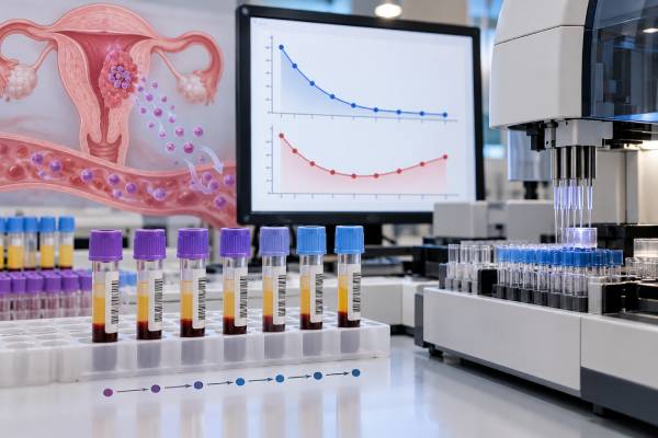
The hCG test is the central tumor-marker test for gestational trophoblastic disease (GTD). Human chorionic gonadotropin is produced by trophoblastic tissue, so blood hCG usually falls after a molar pregnancy is evacuated and after gestational trophoblastic neoplasia (GTN) responds to treatment. Serial values are more informative than one number. A falling trend supports regression; a sustained plateau or confirmed rise can signal persistent GTN and requires specialist review. After hCG normalizes, follow-up continues for a defined period because recurrence is uncommon but possible, and a new pregnancy can make hCG impossible to interpret as a tumor marker. Very low persistent hCG also has several noncancer explanations, including a new pregnancy, pituitary hCG, assay interference from heterophile antibodies, and quiescent trophoblastic disease. The safest interpretation uses the same validated assay when possible, considers the clinical setting, and follows a GTD center’s surveillance schedule rather than relying on a single cutoff.
- Serial quantitative serum hCG is the key marker for monitoring molar pregnancy and GTN, because the direction and rate of change matter more than one isolated value.
- After evacuation of a molar pregnancy, hCG should progressively decline; a sustained plateau or rise can meet criteria for post-molar GTN and needs specialist evaluation.
- Follow-up after hCG normalization depends on whether the pregnancy was a complete or partial mole and on whether chemotherapy was required, so surveillance duration is not identical for every patient.
- A new increase after prior normalization does not automatically mean recurrence; pregnancy and laboratory interference must also be excluded.
- Very low persistent hCG needs careful confirmation before treatment, because “phantom hCG,” pituitary hCG, and quiescent GTD can mimic active disease.
Table of Contents
- Why hCG Is Used as a GTD Tumor Marker
- hCG After a Molar Pregnancy Is Evacuated
- What a Plateau or Rising hCG Level Means
- hCG Monitoring During GTN Treatment
- Follow-Up After hCG Normalization
- Persistent Low-Level hCG and False Positives
- Recurrence, Pregnancy, and Next Steps
Why hCG Is Used as a GTD Tumor Marker
Gestational trophoblastic disease begins in placental trophoblast rather than in the ovary or uterus itself. The spectrum includes complete and partial hydatidiform moles as well as malignant forms grouped under gestational trophoblastic neoplasia, such as invasive mole, choriocarcinoma, and rarer placental-site or epithelioid trophoblastic tumors.
Most trophoblastic tissue produces hCG. That makes quantitative hCG unusually useful: the marker can reflect the amount and activity of disease over time. In a normal pregnancy, hCG rises for physiologic reasons. In GTD surveillance, the same hormone is interpreted after the abnormal pregnancy has ended or during treatment, when the expected direction is downward.
The test most often followed is quantitative serum hCG, reported as a numerical concentration. Urine hCG can provide additional information in selected situations, especially when a serum result may be falsely positive because of assay interference. Laboratories do not all use identical antibodies or detect every molecular form of hCG equally well, so following results in the same laboratory can improve comparability.
The marker is particularly powerful in molar pregnancy and choriocarcinoma because hCG production is usually substantial. Some rarer trophoblastic tumors, especially placental-site trophoblastic tumor and epithelioid trophoblastic tumor, can produce relatively low hCG despite clinically important disease. In those situations, imaging, pathology, symptoms, and other markers can carry more weight than the absolute hCG value.
hCG is also produced in normal pregnancy and can be produced by some germ-cell tumors. That is why clinical context matters. In a nonpregnant patient with an adnexal mass, for example, a different workup for ovarian germ cell tumor beta-hCG may be appropriate. GTD monitoring assumes the clinician knows the preceding pregnancy history and pathology.
hCG After a Molar Pregnancy Is Evacuated
After a suspected molar pregnancy is removed, pathology helps determine whether it was a complete mole, partial mole, or nonmolar gestation. This distinction matters because the risk of post-molar GTN is higher after a complete mole than after a partial mole, and follow-up schedules can differ.
A baseline quantitative serum hCG is obtained around diagnosis and evacuation, then serial testing begins. The expected pattern is a progressive decline toward the laboratory’s normal or negative range. The exact starting value does not predict the whole course. A very high initial hCG can fall normally, while a more modest value can plateau later.
Clinicians therefore focus on the trend across repeated measurements. A steady decline is reassuring. A value that stops declining over serial samples or begins to rise can indicate that trophoblastic tissue remains active. Standardized FIGO-based criteria use defined serial patterns of plateauing or rising hCG to diagnose post-molar GTN; treatment centers apply the current criteria rather than judging a single result in isolation.
Timing matters because small day-to-day changes can reflect assay variation. If one value is unexpectedly higher, the usual response is to confirm the trend and review the clinical context rather than label recurrence from one data point. The hCG series is interpreted alongside symptoms such as heavy bleeding, pelvic pain, shortness of breath, neurologic symptoms, or signs of metastasis.
Ultrasound and pathology are important at diagnosis, but repeated uterine imaging is not a substitute for hCG surveillance. A patient can have persistent trophoblastic disease even when no obvious intrauterine mass is seen. Conversely, a residual ultrasound finding does not automatically prove viable GTN if hCG is falling appropriately.
Complete and partial moles also differ genetically and pathologically. A complete mole usually lacks a normal fetus and is androgenetic, while a partial mole is typically triploid and can contain fetal tissue. When morphology is uncertain, ancillary pathology such as p57 immunostaining and genotyping can help establish the diagnosis, which then informs the expected surveillance pathway.
What a Plateau or Rising hCG Level Means
A plateau means hCG has stopped falling meaningfully across a defined sequence of measurements. A rise means repeated values are increasing rather than regressing. In the correct post-molar setting, either pattern can meet diagnostic criteria for GTN.
The reason serial criteria are used is to avoid overreacting to normal laboratory noise. hCG assays have analytical variation, and blood drawn at different laboratories can produce slightly different values. A clinically significant trend must persist across the required interval and sampling pattern.
When post-molar GTN is diagnosed, the next step is not simply to keep watching indefinitely. The patient is staged and risk-scored using clinical factors such as the preceding pregnancy, interval from pregnancy, hCG level, tumor size, metastatic sites, number of metastases, and prior chemotherapy. Imaging is tailored to the situation. The resulting risk group helps determine whether treatment is likely to succeed with a single chemotherapy drug or should start with multiagent treatment.
A rise can also have other explanations. A new pregnancy is an obvious possibility in a person who can conceive. Retained pregnancy tissue, an ectopic pregnancy, or another pregnancy-related condition may also produce hCG. If the clinical picture is inconsistent with GTN, the care team may repeat serum testing, obtain urine hCG, use a different assay platform, or perform imaging before starting chemotherapy.
Very high hCG at presentation can be associated with complications such as hyperthyroidism, hyperemesis, anemia, ovarian theca-lutein cysts, and early-onset preeclampsia in molar pregnancy. These features can help support the diagnosis, but they do not replace post-evacuation monitoring.
The main rule is that post-molar hCG should be moving toward normal. A confirmed failure to regress deserves specialist evaluation because GTN is highly curable when recognized and treated appropriately.
hCG Monitoring During GTN Treatment
Once GTN treatment begins, quantitative hCG becomes an objective measure of response. Values are checked at regular intervals according to the treatment protocol. Effective therapy should produce a sustained fall.
For low-risk GTN, single-agent chemotherapy is often successful. Methotrexate- or actinomycin-based regimens are commonly used, although the exact schedule varies among centers. If hCG plateaus or rises during treatment, clinicians assess for drug resistance and may change therapy rather than continuing an ineffective regimen.
High-risk GTN generally requires multiagent chemotherapy and care in an experienced center. The widely used EMA-CO regimen is one example, while very high-risk or resistant disease may require induction treatment, alternative combinations, surgery, radiation in selected circumstances, or newer systemic approaches. hCG remains central throughout because it provides rapid feedback about whether the disease is responding.
Treatment usually continues beyond the first normal hCG value with consolidation chemotherapy. The purpose is to reduce relapse risk by treating microscopic disease that may remain after the marker becomes undetectable. The number of consolidation cycles depends on disease risk, protocol, and treating center.
A falling hCG is encouraging, but clinicians also watch the patient rather than the marker alone. New symptoms or known metastatic lesions may require imaging even when hCG is declining. This is particularly important in unusual histologies that can produce less hCG than expected.
If hCG does not behave as expected, the team may review pathology and the original diagnosis. A non-gestational tumor, a placental-site trophoblastic tumor, laboratory interference, or an unusual hCG-producing cancer can occasionally mimic ordinary GTN. Molecular genotyping of tumor tissue can sometimes establish whether a trophoblastic tumor is gestational by identifying paternal genetic contribution.
Because GTN is rare, referral to or consultation with a trophoblastic disease center can be especially valuable for resistant disease, unusual histology, high-risk metastatic disease, or confusing hCG patterns.
Follow-Up After hCG Normalization
Normalization means the quantitative hCG has reached the assay’s negative or normal range and remains appropriately suppressed. This is a major milestone, but the follow-up plan depends on what was treated.
After a molar pregnancy that did not require chemotherapy, surveillance duration differs for complete and partial moles and differs somewhat among national guidelines. Modern protocols have generally shortened follow-up for partial mole after confirmed normalization because recurrence after normalization is very rare. Complete mole typically requires a longer defined period of surveillance. The treating center should specify exactly how often to test and when monitoring can stop.
After GTN treated with chemotherapy, follow-up is typically longer than uncomplicated post-molar surveillance. Regular hCG measurements continue after remission to detect relapse early. The schedule can differ by risk group and regional guideline, so a patient should not stop testing simply because several values are negative.
Reliable contraception is often recommended during the surveillance interval. The reason is primarily diagnostic: a new pregnancy naturally raises hCG and can make it difficult to distinguish normal gestation from recurrent trophoblastic disease. Evidence reviewed in recent systematic analyses indicates that modern hormonal and nonhormonal contraceptive methods generally do not appear to delay hCG regression or increase post-molar GTN risk.
Pregnancy after the recommended surveillance period is usually possible, and many patients retain normal fertility. The appropriate timing depends on whether the patient had a mole only, received chemotherapy, or has another medical consideration. A future pregnancy should be reported early to the obstetric team because prior GTD modestly increases the chance of another molar pregnancy, although most subsequent pregnancies are normal.
Follow-up also includes emotional and reproductive care. Serial testing can create significant anxiety because each hCG result may feel like a cancer test. A clear written schedule, one laboratory when practical, and explicit instructions about whom to contact for an abnormal result can make surveillance easier to manage.
Persistent Low-Level hCG and False Positives
One of the most difficult situations is a very low hCG value that persists without a clear upward or downward trend. Starting chemotherapy solely because any detectable hCG is assumed to be cancer can expose a patient to unnecessary treatment.
Heterophile antibodies can interfere with some serum immunoassays and produce a falsely positive result sometimes called “phantom hCG.” Because these interfering antibodies are generally not filtered into urine in the same way, a positive serum result with a negative urine hCG can be an important clue. Laboratories can also perform dilution studies, blocking procedures, or testing on a different assay platform.
Pituitary hCG is another benign source, especially in perimenopausal or postmenopausal patients. Low concentrations may arise when gonadotropin signaling is increased. Age, menstrual status, follicle-stimulating hormone levels, and suppression testing can help clarify the source when appropriate.
Quiescent gestational trophoblastic disease refers to persistent low-level hCG without clear radiologic or clinical evidence of active proliferating tumor. These patients require expert evaluation and surveillance because some remain stable without immediate chemotherapy while a minority later develop rising hCG. Specialized assays that distinguish hyperglycosylated hCG have been studied in this setting, although availability is limited.
A new intrauterine or ectopic pregnancy must always be considered when biologically possible. The timing of intercourse, contraception, ultrasound, serial hCG kinetics, and symptoms help distinguish pregnancy from recurrent disease.
Rare non-trophoblastic cancers can produce hCG or its subunits. If persistent hCG does not fit the prior GTD history, clinicians may broaden the evaluation instead of repeatedly treating presumed GTN. The correct sequence is to confirm that the laboratory signal is real, identify a plausible source, and then decide whether active trophoblastic neoplasia is present.
Recurrence, Pregnancy, and Next Steps
Recurrence after successful GTN treatment is uncommon, and most relapses are detected through a renewed hCG rise during surveillance. Because hCG can become abnormal before symptoms develop, scheduled testing is valuable even when the patient feels well.
A confirmed rise after prior normalization should trigger prompt review. The team first checks whether the patient could be pregnant and whether the result was obtained with a comparable assay. If recurrence remains likely, staging evaluation is repeated and treatment is chosen according to prior therapy, disease sites, and current risk.
Symptoms that deserve urgent assessment include heavy vaginal bleeding, coughing blood, severe shortness of breath, severe headache, neurologic symptoms, or intense abdominal or pelvic pain. GTN can spread through the bloodstream, including to the lungs or brain, so major new symptoms should not wait for the next scheduled hCG draw.
For patients planning a future pregnancy, the end of surveillance should be confirmed with the treating center rather than estimated from the last normal result. Once pregnancy is considered safe, early ultrasound can confirm a normal intrauterine pregnancy. After delivery or the end of any future pregnancy, some centers recommend a postpartum hCG check because of the prior GTD history.
When reviewing an hCG report, ask four practical questions: Is the value falling, stable, or rising? Was it measured in the same laboratory? Could pregnancy or assay interference explain it? What does the GTD protocol require next? Those questions are more useful than comparing the number with values found online.
The overall message is reassuring despite the seriousness of the diagnosis. Gestational trophoblastic neoplasia is among the most curable gynecologic malignancies, including many metastatic cases. Its exceptional curability depends in part on disciplined hCG monitoring, because the marker allows clinicians to identify persistence, measure treatment response, confirm remission, and detect recurrence early.
References
- Diagnosis and management of gestational trophoblastic disease: 2025 update 2025 (Review)
- Practical Guidelines for the Treatment of Gestational Trophoblastic Disease: Collaboration of the European Organisation for the Treatment of Trophoblastic Disease (EOTTD)-European Society of Gynaecologic Oncology (ESGO)-Gynecologic Cancer InterGroup (GCIG)-International Society for the Study of Trophoblastic Diseases (ISSTD) 2025 (Guideline)
- Diagnosis and Management of Molar Pregnancies 2024 (Review)
- Contraceptive use following gestational trophoblastic disease: A systematic review 2024 (Systematic Review)
- Treatment of High-Risk Gestational Trophoblastic Neoplasia 2024 (Review)
Disclaimer
This article provides general educational information and does not replace care from a gestational trophoblastic disease specialist. hCG trends, surveillance duration, imaging, contraception, and chemotherapy decisions depend on the exact pathology, prior pregnancy, treatment history, laboratory assay, and current guideline. Seek urgent medical care for severe bleeding, breathing difficulty, neurologic symptoms, or other acute complications.