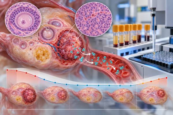
An AMH test can be a useful blood marker for ovarian granulosa cell tumors because granulosa cells naturally produce anti-Müllerian hormone. In a patient whose tumor releases AMH, the level often falls after successful treatment and can rise again when active disease returns. That makes serial testing especially helpful for monitoring, although AMH cannot diagnose a granulosa cell tumor by itself. The result is also influenced by age and normal ovarian function: healthy premenopausal ovaries produce AMH, while levels are usually very low or undetectable after menopause. There is no single tumor-specific AMH cutoff that works across all laboratories and all patients. The most informative comparison is often the patient’s own trend, using the same assay when possible. Clinicians commonly interpret AMH together with inhibin B, imaging, pathology, symptoms, and the original tumor’s marker pattern, particularly because granulosa cell tumors can recur many years after initial treatment.
- AMH can act as a tumor marker for granulosa cell tumors when the tumor produces measurable anti-Müllerian hormone.
- A falling AMH after surgery or therapy usually supports response, while a confirmed plateau or rise can prompt evaluation for persistent or recurrent disease.
- There is no universal AMH “cancer level.” Reference ranges depend strongly on age, menopausal status, assay, and normal ovarian reserve.
- AMH is often paired with inhibin B because using two tumor-related hormones can improve confidence when one marker is less informative.
- Long-term trends matter: adult-type granulosa cell tumors can relapse years or even decades later, so follow-up plans are individualized.
Table of Contents
- What AMH Measures
- Why Granulosa Cell Tumors Produce AMH
- How to Interpret AMH Results
- Monitoring Treatment Response With AMH
- AMH and Recurrence Surveillance
- Limitations and Non-Cancer Factors
- Practical Next Steps After an Abnormal Result
What AMH Measures
AMH stands for anti-Müllerian hormone. In females, it is produced mainly by granulosa cells surrounding small developing ovarian follicles. In everyday reproductive medicine, AMH is commonly used as a marker of the remaining pool of recruitable follicles, often called ovarian reserve. In gynecologic oncology, the same hormone can serve a different purpose: some ovarian granulosa cell tumors produce enough AMH to become a measurable marker of tumor activity.
The test is performed on blood. Laboratories may report AMH in ng/mL or pmol/L, and values are not perfectly interchangeable across assay platforms. A fertility-oriented reference interval should not be treated as a cancer threshold. AMH normally varies with age, falls as the follicle pool declines, and becomes very low around and after menopause.
That age relationship is central to interpretation. An AMH of several ng/mL might be expected in a healthy younger adult with active ovaries, while the same value could be highly unusual in someone who is postmenopausal or has had both ovaries removed. The clinical question is therefore not simply, “Is AMH high?” It is, “Is this level plausible for this patient’s normal ovarian physiology, and how does it compare with prior tumor-associated values?”
For granulosa cell tumors, clinicians may also order inhibin B and sometimes inhibin A. These hormones arise from the same general cell lineage and can provide complementary information.
Why Granulosa Cell Tumors Produce AMH
Granulosa cell tumors are sex cord-stromal ovarian tumors, not epithelial ovarian cancers. Adult-type granulosa cell tumor is the more common subtype; juvenile granulosa cell tumor is biologically distinct and occurs more often in younger patients. Because the tumor cells retain features of normal granulosa cells, they may continue to produce hormones such as inhibin, estradiol, and AMH.
AMH is attractive as a biomarker because it is closely tied to granulosa-cell biology. Research has shown that serum AMH can be elevated in active granulosa cell tumors and may correlate with macroscopic tumor burden. In a large longitudinal study, AMH and inhibin B performed similarly for detecting adult-type granulosa cell tumor, and combining the markers improved detection compared with inhibin B alone.
However, not every granulosa cell tumor produces a useful AMH signal. A person can have active disease with an AMH that is normal for age or below the assay’s detection limit. Tumor-marker secretion can also change as disease evolves. Pathology, imaging, and clinical assessment therefore remain essential even when AMH has historically been reliable.
Adult-type granulosa cell tumors are also known for their potential for late recurrence. The biology is often relatively slow-growing compared with many high-grade ovarian carcinomas. This makes a repeatable serum marker appealing because it can provide a low-burden way to track change over long periods. Still, the optimal duration and intensity of surveillance continue to be studied, and follow-up should be individualized rather than based on a single universal schedule.
The tumor’s molecular identity can help confirm diagnosis. Most adult-type granulosa cell tumors carry a characteristic FOXL2 alteration, while juvenile tumors have different molecular patterns. FOXL2 testing is a tissue diagnostic tool, not a replacement for serum AMH monitoring.
How to Interpret AMH Results
There is no single AMH value that proves or excludes a granulosa cell tumor. Interpretation depends on the patient’s age, menopausal status, ovarian anatomy, treatment history, assay, and prior results.
| Pattern | Possible interpretation | What usually matters next |
|---|---|---|
| High before treatment | The tumor may be secreting AMH | Establish the value as a baseline and compare with inhibin and imaging. |
| Falls after complete surgery | Supports reduction in AMH-producing tumor burden | Confirm continued decline or normalization at follow-up. |
| Remains elevated | Could reflect residual disease or normal ovarian production | Interpret by age, remaining ovary, imaging, and pre-treatment level. |
| Rises after prior low/undetectable level | Can suggest recurrent activity | Repeat to confirm and correlate with inhibin, symptoms, and imaging. |
| Normal despite known tumor | The tumor may not secrete measurable AMH | Use other markers and clinical surveillance rather than relying on AMH. |
In premenopausal patients with an intact ovary, normal follicular production can make low-to-moderate AMH values difficult to interpret as tumor-specific. The strongest evidence often comes from within-person change. For example, if AMH was markedly elevated before surgery, became very low afterward, and then begins to rise reproducibly years later, the trend is more meaningful than comparison with a broad population reference range.
In postmenopausal patients, or after bilateral removal of ovarian tissue, any clearly measurable or rising AMH can attract more attention because physiologic production should be minimal. Even then, clinicians confirm the laboratory result and consider assay limitations before concluding that cancer has returned.
It is also important to separate two meanings of a “low AMH.” In fertility medicine, a low value may indicate reduced ovarian reserve. In granulosa cell tumor follow-up, a low or undetectable AMH after treatment may be exactly what is hoped for if the tumor had previously secreted AMH. The same number can therefore have different implications depending on why the test was ordered.
Monitoring Treatment Response With AMH
The best use of AMH in granulosa cell tumor care is often serial monitoring. A baseline is ideally measured near diagnosis before definitive treatment. If elevated, it provides a personalized marker that can be followed after surgery and, when needed, during systemic therapy.
After successful removal of an AMH-secreting tumor, levels should decline. The exact time to reach a low or undetectable concentration depends on the starting value, assay, remaining normal ovarian function, and treatment context. Clinicians therefore avoid rigid expectations based on a single timetable.
During treatment for recurrent or advanced disease, AMH can provide an additional signal of whether tumor burden is changing. A consistent fall may support response; a persistent rise can suggest progression. The marker is most reliable when it moves in the same direction as other evidence such as imaging or inhibin B.
Discordant results require more thought. If scans show improvement but AMH rises slightly, the test may be repeated before conclusions are drawn. If AMH falls while a mass remains visible, the mass may contain treatment-related tissue or tumor that no longer secretes much AMH. Imaging and pathology answer questions that a blood marker cannot.
An important practical principle is to use the same laboratory or assay when possible. AMH methods have changed over time, and different platforms can produce somewhat different values. For a tumor marker followed over years, consistent measurement reduces noise and makes trend interpretation easier.
Treatment can also affect normal ovarian AMH production. Surgery involving ovarian tissue, chemotherapy, aging, and menopause can lower AMH independently of tumor response. This is one reason clinicians may follow both AMH and inhibin B rather than viewing either as a perfect stand-alone test.
AMH and Recurrence Surveillance
Granulosa cell tumors have a distinctive recurrence pattern. Many are diagnosed at an early stage and have favorable outcomes, yet recurrence can occur long after the original diagnosis. Studies of adult-type granulosa cell tumor include relapses more than a decade later, which has traditionally supported prolonged follow-up.
AMH can be valuable in this setting when three conditions are met:
- it was elevated when active tumor was present;
- it fell substantially after successful treatment; and
- later measurements use a reasonably comparable assay.
A confirmed upward trend may trigger closer evaluation before symptoms become obvious. The next step can include repeat AMH, inhibin B, pelvic or abdominal imaging, and clinical review. The exact response depends on the magnitude and persistence of the change and the patient’s previous recurrence pattern.
Surveillance is not only about laboratory numbers. Patients should report new persistent abdominal or pelvic pain, increasing abdominal size, early satiety, abnormal uterine bleeding, shortness of breath, or other symptoms that could indicate recurrence or another condition. Hormone-related symptoms may also matter because some granulosa cell tumors produce estrogen.
The best follow-up schedule remains an area of debate. Recent multicenter data have questioned whether very prolonged standardized surveillance benefits every patient in the same way. That does not make AMH unimportant; rather, it reinforces that follow-up should consider stage, tumor rupture, prior recurrence, pathology, treatment, age, symptoms, and the reliability of each patient’s biomarkers.
For someone whose original tumor had a strong AMH signal, periodic testing may remain a useful component of individualized long-term care. For someone whose AMH was never elevated, repeated testing is less likely to add value.
Limitations and Non-Cancer Factors
AMH is more biologically specific to granulosa cells than many general tumor markers, but it is not cancer-specific. Healthy ovaries produce AMH, and several non-cancer factors affect the number.
Age is the biggest physiologic factor. AMH is highest earlier in reproductive life and generally declines as the ovarian follicle pool falls. Population ranges are wide at every age, so one person’s result cannot be interpreted from an age chart alone.
Polycystic ovary syndrome can be associated with higher AMH because the ovaries often contain a larger number of small follicles. A high AMH in a young patient with PCOS is not automatically suspicious for a granulosa cell tumor.
Hormonal contraception, ovarian surgery, and gonadotoxic treatment can lower AMH. The size of these effects varies. Chemotherapy may reduce normal follicular production, which can make a post-treatment decline partly reflect ovarian reserve loss as well as tumor response.
Menopause lowers AMH to very low or undetectable levels. In a postmenopausal patient, a measurable rise can therefore be more unusual, but laboratory error and assay interference still need consideration.
AMH should also not be used as a substitute for tissue diagnosis. A suspicious ovarian mass requires imaging and, when appropriate, surgery or biopsy with expert pathology. Serum markers can help classify and monitor disease, but they do not reveal stage, tumor rupture, microscopic spread, or every histologic feature relevant to treatment.
Finally, AMH expression seen by immunohistochemistry in tumor tissue is not the same thing as serum AMH concentration. Tissue staining can support lineage in some contexts, while the blood test is used as a circulating marker. The two tests answer different questions.
Practical Next Steps After an Abnormal Result
If AMH is unexpectedly high during evaluation of an ovarian mass, the next step is to put the result into context. A clinician may review age, menopausal status, fertility history, ovarian reserve factors, ultrasound or MRI findings, and other tumor markers. If a granulosa cell tumor is suspected, referral to a gynecologic oncologist and expert pathologic review may be appropriate.
If the patient has already been treated for a granulosa cell tumor, interpretation should start with the original marker pattern. Bring prior AMH values, dates, units, and laboratory names to the visit. A simple timeline often reveals whether the change is meaningful.
For a new rise, clinicians may:
- repeat AMH to confirm that the change is real;
- measure inhibin B or another marker that was informative before;
- review menstrual or menopausal status and medications;
- assess symptoms and perform a focused examination; and
- obtain imaging when the marker trend or clinical picture warrants it.
The broader ovarian cancer biomarker profile used for epithelial tumors is not interchangeable with granulosa cell tumor surveillance. CA-125, HE4, and related algorithms answer different questions and are not substitutes for AMH or inhibin in a known sex cord-stromal tumor.
The most informative surveillance plan often uses more than one line of evidence. AMH and inhibin B can be complementary because some granulosa cell tumors secrete one marker more consistently than the other. If both were elevated before treatment and both normalize, a later concordant rise is more persuasive than a small isolated change in only one assay. If the markers disagree, repeating the test and checking imaging or symptoms can prevent premature conclusions.
Long-term follow-up matters because adult-type granulosa cell tumors can recur many years after initial treatment. A normal AMH today is reassuring but does not permanently eliminate recurrence risk. Patients should know the planned duration of follow-up and report new abdominal distension, pelvic pressure, pain, abnormal uterine bleeding, or other persistent symptoms rather than waiting for the next marker draw. In postmenopausal patients, a newly detectable or rising AMH can be more unusual than in reproductive-age patients, but it still requires confirmation and clinical correlation rather than an automatic cancer diagnosis.
The most useful question to ask about AMH is therefore not “What number means cancer?” but “How does this result fit this patient’s tumor biology and previous trend?” When AMH clearly tracked the original granulosa cell tumor, it can become a highly practical marker of treatment response and possible recurrence. When it did not, surveillance should lean more heavily on other biomarkers, imaging, symptoms, and clinical follow-up.
For patients who move between oncology centers, preserving the original pathology report and pretreatment AMH value is especially useful. Different laboratories can use different platforms and reference intervals, so a small numerical shift after a laboratory change may not represent a biologic change. When possible, a new baseline can be established before interpreting later surveillance values.
References
- Advanced Granulosa Cell Tumors of the Ovary: A Review with a Focus on Current and Novel Therapeutic Approaches 2024 (Review)
- Adult ovarian granulosa cell tumors: analysis of outcomes and risk factors for recurrence 2023
- Immunohistochemical markers of prognosis in adult granulosa cell tumors of the ovary – a review 2023 (Review)
- Is it time to abandon staging surgery and prolonged follow-up in patients with primary adult-type granulosa cell tumor? 2025
- The clinical utility of serum anti-Müllerian hormone in the follow-up of ovarian adult-type granulosa cell tumors–A comparative study with inhibin B 2015
Disclaimer
This article provides general education about AMH as a marker for ovarian granulosa cell tumors and does not replace individualized medical advice. AMH should be interpreted with age, menopausal status, ovarian function, pathology, imaging, treatment history, and other tumor markers. Contact your oncology team about a new persistent marker rise or concerning symptoms rather than changing follow-up or treatment based on one AMH result.