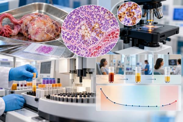
An AFP test can help identify and monitor certain ovarian germ cell tumors, especially yolk sac tumors and mixed tumors that contain a yolk sac component. AFP, or alpha-fetoprotein, is a protein made in large amounts during fetal development. In most nonpregnant adults, blood levels are very low. When an ovarian germ cell tumor produces AFP, the number can rise dramatically and often falls as the tumor responds to surgery or chemotherapy. The most useful information usually comes from the pattern over time rather than from a single result. A high AFP level does not prove that an ovarian mass is cancer, and a normal result does not rule out every germ cell tumor. Pregnancy, liver disease, age, laboratory method, and tumor subtype all affect interpretation. For that reason, AFP is used alongside imaging, pathology, other tumor markers, and the clinical picture.
- AFP is most strongly associated with ovarian yolk sac tumors and mixed germ cell tumors that contain yolk sac elements.
- Most nonpregnant adult laboratories use an upper reference limit around 7–10 ng/mL, but the exact range is assay-specific.
- A falling AFP after treatment is generally reassuring; persistent elevation, a plateau, or a new rise can signal residual or recurrent AFP-producing disease.
- AFP normally has a blood half-life of about 5–7 days, so clinicians often judge whether the decline is compatible with expected clearance.
- Pregnancy and liver conditions can also raise AFP, so an abnormal result must be interpreted in context rather than treated as a cancer diagnosis by itself.
Table of Contents
- What the AFP Test Measures
- AFP and Ovarian Germ Cell Tumor Types
- Understanding AFP Results and High Levels
- How AFP Is Used to Monitor Treatment
- AFP in Recurrence and Long-Term Follow-Up
- Non-Cancer Causes and Confusing Results
- What to Do With an Abnormal AFP Result
What the AFP Test Measures
AFP stands for alpha-fetoprotein, a protein normally produced by the fetal yolk sac and liver. It circulates at high levels during fetal development, crosses into maternal blood during pregnancy, and then falls after birth. In healthy nonpregnant adults, only a small amount is usually detectable.
The AFP tumor marker test is a blood test, usually reported in nanograms per milliliter (ng/mL). Laboratories do not all use the same assay or reference interval, but many adult reference ranges place the upper limit near 7–10 ng/mL. The laboratory’s own reference range should always be used when interpreting a result.
AFP is not a general ovarian cancer screening test. It is most useful when the clinical situation suggests a germ cell tumor, a group of uncommon ovarian tumors that arise from cells related to egg-cell development. These tumors are different from the much more common epithelial ovarian cancers. They tend to occur in children, adolescents, and young adults, although they can occur outside those age groups.
Clinicians often order AFP with other markers when evaluating a suspicious ovarian mass in a younger patient. Depending on the differential diagnosis, the workup may also include beta-hCG testing and LDH testing. The marker pattern can suggest a tumor subtype, but pathology remains central to the final diagnosis.
A useful way to think about AFP is as a biologic signal from AFP-producing tumor cells. If a tumor makes AFP, the blood level can provide a measurable marker of disease activity. If that same patient’s AFP falls after effective treatment and later rises again, the change can be clinically meaningful. If the tumor never produced AFP, however, the test may offer little or no value for surveillance.
AFP and Ovarian Germ Cell Tumor Types
AFP is strongly linked to some ovarian germ cell tumor subtypes and usually absent in others. That distinction is one of the reasons the marker can be so helpful before and after surgery.
| Tumor type | Typical AFP relationship | Practical meaning |
|---|---|---|
| Yolk sac tumor | Often markedly elevated | AFP is a major diagnostic and monitoring marker. |
| Mixed germ cell tumor | May be elevated | An elevated AFP can indicate a yolk sac or other AFP-producing component. |
| Embryonal carcinoma | Can be elevated | AFP may rise, often with other germ cell markers depending on tumor composition. |
| Immature teratoma | Usually normal or only modestly increased | A clearly abnormal AFP raises concern for an associated yolk sac component or another source. |
| Pure dysgerminoma | Usually normal | LDH can be more useful; elevated AFP should prompt reconsideration of a pure dysgerminoma diagnosis. |
| Mature teratoma | Usually normal | AFP is not expected to act as a tumor marker for a benign mature teratoma. |
The strongest association is with yolk sac tumor, historically called endodermal sinus tumor. These tumors can produce very high AFP concentrations, sometimes in the hundreds, thousands, or far higher. There is no single AFP cutoff that separates cancer from non-cancer in every patient. A result must be interpreted with age, pregnancy status, imaging, pathology, liver tests, and the trend in mind.
Mixed tumors deserve special attention. An ovarian mass may contain more than one germ cell tumor type. A biopsy or surgical specimen can therefore show a combination of dysgerminoma, yolk sac tumor, embryonal carcinoma, immature teratoma, or other components. An elevated AFP may provide an early clue that an AFP-producing component is present even when the imaging appearance is nonspecific.
For immature teratoma, the interpretation can be more nuanced. Mild AFP elevations have been described, but a substantial or persistent increase should make clinicians consider whether microscopic yolk sac tumor is present, whether the original pathology needs expert review, or whether another condition is contributing. This is one reason complex ovarian germ cell tumors are often reviewed by gynecologic pathologists or specialized germ cell tumor teams.
AFP is only one part of a broader ovarian tumor biomarker assessment. The most informative marker depends on tumor biology, not simply on the fact that a mass is located in the ovary.
Understanding AFP Results and High Levels
The first question is whether the result is actually above the laboratory’s reference limit. In a nonpregnant adult, many laboratories consider values below roughly 7–10 ng/mL normal, but methods vary. Small differences near the upper limit can reflect assay variation and do not carry the same meaning as a level that is hundreds or thousands of ng/mL.
A high AFP result is interpreted in layers:
- Magnitude: How far above the reference range is it?
- Direction: Is the level rising, falling, or stable?
- Timing: Was it measured before surgery, immediately after surgery, during chemotherapy, or months later?
- Tumor subtype: Is the known pathology expected to produce AFP?
- Other explanations: Is the patient pregnant, a newborn or infant, or affected by liver inflammation or another AFP-producing cancer?
For ovarian germ cell tumors, a single pre-treatment AFP value can support the working diagnosis and provides a baseline for later comparison. Very high levels make an AFP-producing germ cell tumor more likely in the right clinical setting, but they are not a substitute for histologic diagnosis.
The rate of change often matters more than the absolute number. AFP has an approximate serum half-life of 5–7 days. If all AFP-producing tumor tissue has been removed or effectively treated, clinicians expect the concentration to decline in a roughly compatible pattern. Real-world declines are not perfectly mathematical because tumor cells may still be dying, treatment is ongoing, assays vary, and disease burden can be complex.
For example, a level of 10,000 ng/mL would not be expected to become normal within a few days simply because surgery was successful. Multiple half-lives are needed for a large concentration to clear. Conversely, if the level stops falling well before it reaches the reference range, or begins to increase, the result deserves prompt review.
Do not compare numbers from different laboratories as though they were identical when assays or units differ. When possible, serial monitoring is easiest to interpret when performed by the same laboratory using the same method.
How AFP Is Used to Monitor Treatment
For a confirmed AFP-producing ovarian germ cell tumor, serial AFP measurement is one of the most useful ways to track response. Clinicians commonly obtain a baseline before treatment and repeat testing at clinically appropriate intervals after surgery and during systemic therapy.
A favorable pattern is a consistent decline toward the normal range. The marker can sometimes show response before a mass has fully changed on imaging because blood proteins can fall as viable AFP-producing cells disappear. Imaging is still necessary when indicated because residual masses may contain scar tissue, mature teratoma, viable tumor, or other components that AFP alone cannot distinguish.
A concerning pattern includes:
- AFP that remains clearly elevated longer than expected after treatment;
- an inadequate decline compared with the expected biologic half-life;
- repeated values that flatten into a plateau;
- a confirmed rise on serial testing; or
- a new elevation after prior normalization.
None of these patterns should be interpreted from one isolated measurement. A clinician may repeat the test to exclude laboratory error, confirm the units, review liver function, check pregnancy status when relevant, and correlate the number with symptoms, imaging, and treatment timing.
The marker is also valuable because ovarian germ cell tumors frequently affect people for whom fertility preservation matters. Treatment often aims to cure the cancer while preserving the uterus and an unaffected ovary when oncologically appropriate. A reliable serum marker can strengthen surveillance by adding a low-burden way to look for disease activity between imaging assessments. The broader management strategy, however, depends on stage, pathology, surgical findings, and treatment response rather than on AFP alone.
Another practical point is that normalization is not always equivalent to complete cure. Tumor components that do not secrete AFP can remain even when the blood test is normal. This is particularly relevant in mixed tumors and tumors containing teratoma. Clinicians therefore interpret AFP together with the rest of the disease picture.
AFP in Recurrence and Long-Term Follow-Up
After treatment, AFP is most useful for follow-up when it was elevated at diagnosis and normalized with successful therapy. In that setting, each subsequent measurement can be compared with an established personal baseline.
A newly rising AFP can precede obvious clinical progression in some patients. The response is usually not to assume recurrence immediately, but to confirm the abnormality and investigate the cause. This may include repeat AFP, other tumor markers, examination, imaging, and review by the oncology team.
Follow-up schedules vary by tumor subtype, stage, treatment, age, and institutional protocol. Malignant ovarian germ cell tumors have high cure rates, but recurrences can occur, and survivors also need follow-up for treatment-related effects. Surveillance may include clinical visits, tumor markers that were informative at diagnosis, and imaging at selected intervals.
The value of AFP during follow-up is highly individualized. Consider two examples:
- A patient had a yolk sac tumor with an AFP of 25,000 ng/mL before treatment, followed by normalization. A later reproducible increase is potentially important because the tumor clearly demonstrated AFP secretion.
- A patient had a pure dysgerminoma with normal AFP at diagnosis. Repeating AFP indefinitely is less informative because there is no evidence that the original tumor produced it. Other clinical tools and markers may be more relevant.
This distinction prevents a common mistake: assuming that every ovarian cancer survivor needs the same blood tumor markers. Biomarker surveillance works best when it is matched to the biology of the original tumor.
Non-Cancer Causes and Confusing Results
AFP is not cancer-specific. Several situations can produce an elevated result without an ovarian germ cell malignancy.
Pregnancy is a major physiologic cause. Maternal serum AFP rises because the developing fetus produces AFP. Interpretation in pregnancy uses gestational-age-specific methods and is completely different from the nonpregnant adult tumor-marker reference range. A pregnancy test is often part of the evaluation of a reproductive-age patient with an ovarian mass for this and many other reasons.
Liver injury can also increase AFP. Hepatitis, cirrhosis, liver regeneration, and hepatocellular carcinoma can produce elevated levels. When the ovarian diagnosis does not fit the marker pattern, liver history and liver tests may be important.
Infants normally have much higher AFP than adults. Levels decline after birth over many months, so adult reference ranges cannot be applied to babies or very young children. Pediatric interpretation requires age-adjusted reference data.
Other possible explanations include another AFP-producing malignancy, assay interference, or simple laboratory variation near the cutoff. A low-level unexpected elevation should therefore be repeated and placed in clinical context before major decisions are made.
It is also possible for a real ovarian germ cell tumor to have normal AFP. Pure dysgerminoma, mature teratoma, and some immature teratomas generally do not produce significant AFP. The absence of elevation does not make a suspicious ovarian mass benign.
What to Do With an Abnormal AFP Result
An abnormal AFP result should trigger interpretation, not panic. The useful next steps depend on why the test was ordered.
If AFP was measured during evaluation of an ovarian mass, the clinician may review pelvic imaging, pregnancy status, age, symptoms, liver tests, and other germ cell markers. In a young patient with a complex ovarian mass and substantially elevated AFP, prompt evaluation by a gynecologic oncologist or team experienced in ovarian germ cell tumors is often appropriate because treatment planning may involve both cancer control and fertility preservation.
If AFP is being monitored after treatment, ask three practical questions:
- Is the value changing in the expected direction?
- Does the timing fit the expected 5–7 day half-life and the treatment that was given?
- Is there another credible explanation for the result?
A small unexpected rise may be repeated before additional testing. A persistent increase, plateau, or major elevation usually deserves faster assessment. The oncology team may add imaging or repeat pathology review depending on the situation.
Patients should also keep a simple record of AFP values, dates, units, and laboratories. A graph can make the trend easier to see than a list of isolated numbers. The same principle applies to other germ cell tumor markers: the pattern across time is often more informative than whether one value is technically above or below a cutoff.
Age and treatment setting also change how aggressively an abnormal result is investigated. Ovarian germ cell tumors often occur in adolescents and young adults, so clinicians commonly consider fertility goals, menstrual history, and the possibility of pregnancy while evaluating the marker. After fertility-sparing surgery, the presence of a remaining ovary does not make AFP unreliable; the marker still reflects AFP-producing tumor tissue rather than normal ovarian function. During chemotherapy, liver injury can occasionally complicate interpretation because AFP may rise with hepatic regeneration, making liver tests and the overall trajectory useful context.
If AFP was normal at the original diagnosis, it may not be a sensitive recurrence marker for that patient. Follow-up should then rely more heavily on the tumor’s other markers, examination, imaging, and symptoms. This individualized baseline is important: a marker that was never produced by the cancer should not be expected to become a reliable surveillance test later.
The central point is that AFP is a tumor-behavior marker, not a stand-alone diagnosis. Used correctly, it can help identify AFP-producing ovarian germ cell tumors, measure treatment response, and detect possible recurrence while keeping the interpretation grounded in pathology, imaging, and the patient’s overall clinical picture.
References
- Malignant germ cells tumor of the ovary 2025 (Review)
- Updates in the Management of Malignant Ovarian Germ Cell Tumors 2025 (Review)
- Clinical Challenges in the Management of Malignant Ovarian Germ Cell Tumours 2023 (Review)
- Diagnostic, Prognostic and Predictive Markers in Pediatric Germ Cell Tumors-Past, Present and Future 2022 (Review)
- Tumor Markers and Their Diagnostic Significance in Ovarian Cancer 2023 (Review)
Disclaimer
This article provides general information about AFP testing in ovarian germ cell tumors and is not a diagnosis or treatment plan. AFP results should be interpreted by a qualified clinician using the laboratory’s reference range, pregnancy status, pathology, imaging, treatment history, and other relevant tests. Seek prompt medical care for a rapidly enlarging abdominal mass, severe pain, heavy bleeding, fainting, or other urgent symptoms.