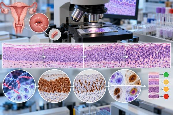
A cervical cancer biomarker panel is not one universal blood test. It is a group of laboratory tools used at different points in cervical cancer prevention, diagnosis, and treatment. High-risk HPV testing identifies the viral infection that causes nearly all cervical cancers, while p16 and Ki-67 help show whether HPV is disrupting normal cell-cycle control and driving precancerous change. A p16/Ki-67 dual-stain test can be used to triage some HPV-positive screening results and decide who needs colposcopy sooner. Tissue p16 immunohistochemistry can support the interpretation of cervical biopsies, but it does not replace morphology. In established invasive cancer, other biomarkers such as PD-L1, mismatch-repair status, microsatellite instability, HER2 in selected tumors, and serum SCC antigen may answer treatment or monitoring questions. The most important point is that each marker has a specific job. A positive HPV result is not a cancer diagnosis, and a negative tumor marker cannot rule out cervical cancer when symptoms or biopsy findings are concerning.
- High-risk HPV is the primary causal biomarker for cervical cancer risk, especially persistent infection with oncogenic types such as HPV16 and HPV18.
- p16 overexpression signals disrupted cell-cycle control from transforming HPV activity and is widely used as an adjunct in cervical tissue diagnosis.
- Ki-67 marks cell proliferation; p16/Ki-67 dual staining is abnormal because the same cervical cell should not normally express both markers together.
- For many HPV-positive patients, dual-stain positivity supports colposcopy while dual-stain negativity can support 1-year HPV-based follow-up, with important exceptions such as HPV16/18 positivity.
- Blood tumor markers are not cervical cancer screening tests. Markers such as SCC antigen are mainly considered in selected patients with established disease.
Table of Contents
- What a Cervical Biomarker Panel Measures
- High-Risk HPV and Genotype Meaning
- p16, Ki-67, and Dual-Stain Testing
- How Biomarkers Refine Dysplasia Risk
- Biomarkers in Biopsy and Cancer Diagnosis
- Tumor Markers in Established Cervical Cancer
- How to Act on Common Result Combinations
What a Cervical Biomarker Panel Measures
Cervical biomarker testing spans three different clinical questions: Is high-risk HPV present? Is the infection causing transforming cellular changes? If cancer is already present, does the tumor have markers that influence treatment or monitoring? Mixing these questions together can make results seem more confusing than they are.
High-risk HPV testing is a viral nucleic-acid test. It detects DNA or RNA from oncogenic human papillomavirus types. It does not measure cancer cells. A positive result means a carcinogenic HPV type is present, but most HPV infections clear or become undetectable without progressing to cancer.
p16 and Ki-67 are host-cell proteins. p16 is a cyclin-dependent kinase inhibitor that becomes strongly overexpressed when high-risk HPV E7 disrupts the retinoblastoma protein pathway. Ki-67 is a nuclear marker of active cell proliferation. Their expression patterns can help distinguish transient infection or reactive change from biologically transforming HPV infection.
Cytology, often called a Pap test, examines cell appearance rather than a single molecular marker. Histology examines tissue architecture from biopsy. These methods remain essential because biomarkers work best when they refine, not replace, morphologic assessment.
In invasive cervical cancer, a separate set of biomarkers may become relevant. These can include PD-L1 expression, mismatch-repair deficiency or microsatellite instability, HER2 in selected histologies, and serum SCC antigen. They answer different questions from screening HPV and dual stain.
A useful way to view the “panel” is therefore as a sequence of decision tools, not a bundle in which every patient needs every marker.
High-Risk HPV and Genotype Meaning
Persistent infection with a carcinogenic HPV type is the necessary driver of nearly all cervical squamous cancers and most cervical adenocarcinomas. Modern screening increasingly starts with high-risk HPV testing because it is more sensitive for identifying people at risk of cervical precancer than cytology alone.
A positive high-risk HPV test does not mean cancer is present. It means the patient has a viral type associated with a higher probability of precancer if infection persists. Risk rises with persistence, certain genotypes, age, immune status, prior abnormal results, and cytology or biomarker findings.
HPV16 and HPV18 deserve special attention. HPV16 causes the largest share of cervical cancers, while HPV18 is particularly important in adenocarcinoma. Many screening assays report these genotypes separately. A dedicated HPV16 and HPV18 result can therefore change management even when other triage markers are negative.
Other high-risk types are often grouped together. Their individual risk is generally lower than HPV16 but still clinically important. Screening algorithms may use reflex cytology, dual staining, or repeat HPV testing to separate patients who need immediate colposcopy from those who can be followed safely.
A negative high-risk HPV test is strongly reassuring in routine screening, but no test is perfect. Sampling quality, recent treatment, rare HPV-independent cervical cancers, and symptoms matter. A patient with unexplained postcoital bleeding, visible cervical abnormality, or another concerning clinical finding may need diagnostic evaluation regardless of a recent screening result.
HPV testing also differs from HPV antibody testing. Routine cervical screening detects viral nucleic acid from cervical or vaginal samples; blood antibody tests are not used to decide whether cervical precancer is present.
p16, Ki-67, and Dual-Stain Testing
p16 and Ki-67 are useful because they reflect two different parts of the cell cycle. In normal cells, strong p16-mediated growth arrest and active Ki-67 proliferation should not occur in the same cell. High-risk HPV can break this normal relationship.
p16 immunohistochemistry is commonly used on cervical biopsy tissue. In HPV-associated high-grade squamous lesions, p16 often shows a strong, continuous “block-positive” staining pattern. Pathologists use this pattern as an adjunct when morphology is borderline or when deciding whether a lesion fits a high-grade squamous intraepithelial lesion. Focal or patchy staining is less specific. A p16 IHC result must therefore be interpreted by pattern, not merely as “positive” or “negative.”
Ki-67 labels nuclei in cells that are actively cycling. Normal cervical epithelium has proliferation mainly in the basal and parabasal layers. Increasing Ki-67 expression higher into the epithelium can support dysplasia, but Ki-67 alone is not specific enough to diagnose a grade of cervical lesion. A Ki-67 assessment is most useful in context.
p16/Ki-67 dual stain is performed on cervical cytology material and asks whether one cell expresses both proteins at the same time. That co-expression is a marker of abnormal HPV-driven cell-cycle deregulation. The test does not require the cytologist to classify the cell morphologically as ASC-US, LSIL, or HSIL in the same way as conventional cytology; instead, the presence of a dual-stained cell makes the assay positive.
Recent U.S. consensus recommendations support dual stain as an acceptable triage method for HPV-positive screening results. In general, dual-stain-positive patients are referred to colposcopy, while many dual-stain-negative patients can return in one year for HPV-based testing. HPV16- or HPV18-positive results remain important exceptions, where colposcopy is recommended even if dual stain is negative in the relevant algorithms.
How Biomarkers Refine Dysplasia Risk
Cervical dysplasia is graded by the likelihood that abnormal cells represent a true precancer and by the depth of epithelial involvement. In modern terminology, biopsy diagnoses often use low-grade squamous intraepithelial lesion for HPV effect/CIN1 and high-grade squamous intraepithelial lesion for CIN2 or CIN3, with p16 used selectively to clarify ambiguous cases.
Biomarkers help because an HPV-positive result alone is very common relative to the number of people who develop CIN3 or cancer. A good triage test should identify the smaller group with transforming infection while allowing the majority with lower short-term risk to avoid unnecessary colposcopy.
Recent evidence supports p16/Ki-67 dual stain as a stronger risk separator than cytology alone in many HPV-positive populations. Meta-analyses report high sensitivity for CIN2+ and CIN3+, with moderate specificity. That balance means dual stain is good at finding many clinically important lesions while still producing some positive results in patients who do not have high-grade disease.
| Marker or test | What it detects | Main role |
|---|---|---|
| High-risk HPV DNA/RNA | Oncogenic viral infection | Primary screening or cotesting; establishes infection-related risk. |
| HPV16/18 genotyping | Highest-risk common genotypes | Identifies patients who may need direct colposcopy. |
| p16/Ki-67 dual stain | Transforming cell-cycle abnormality in the same cell | Triage of selected HPV-positive results. |
| p16 tissue IHC | Pattern of p16 overexpression | Adjunct to biopsy interpretation, especially equivocal CIN2-like lesions. |
| Ki-67 tissue IHC | Proliferation pattern | Supportive morphologic information; not a stand-alone grade. |
Risk is cumulative. A persistent HPV16 infection plus abnormal cytology and dual-stain positivity is more concerning than a first-time pooled high-risk HPV positive result with negative dual stain. Clinical management systems increasingly calculate risk from current and prior results together, rather than treating each test in isolation.
Pregnancy, immune suppression, prior treatment for CIN2+, age, and screening history can alter recommended follow-up. A biomarker table from the internet cannot replace the patient’s actual risk-based management pathway.
Biomarkers in Biopsy and Cancer Diagnosis
Once colposcopy identifies a suspicious area, biopsy establishes whether precancer or invasive cancer is present. Biomarkers can help the pathologist, but tissue architecture and cellular morphology remain the foundation of diagnosis.
p16 is especially helpful when a lesion sits near the boundary between a benign mimic, CIN1, and CIN2. Strong block staining can support a high-grade HPV-associated lesion when the morphology warrants it. Overusing p16 is a mistake because some benign or metaplastic tissues can show staining, and not every cervical cancer is HPV-associated.
Ki-67 can show an expanded proliferation zone in dysplastic epithelium, but inflammation and repair can also increase proliferation. That is why the marker is supportive rather than definitive.
For glandular lesions, p16 and Ki-67 may also assist the distinction between HPV-associated adenocarcinoma or adenocarcinoma in situ and benign glandular mimics. Interpretation can be more complex because some cervical adenocarcinoma subtypes are HPV-independent and may not show the classic diffuse p16 pattern.
Once invasive cancer is diagnosed, p16 positivity can support HPV association but does not determine stage. Cervical cancer stage depends on tumor extent, imaging, examination, and pathologic findings rather than biomarker intensity.
Tumor Markers in Established Cervical Cancer
The biomarker focus changes after invasive cervical cancer is confirmed. Screening markers become less important than markers that help select therapy or monitor known disease.
PD-L1 immunohistochemistry can influence immunotherapy choices in advanced or recurrent cervical cancer, commonly using a combined positive score. The exact drug eligibility criteria depend on current approvals and treatment setting.
Mismatch-repair deficiency and microsatellite instability can identify a small subset of tumors with impaired DNA mismatch repair and may support immune-checkpoint therapy in appropriate settings. Tumor mutational burden and other molecular features can also be assessed on broader profiling panels.
HER2 may be relevant in selected cervical cancers, particularly certain adenocarcinoma subtypes or advanced tumors being considered for HER2-directed strategies. Testing methods and treatment implications are evolving.
Squamous cell carcinoma antigen (SCC-Ag) is a serum marker that can correlate with tumor burden in some squamous cervical cancers. It may be followed in selected patients, but it is neither sensitive nor specific enough for population screening or for ruling out recurrence by itself. A rising value can prompt closer evaluation when the marker was elevated before treatment, yet imaging, examination, and biopsy remain more important for confirming persistent or recurrent disease. Benign conditions and assay differences can also affect the number, so trends are more useful than a single isolated value.
Other blood tests sometimes ordered during cancer care—such as CEA or CA-125—are not validated cervical screening markers. They may be abnormal in some advanced tumors or used in selected histologic subtypes, but normal results do not exclude cervical cancer. Likewise, a broad tumor-sequencing report can list dozens of mutations without proving that any one of them should change therapy. The clinically useful part of profiling is the subset of findings tied to a validated treatment, trial, hereditary-risk question, or established prognostic purpose.
These markers illustrate why “cervical cancer biomarker panel” can mean very different things before and after diagnosis. The right test depends on the decision that needs to be made.
How to Act on Common Result Combinations
The safest way to interpret cervical biomarkers is to start with the clinical stage of the process: routine screening, abnormal-screen follow-up, biopsy interpretation, or established cancer.
For common screening scenarios:
- HPV negative: usually reassuring in routine screening, with interval determined by age and screening method.
- HPV positive, dual stain positive: generally supports colposcopy in current dual-stain triage pathways.
- HPV positive, dual stain negative: often supports 1-year HPV-based follow-up, except when genotype or cytology places the patient in a higher-risk category.
- HPV16 or HPV18 positive: typically warrants colposcopy even when some other triage findings are reassuring.
- Persistent HPV positivity: becomes more concerning over time and should be managed using the full risk history.
If a biopsy report mentions p16, ask whether staining was block-positive and how it changed the morphologic diagnosis. If the report says only “p16 positive,” the pattern and pathology context are still needed.
For invasive cancer, ask which biomarkers have a direct treatment purpose. A broad next-generation sequencing panel can generate many findings, but not every alteration is actionable. The oncology team should distinguish validated predictive biomarkers from exploratory findings.
Most importantly, do not use a negative biomarker to dismiss symptoms. Persistent postcoital bleeding, unexplained vaginal bleeding, watery or bloody discharge, pelvic pain, or a visible cervical lesion requires clinical assessment even if a recent HPV or Pap test was reassuring.
Before leaving a follow-up visit, it is useful to know the exact next test and interval rather than simply being told that a result is “abnormal.” For screening, that may mean colposcopy now, repeat HPV-based testing in one year, or another risk-based interval. For a biopsy, it may mean observation, excisional treatment, or further pathology review. For invasive cancer, the next biomarker may be ordered specifically because it has a treatment consequence. Clear documentation of the HPV genotype, cytology category, dual-stain result, biopsy diagnosis, and prior treatment prevents a later test from being interpreted without the history that gives it meaning.
Cervical biomarkers work best as a layered risk system: HPV identifies carcinogenic infection, p16 and Ki-67 identify transforming cellular effects, biopsy establishes disease, and tumor profiling refines treatment after cancer is confirmed. Keeping those roles separate makes the results far easier to understand and act on.
Vaccination history does not make screening or diagnostic biomarkers unnecessary. HPV vaccination greatly reduces the risk of infection and precancer from vaccine-covered types, but vaccinated people can still have prior infection or disease caused by other oncogenic types. Screening and abnormal-result follow-up should therefore follow the age- and risk-appropriate program rather than being stopped because vaccination was completed.
References
- Recommendations for Use of p16/Ki67 Dual Stain for Management of Individuals Testing Positive for Human Papillomavirus 2024 (Guideline)
- WHO guideline for screening and treatment of cervical pre-cancer lesions for cervical cancer prevention: Use of dual-stain cytology to triage women after a positive test for human papillomavirus (HPV) [Internet] 2024 (Guideline)
- p16/ki-67 dual stain triage of individuals positive for HPV to detect cervical precancerous lesions 2025 (Review)
- P16/Ki67 dual staining versus cytology for identifying high-grade cervical intraepithelial neoplasia (CIN2+/CIN3+) in triage management of HR-HPV-positive women: a systematic review and meta-analysis 2026 (Systematic Review)
- Integrating Biomarkers into Cervical Cancer Screening-Advances in Diagnosis and Risk Prediction: A Narrative Review 2026 (Review)
- Diagnostic value of p16 and Ki-67 expression in cervical glandular intraepithelial disease: A review 2022 (Review)
Disclaimer
This article provides general information about cervical cancer biomarkers and is not a substitute for individualized screening or treatment recommendations. HPV, p16, Ki-67, cytology, biopsy, and tumor-marker results must be interpreted together with age, prior screening history, immune status, pregnancy, symptoms, and current guidelines. Seek prompt medical evaluation for unexplained vaginal bleeding, postcoital bleeding, or a visible cervical abnormality.