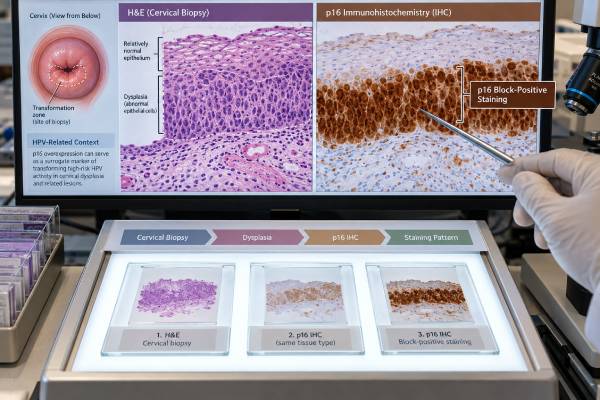
The p16 immunohistochemistry (IHC) test is a tissue stain that helps pathologists recognize cervical cells affected by transforming high-risk human papillomavirus (HPV) activity. In the cervix, strong, continuous “block-positive” p16 staining can support a diagnosis of high-grade squamous intraepithelial lesion (HSIL) when the microscopic appearance is equivocal, and it is also common in HPV-associated cervical cancers. p16 is not a direct HPV test: it detects overexpression of the p16INK4a protein, which often occurs when oncogenic HPV disrupts normal cell-cycle control. The pattern matters more than simply whether a few cells stain. Patchy, focal, or weak staining is not interpreted the same way as diffuse block staining. p16 is most useful as an adjunct to routine histology, especially in selected CIN2-like lesions or difficult differentials. It should not be used to upgrade clearly low-grade disease merely because some staining is present, and it does not replace HPV testing, cytology, colposcopy, or biopsy interpretation.
- Block-positive p16 supports transforming high-risk HPV activity: the classic pattern is strong, continuous nuclear and cytoplasmic staining across a broad area of abnormal epithelium.
- p16 is an adjunct, not a stand-alone diagnosis: the stain must be interpreted with cell morphology, lesion location, and adequate internal controls.
- p16 is especially useful in equivocal high-grade dysplasia: it can help distinguish true HSIL from reactive or immature changes that resemble CIN2.
- Patchy or focal staining is not the same as block positivity: scattered positive cells can occur in benign or low-grade tissue.
- p16 IHC is different from p16/Ki-67 dual-stain cytology: IHC is usually performed on tissue sections, while dual stain is a separate cervical screening/triage method on cytology specimens.
Table of Contents
- What p16 IHC Measures in Cervical Tissue
- What Block-Positive p16 Staining Looks Like
- How p16 Helps Diagnose Cervical Dysplasia
- p16 in Cervical Cancer and HPV-Related Tumors
- Negative, Patchy, and Unusual p16 Patterns
- p16 IHC vs HPV Testing and p16/Ki-67 Dual Stain
- How to Read a p16 Pathology Report and Next Steps
What p16 IHC Measures in Cervical Tissue
p16 IHC uses an antibody stain on a thin tissue section to show where the p16INK4a protein is overexpressed. The test is usually performed by a pathology laboratory on a cervical biopsy, loop excision, cone specimen, or tumor tissue rather than on a routine blood sample.
To understand why p16 becomes useful, it helps to know what high-risk HPV does. In a transforming HPV infection, the viral E7 protein interferes with the retinoblastoma protein, or RB, which normally helps control progression through the cell cycle. When RB signaling is disrupted, cervical cells respond by producing much more p16. That overexpression can create a characteristic staining pattern visible under the microscope.
This is why p16 is often described as a surrogate marker of transforming high-risk HPV activity. “Surrogate” is important: the stain does not detect HPV DNA or RNA itself. A direct high-risk HPV test for cervical cancer screening asks whether viral genetic material is present in the sampled cells; p16 IHC asks whether a tissue protein pattern suggests that an oncogenic HPV pathway is affecting cell-cycle control.
Pathologists do not interpret p16 in isolation. They first examine the hematoxylin-and-eosin-stained tissue for architecture and cell abnormalities. The p16 stain can then answer a focused question, such as whether an equivocal lesion behaves immunohistochemically like HSIL.
The test needs technical controls. Normal cells or other expected structures can help confirm that the stain worked. Complete absence of staining is meaningful only when the slide’s internal and external controls are adequate. Poor fixation, cautery, scant tissue, or damaged epithelium can make interpretation more difficult.
p16 can be used in other body sites and cancers as well, but the meaning is site-specific. In cervical pathology, its strongest role is tied to HPV-related squamous and glandular neoplasia and to standard histologic criteria for lower- versus higher-grade disease.
What Block-Positive p16 Staining Looks Like
The most important p16 pattern in cervical squamous lesions is block positivity. This term does not simply mean “many cells are brown.” It refers to a strong, continuous staining pattern that involves a broad region of the abnormal epithelium.
In a typical block-positive lesion, staining is:
- strong enough to be obvious at low magnification;
- continuous across groups of adjacent abnormal cells rather than scattered randomly;
- present in both the nucleus and cytoplasm;
- starts in the basal or parabasal portion of the involved squamous epithelium;
- extends upward through a meaningful thickness of the epithelial lesion.
The exact visual pattern depends on the biopsy orientation and lesion. A tangential section can make the thickness harder to judge, which is one reason morphology remains essential.
By contrast, non-block-positive staining may be focal, patchy, discontinuous, weak, or limited to isolated cells. Such staining can occur in low-grade HPV effects, immature squamous metaplasia, inflammation, and otherwise non-neoplastic tissue. It should not be interpreted as proof of HSIL.
The distinction matters because p16 is sometimes misunderstood as a binary positive/negative cancer stain. It is not. The clinically useful question is usually whether the staining meets criteria for a block-positive pattern in the morphologic setting being evaluated.
A report may use phrases such as “diffuse block-positive p16,” “strong continuous p16 staining,” or “p16 supports HSIL.” These descriptions generally indicate that the immunostain supports a transforming HPV-associated high-grade lesion. A report that says “patchy,” “focal,” “non-block,” or “mosaic” staining usually carries less support for HSIL.
A pathologist may also use Ki-67 in cervical dysplasia as a proliferation marker in selected difficult cases. Ki-67 and p16 provide different information and should be interpreted as part of the microscopic pattern rather than as isolated numerical tests.
How p16 Helps Diagnose Cervical Dysplasia
The best-established practical use of p16 IHC is helping resolve certain equivocal cervical squamous lesions. Cervical intraepithelial neoplasia (CIN) and the Lower Anogenital Squamous Terminology framework group lesions broadly into low-grade squamous intraepithelial lesion (LSIL) and high-grade squamous intraepithelial lesion (HSIL).
Morphology usually provides the diagnosis. Problems arise when a lesion looks intermediate—especially a possible CIN2—or when benign mimics resemble high-grade dysplasia. Immature squamous metaplasia, repair, atrophy, inflammation, and tangentially sectioned epithelium can all create diagnostic uncertainty.
In an appropriate equivocal lesion, block-positive p16 supports classification as HSIL. A negative or non-block stain can support a lower-grade or benign interpretation when the morphology is also compatible. This use can improve reproducibility between pathologists, particularly for the historically less reproducible CIN2 category.
However, p16 should not be used mechanically. One important principle is that an unequivocal morphologic LSIL/CIN1 should not simply be upgraded to HSIL because it happens to be p16-positive. Productive HPV infection can sometimes produce substantial p16 expression without the full biologic and morphologic features of a high-grade precancer.
Likewise, a pathologist should not use p16 to override a clearly malignant morphology. If invasive carcinoma is evident, the stain may help with classification or HPV association, but the diagnosis of invasion rests on histologic features such as destructive stromal growth.
The value of p16 is therefore greatest when it answers a specific diagnostic uncertainty. In practice, a pathology report may say that p16 “supports the interpretation of HSIL” rather than declaring that “p16 is positive, therefore HSIL.” That wording reflects the proper relationship between the stain and morphology.
Patients may encounter older terminology such as CIN1, CIN2, or CIN3 alongside LSIL and HSIL. CIN2 and CIN3 generally fall within the HSIL spectrum, while CIN1 corresponds to LSIL. Management depends on age, pregnancy status, lesion grade, transformation-zone findings, prior results, and current cervical screening guidelines—not on the p16 stain alone.
p16 in Cervical Cancer and HPV-Related Tumors
Most cervical squamous cell carcinomas and many usual-type cervical adenocarcinomas are caused by persistent high-risk HPV infection. In these tumors, p16 is commonly strongly and diffusely block-positive, reflecting the same RB-pathway disruption seen in high-grade precursors.
For an invasive cervical tumor, p16 can help support HPV-associated classification, especially when combined with morphology and, when needed, direct HPV testing. The stain can be useful when distinguishing a cervical primary from a tumor involving the cervix from another site, although no single immunostain should carry that entire distinction.
Cervical adenocarcinoma deserves particular nuance. Modern classification separates many adenocarcinomas according to whether they are HPV-associated or HPV-independent. Usual-type HPV-associated adenocarcinoma often shows diffuse p16 staining, while some HPV-independent subtypes—such as gastric-type adenocarcinoma—may show absent, patchy, or non-block staining. There are exceptions, so p16 alone cannot define the subtype.
A positive p16 stain also does not identify the HPV genotype. HPV16 and HPV18 are major oncogenic types, but block-positive p16 does not tell whether either is present. Direct HPV16 and HPV18 genotyping is required when genotype-specific information is clinically needed.
Prognostic studies have explored whether p16 expression predicts survival in cervical cancer. Results are heterogeneous because tumor stage, histology, HPV status, treatment, and staining definitions differ. p16 status should not be used as a simple independent prognosis score for an individual patient.
For treatment planning, stage, tumor size, lymph-node status, histologic type, imaging, surgical findings, and established predictive biomarkers generally matter more than the degree of p16 staining. p16’s main contribution remains diagnostic and classification support, not choosing chemotherapy based on how intense the stain looks.
Negative, Patchy, and Unusual p16 Patterns
A negative p16 result means the expected block pattern is absent, provided the stain worked technically. In an equivocal squamous lesion, this can argue against HSIL, but it does not automatically prove normal tissue. Morphology can still show LSIL, benign reactive change, or another process.
A patchy or focal result is also not equivalent to block positivity. Individual positive cells or small discontinuous groups are common enough that pathologists specifically distinguish them from the continuous high-grade pattern.
Several situations can produce unexpected results:
- A true high-grade lesion may be difficult to interpret because the biopsy is tiny, cauterized, poorly oriented, or partly detached.
- Some HPV-associated lesions can show less classic staining than expected.
- Some HPV-independent cancers may show p16 overexpression for mechanisms unrelated to HPV.
- Tissue from a previously treated area may have altered morphology.
- Very inflamed or atrophic epithelium can create mimics that require careful correlation.
Because p16 is an indirect marker, discordance with HPV testing can occur. For example, p16 may be block-positive while a particular HPV DNA test is negative because of assay coverage, low viral load, sampling differences, or a non-HPV mechanism of p16 overexpression. Conversely, high-risk HPV can be detected in a transient or productive infection without the block-positive p16 pattern associated with transforming disease.
When results conflict, the pathologist may review deeper tissue levels, repeat the stain, order an RNA-based HPV test, use additional immunostains, or seek expert gynecologic pathology review. The right response depends on whether the uncertainty would change diagnosis or management.
The phrase “p16 positive” by itself is therefore incomplete. A useful report identifies the pattern, the tissue compartment, and how the finding affects the morphologic diagnosis.
p16 IHC vs HPV Testing and p16/Ki-67 Dual Stain
Three tests with similar names can easily be confused: tissue p16 IHC, high-risk HPV testing, and p16/Ki-67 dual-stain cytology.
p16 IHC on tissue is a pathology tool. It is usually performed after a biopsy or excision and shows where p16 protein is expressed within tissue architecture. This allows the pathologist to judge whether staining is block-like and whether it corresponds to the suspicious microscopic lesion.
High-risk HPV testing is commonly performed on cervical screening specimens. It detects viral DNA or RNA from carcinogenic HPV types and helps estimate future risk of cervical precancer. A positive HPV test does not mean cancer is present; most HPV infections do not progress to cancer.
p16/Ki-67 dual-stain cytology is a separate laboratory method performed on cervical cells. It looks for cells that simultaneously express p16 and Ki-67, a combination that is biologically unusual because p16 normally restrains cell-cycle progression while Ki-67 marks proliferation. Dual-stain testing can be used in screening triage according to approved indications and guidelines.
These tools answer different questions:
- HPV test: Is oncogenic HPV present?
- p16 tissue IHC: Does this biopsy show a protein pattern supporting transforming HPV-associated neoplasia?
- p16/Ki-67 dual stain: Are screened cervical cells showing a high-risk transformation-associated protein combination?
A cervical cancer biomarker panel may discuss all three, but a result from one test should not be substituted for another. This is especially important when patients see “p16 positive” in a pathology report and assume it is the same as an HPV screening result.
How to Read a p16 Pathology Report and Next Steps
Start with the final diagnosis line, not the stain result. The final diagnosis may say benign squamous mucosa, LSIL/CIN1, HSIL/CIN2, HSIL/CIN3, adenocarcinoma in situ, squamous cell carcinoma, or another entity. The p16 result usually appears in a comment explaining how the stain supported that diagnosis.
Helpful report phrases include:
- “block-positive” or “diffuse strong staining” — typically supports transforming HPV-associated high-grade disease in the right morphology;
- “patchy,” “focal,” or “non-block” — does not provide the same support for HSIL;
- “negative with intact internal control” — a technically interpretable negative stain;
- “supports HSIL” — p16 helped resolve an equivocal lesion toward a high-grade diagnosis;
- “HPV-associated” — a broader classification that may incorporate morphology, p16, and direct HPV testing.
Next steps depend on the diagnosis, not simply on p16. HSIL may lead to excisional treatment, observation in selected younger patients, or other guideline-based management. Invasive cancer requires staging and gynecologic oncology care. A benign or low-grade result may lead to surveillance based on prior HPV and cytology history.
Useful questions for the clinician or pathologist are: Was the stain block-positive or only focal? What was the original microscopic differential? Did p16 change the final diagnosis? Was direct HPV testing also performed? Are the biopsy margins or transformation zone relevant? What follow-up is recommended for my age and result history?
p16 is valuable precisely because it adds context, not certainty. When used according to accepted pathology criteria, it can reduce ambiguity in difficult cervical biopsies and help classify HPV-related tumors. When treated as a stand-alone positive/negative cancer marker, it can create confusion.
Specimen type can also explain why results appear different across visits. A tiny punch biopsy samples only part of a lesion, while a loop excision or cone includes a broader portion of the transformation zone. If the first biopsy is superficial, inflamed, or poorly oriented, a later excision can reveal a clearer high-grade pattern even when the original p16 result was equivocal. That does not necessarily mean the disease changed rapidly; it may reflect better sampling. For this reason, clinicians base management on the complete sequence of HPV results, cytology, colposcopic impression, biopsy diagnosis, and excisional pathology rather than one immunostain in isolation.
References
- Adjunctive use of p16 immunohistochemistry for optimizing management of CIN lesions in a high-risk human papillomavirus-positive population 2022
- The Role of p16/Ki-67 Immunostaining, hTERC Amplification and Fibronectin in Predicting Cervical Cancer Progression: A Systematic Review 2022 (Systematic Review)
- Diagnostic value of p16 and Ki-67 expression in cervical glandular intraepithelial disease: A review 2023 (Review)
- Molecular markers predicting the progression and prognosis of human papillomavirus-induced cervical lesions to cervical cancer 2023 (Review)
- p16 expression and its correlation with the clinical pathological characteristics of patients with cervical cancer: a systematic review and meta-analysis 2025 (Systematic Review)
Disclaimer
This article provides general information about p16 immunohistochemistry and does not interpret an individual cervical biopsy. p16 must be read by a pathologist together with tissue morphology, stain controls, HPV-related findings, and the full pathology specimen. Follow-up and treatment should be based on the final diagnosis and current cervical management guidelines, not on the word “positive” alone.