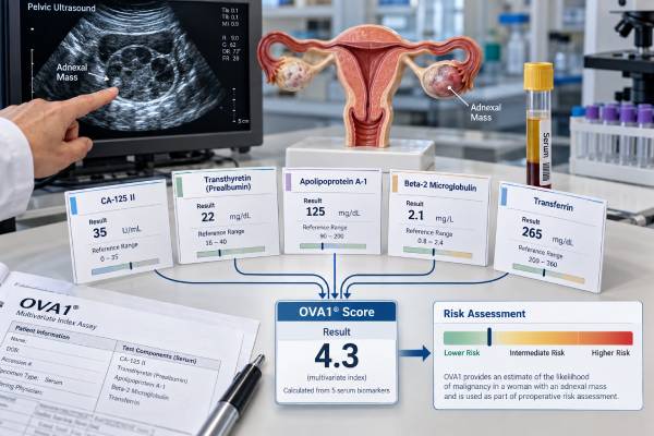
The OVA1 test is a blood-based risk assessment used before surgery in certain adults who already have an ovarian or adnexal mass. It combines five serum proteins into one score to help estimate whether a mass has a higher or lower likelihood of being malignant. The main purpose is not to diagnose ovarian cancer. Instead, OVA1 can add information to imaging, examination findings, menopausal status, and a clinician’s judgment when deciding whether a gynecologic oncologist should be involved in the planned operation. That distinction matters because ovarian cancer surgery is most effective when appropriate staging and tumor-removal procedures are available from the start. A high OVA1 result does not prove cancer, and a low result does not completely exclude it. Benign conditions, other cancers, and non-ovarian illnesses can affect the proteins in the score. The result therefore makes the most sense when read as one part of a preoperative risk assessment rather than as a stand-alone cancer test.
- OVA1 is for preoperative risk assessment, not screening: it is used when an adnexal mass is already known and surgery is planned.
- The score runs from 0.0 to 10.0: higher values are classified as higher likelihood of malignancy using menopause-specific cutoffs.
- Classic OVA1 uses five proteins: CA-125, transferrin, apolipoprotein A-1, beta-2 microglobulin, and transthyretin (prealbumin).
- High-risk cutoffs are 5.0 or higher before menopause and 4.4 or higher after menopause for the original FDA-cleared OVA1 test.
- A high score does not diagnose ovarian cancer: imaging, symptoms, examination, and ultimately surgical pathology determine what the mass is.
Table of Contents
- What OVA1 Is and When It Is Used
- The Five Biomarkers in the OVA1 Score
- OVA1 Score Meaning and Cutoffs
- Accuracy, Sensitivity, and False-Positive Results
- Important Limitations and Factors That Affect Interpretation
- What Usually Happens After an OVA1 Result
- OVA1 Compared With CA-125, ROMA, and Overa
What OVA1 Is and When It Is Used
OVA1 is an in vitro diagnostic multivariate index assay. In everyday terms, that means the laboratory measures several blood proteins and then uses a proprietary mathematical algorithm to combine them into one risk score. The FDA-cleared intended-use population is narrow: adults with an ovarian or adnexal mass for which surgery is already planned and who have not yet been referred to a gynecologic oncologist.
That narrow use is important. OVA1 is not designed to answer, “Do I have ovarian cancer?” in a person without a known mass. It is also not intended to decide whether surgery is needed. Its role is to improve preoperative triage when the clinician’s independent examination and imaging assessment have not already established a clear high-risk picture.
The practical question is often whether the planned operation should involve a gynecologic oncologist. Specialists in gynecologic oncology are trained to perform comprehensive staging and cytoreductive procedures when malignancy is found. If a mass turns out to be cancer, having the appropriate surgical expertise available during the first operation can avoid delays, repeat surgery, or incomplete staging.
OVA1 is generally ordered after a pelvic mass is identified by ultrasound, CT, MRI, physical examination, or another clinical evaluation. It should be interpreted alongside the imaging appearance of the mass, age, menopausal status, symptoms, personal and family history, and other laboratory findings. For readers trying to understand individual tumor markers, a CA-125 ovarian cancer test is only one part of this broader risk assessment.
The test does not replace pathology. A definitive diagnosis usually requires examination of tissue removed at surgery or biopsy. Even a strongly elevated risk score still represents a probability classification rather than histologic proof of cancer.
The Five Biomarkers in the OVA1 Score
The original OVA1 assay combines five serum proteins: CA-125, transferrin, apolipoprotein A-1, beta-2 microglobulin, and transthyretin, which is also called prealbumin. The algorithm does not simply count how many are “abnormal.” It weighs their values together to identify a pattern associated with a greater or lesser likelihood of malignancy.
CA-125 is the best-known component. It can rise in epithelial ovarian cancer, especially advanced high-grade serous disease, but it is not specific to cancer. Endometriosis, menstruation, pregnancy, pelvic inflammation, liver disease, and other benign conditions may increase it. Some ovarian cancers, especially at an early stage or of certain histologic types, produce little CA-125.
Beta-2 microglobulin may rise with increased immune-cell turnover, kidney dysfunction, inflammation, and several malignancies. Transferrin, apolipoprotein A-1, and transthyretin are transport or nutritional proteins whose concentrations may fall or shift with systemic illness, inflammation, liver function changes, and nutritional state. Those broader biological signals are part of why the panel may detect risk patterns that CA-125 alone misses, but they also help explain why false-positive results occur.
The laboratory enters the five measured values into the OVA1 software, which produces a unitless score from 0.0 to 10.0. The individual analyte values are not meant to be interpreted independently as five separate cancer tests. A person may have a CA-125 value within the laboratory reference range and still have a high OVA1 score because the combination of all five proteins changes the overall pattern.
This multianalyte approach is one reason OVA1 can show higher sensitivity than a single marker. It is also why a clinician should not try to reconstruct or reinterpret the score using unrelated reference ranges. The validated result is the algorithm-generated OVA1 classification produced by the designated laboratory method.
OVA1 Score Meaning and Cutoffs
The original OVA1 score ranges from 0.0 to 10.0. Interpretation depends on menopausal status.
| Menopausal group | Lower-likelihood result | Higher-likelihood result |
|---|---|---|
| Premenopausal | Below 5.0 | 5.0 or higher |
| Postmenopausal | Below 4.4 | 4.4 or higher |
These are classification cutoffs, not “normal laboratory ranges” in the same sense as sodium or hemoglobin. A score just above a cutoff does not mean the same thing as a confirmed cancer diagnosis, and a score just below it does not establish that a mass is benign.
What a high OVA1 score means
A high-risk result means the combination of serum biomarkers places the patient in the higher-likelihood category for malignancy within the population for which the test was designed. The usual clinical implication is to reconsider the surgical plan and determine whether referral to or involvement of a gynecologic oncologist is appropriate.
The result should be viewed in context. A complex solid-and-cystic mass with ascites and a high OVA1 score is a different clinical situation from a simple-appearing cyst with a borderline-high score. Imaging features may be more immediately concerning in the first scenario. Conversely, a high OVA1 result can add concern when imaging findings are indeterminate.
What a low OVA1 score means
A low-risk result decreases concern but does not reduce the chance of malignancy to zero. No biomarker panel detects every ovarian cancer. Early-stage tumors, uncommon histologic types, tumors that shed few circulating proteins, and biological variability can all contribute to false-negative results.
A low score should never be used to dismiss concerning symptoms or imaging. Rapid mass growth, nodularity, papillary projections, significant ascites, unexplained weight loss, persistent abdominal distention, or other high-risk findings may still justify specialist evaluation regardless of the OVA1 classification.
Menopausal status matters because the original assay uses a lower threshold after menopause. A person’s laboratory report should identify the category applied. If the menopause designation appears incorrect or is unclear, the ordering clinician should verify it before making decisions based on the score.
Accuracy, Sensitivity, and False-Positive Results
OVA1 was developed to favor sensitivity: it is designed to identify as many malignancies as practical among patients already proceeding to surgery for an adnexal mass. That design helps explain both its strength and its main tradeoff.
Clinical studies have shown that adding a multivariate index assay to a clinician’s preoperative assessment can identify ovarian malignancies that might be missed by clinical assessment or CA-125 alone. Performance has been particularly notable for sensitivity and negative predictive value in intended-use populations. However, specificity is lower, which means many benign masses can receive a high-risk result.
A false-positive result occurs when OVA1 classifies a patient as high likelihood but surgery ultimately shows benign pathology. This can happen because several proteins in the algorithm respond to inflammation, nutritional changes, kidney function, liver function, or other diseases. The assay intentionally accepts more false positives in exchange for reducing the chance that an ovarian malignancy is overlooked before surgery.
A false-negative result is also possible. A mass may be malignant even if OVA1 is below the cutoff. This is why the FDA-cleared labeling requires independent clinical and imaging assessment rather than allowing the score to stand alone.
Sensitivity and specificity should also not be confused with a person’s individual probability of cancer. Positive predictive value changes with the prevalence of malignancy in the tested population. For example, a high-risk result in a population with many benign masses will produce more false positives than the same test used in a population where cancer is more common. Age, referral setting, imaging characteristics, and histology mix therefore influence how the statistics translate to an individual patient.
The same principle applies to newer ovarian mass algorithms. A test can be very sensitive yet still generate substantial anxiety and additional specialist referrals because its specificity is modest. That is a design tradeoff, not necessarily a laboratory error.
Important Limitations and Factors That Affect Interpretation
The most important limitation is that OVA1 is not an ovarian cancer screening test. It should not be ordered in an asymptomatic person simply because of family history, age, or fear of ovarian cancer. People with hereditary risk need genetic counseling and appropriate risk-management strategies rather than serial OVA1 testing. When inherited susceptibility is a concern, testing such as BRCA1 and BRCA2 genetic testing addresses a different clinical question.
OVA1 also cannot determine the tumor type. A high result does not distinguish epithelial ovarian cancer from a borderline tumor, metastatic disease involving the ovary, or another malignancy. It does not provide stage, grade, BRCA status, homologous recombination deficiency, or treatment sensitivity. Those questions require pathology and molecular testing after tissue is available.
Several conditions can influence one or more component proteins. Kidney dysfunction can affect beta-2 microglobulin. Liver disease, inflammation, malnutrition, and systemic illness may influence transferrin, apolipoprotein A-1, or transthyretin. CA-125 may rise with endometriosis, fibroids, menstruation, pregnancy, pelvic inflammatory disease, pleural or peritoneal irritation, and other benign causes.
Timing also matters. The test is designed for a preoperative serum sample. It is not validated as a routine marker for tracking chemotherapy response, measuring minimal residual disease, or monitoring recurrence. After ovarian cancer is diagnosed, clinicians usually use tumor-specific follow-up strategies such as symptoms, examination, imaging when indicated, and sometimes serial CA-125 in patients whose cancer produced CA-125 at baseline.
Another common mistake is comparing an OVA1 result directly with a different multivariate test as though the numbers share the same scale. Although several tests may produce values from 0 to 10 or risk percentages, their biomarker combinations, algorithms, cutoffs, and validation populations differ. The number only has meaning within the specific assay that generated it.
Finally, the result can be clinically unnecessary when imaging and examination already show a clearly suspicious malignancy requiring gynecologic oncology referral. Biomarkers are most useful when they change a decision, not when they simply confirm a referral that is already indicated.
What Usually Happens After an OVA1 Result
The next step depends on the entire preoperative picture, not the score alone.
For a high-risk OVA1 result, the ordering clinician typically reviews the imaging again, confirms the planned surgical approach, and considers consultation with a gynecologic oncologist. If the patient is already seeing a gynecologic oncologist, the test may add little because the referral question has already been answered.
For a low-risk OVA1 result, the clinician still asks whether the mass looks benign enough for the planned approach. A low score can be reassuring when imaging, examination, and symptoms are also low risk. It should not override a highly suspicious ultrasound or CT appearance.
Useful questions for a preoperative visit include:
- Which OVA1 cutoff was applied based on my menopausal status?
- What features on ultrasound or other imaging raise or lower concern?
- Does my personal or family history suggest hereditary cancer testing?
- Would the result change who should perform the operation or where it should be done?
- If pathology shows cancer, what additional tumor testing would be performed?
Pathology after surgery remains the decisive step. If cancer is found, the tissue report identifies histologic type, grade, stage-related findings, and other features. Depending on the cancer type, clinicians may then order a broader ovarian cancer biomarker panel, including germline and tumor genetic testing when appropriate.
Urgent assessment is not based on OVA1 alone. Seek prompt medical evaluation for severe or rapidly worsening pelvic or abdominal pain, fainting, significant bleeding, persistent vomiting, or symptoms suggesting torsion, rupture, bowel obstruction, or another acute complication of an adnexal mass.
OVA1 Compared With CA-125, ROMA, and Overa
OVA1 is one of several tools that can be used to characterize malignancy risk before surgery, but the tests are not interchangeable.
CA-125 is a single serum marker. It is widely used in ovarian cancer care and may help monitor known disease when elevated at baseline, but it has limited specificity for diagnosing a new adnexal mass. A normal CA-125 also cannot exclude ovarian cancer.
ROMA combines CA-125, HE4, and menopausal status in an algorithm. Like OVA1, it is intended for selected patients with an adnexal mass who are already being considered for surgery, not for population screening. The ROMA ovarian cancer risk score uses different formulas and assay-specific cutoffs, so its percentage or numerical result should not be translated into an OVA1 category.
Overa, also known in FDA documents as OVA1 Next Generation, uses five proteins: CA-125, HE4, apolipoprotein A-1, transferrin, and follicle-stimulating hormone. It replaced beta-2 microglobulin and transthyretin from the original OVA1 panel and uses a single cutoff of 5.0 in the FDA-cleared version. The Overa ovarian mass risk assessment was developed in part to improve specificity while retaining high sensitivity.
A clinician may choose among these approaches based on availability, local practice, patient characteristics, and whether the result is likely to change referral or surgical planning. No one serum algorithm replaces expert ultrasound interpretation, clinical judgment, or pathology.
The most useful way to read an OVA1 result is therefore as a triage signal: high means the surgical team should take malignancy risk seriously and consider gynecologic oncology involvement; low means risk is reduced but not eliminated. The score becomes meaningful only when it is combined with the rest of the patient’s clinical picture.
When comparing reports over time, remember that OVA1 is not intended for serial cancer surveillance. A later score is not a validated measure of tumor growth or treatment response, and small numerical changes should not be interpreted as improvement or progression. If the clinical question changes after surgery—from preoperative triage to confirmed-cancer management—the testing strategy should change as well. Pathology, stage, tumor genetics, treatment response, and established follow-up markers become more relevant than repeating the presurgical algorithm.
References
- Diagnostic biomarkers in ovarian cancer: advances beyond CA125 and HE4 2024 (Review)
- Current and Emerging Methods for Ovarian Cancer Screening and Diagnostics: A Comprehensive Review 2022 (Review)
- A reflex testing protocol using two multivariate index assays improves the risk assessment for ovarian cancer in patients with an adnexal mass 2023
- Current clinical application of serum biomarkers to detect and monitor ovarian cancer – update 2022 (Review)
- REVIEW MEMORANDUM 2009 (FDA)
Disclaimer
OVA1 results should be interpreted by a clinician who knows the imaging findings, menopausal status, symptoms, and planned surgery. The test does not diagnose or rule out ovarian cancer and is not intended for screening. Decisions about referral, surgery, or additional testing should not be made from the OVA1 score alone.