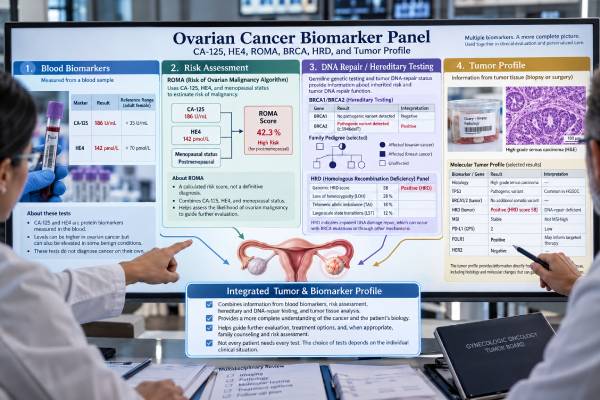
An ovarian cancer biomarker panel is not one fixed laboratory test. It is a group of blood, inherited-gene, and tumor tests chosen for different questions: how suspicious an ovarian or adnexal mass is, how a known cancer is responding, whether a hereditary cancer syndrome is present, and which treatments may fit the tumor’s biology. CA-125 and HE4 are blood proteins; ROMA combines those markers with menopause status for preoperative risk assessment. BRCA1 and BRCA2 testing can look for inherited or tumor-acquired DNA-repair changes, while homologous recombination deficiency (HRD) testing examines a broader pattern of impaired DNA repair. Tumor profiling may add other biomarkers depending on histology, stage, prior treatment, and available therapies. No single result can diagnose ovarian cancer by itself. The most useful interpretation comes from matching each biomarker to the clinical question, the tumor type, imaging findings, pathology, and the laboratory method used.
- CA-125 is mainly useful for monitoring and risk assessment, not population screening: many benign conditions can raise it, and some ovarian cancers do not produce much CA-125.
- HE4 can complement CA-125: it is used in some adnexal-mass risk tools, but kidney function, age, smoking, and assay differences can affect interpretation.
- ROMA is a triage score: it combines CA-125, HE4, and menopause status to classify preoperative risk in women with an adnexal mass; it does not diagnose cancer.
- BRCA and HRD results can affect treatment and family counseling: germline and tumor testing answer related but different questions.
- Tumor profiling should be histology- and treatment-specific: the useful panel for high-grade serous ovarian cancer is not identical to that for clear-cell, endometrioid, mucinous, or sex-cord stromal tumors.
Table of Contents
- What an Ovarian Cancer Biomarker Panel Measures
- CA-125, HE4, and ROMA for Blood-Based Risk Assessment
- BRCA and Hereditary Testing
- HRD and DNA-Repair Testing
- Tumor Profile and Treatment-Selection Biomarkers
- How to Interpret Results Together
- Testing Timing, Limitations, and Next Steps
What an Ovarian Cancer Biomarker Panel Measures
The phrase ovarian cancer biomarker panel can refer to several different categories of testing. They should not be treated as interchangeable because each category answers a different clinical question.
A practical way to separate them is:
- Serum biomarkers: proteins measured in blood, especially CA-125 and HE4. These can support risk assessment for an adnexal mass or help monitor a diagnosed cancer.
- Risk algorithms: calculations such as ROMA that combine laboratory values with clinical information, usually menopause status.
- Germline genetic testing: testing blood or saliva for inherited pathogenic variants such as BRCA1, BRCA2, RAD51C, RAD51D, BRIP1, and Lynch syndrome genes.
- Tumor genomic testing: testing cancer tissue for somatic mutations and other molecular features that arose in the tumor.
- HRD testing: evaluating whether a tumor has defective homologous recombination DNA repair, often through BRCA status and/or genomic-scarring measures.
- Histology-specific biomarkers: tests selected because a particular ovarian cancer subtype may have a targetable protein or molecular alteration.
These tests are usually ordered at different points. A person with a newly found pelvic mass may first have imaging and serum markers. Once cancer is confirmed by pathology, molecular testing becomes more important for treatment planning. Germline testing can be appropriate even when there is no striking family history because a meaningful fraction of epithelial ovarian cancers are associated with inherited susceptibility.
This distinction prevents a common mistake: assuming that a “normal panel” rules out ovarian cancer. It does not. A normal CA-125 result in ovarian cancer, for example, cannot exclude malignancy when imaging or symptoms remain concerning. Likewise, a negative hereditary panel does not mean the tumor lacks actionable somatic changes.
The panel should therefore be built around a decision. Before testing, it is reasonable to ask: Are we assessing a mass, confirming inherited risk, choosing therapy, or monitoring disease? The answer determines which biomarkers are useful.
CA-125, HE4, and ROMA for Blood-Based Risk Assessment
CA-125 and HE4 are the best-known serum markers used in epithelial ovarian cancer, but they are most useful when interpreted in context rather than as stand-alone cancer tests.
CA-125 is a glycoprotein that can rise in many epithelial ovarian cancers, especially advanced high-grade serous disease. It can also rise with endometriosis, menstruation, fibroids, pregnancy, pelvic inflammation, liver disease, and other cancers. Early-stage ovarian cancer may produce a normal CA-125. For these reasons, CA-125 is not sufficiently specific or sensitive to screen average-risk people by itself.
After ovarian cancer is diagnosed, serial CA-125 values can be more informative than one isolated number. A falling level during treatment may support a response, while a sustained rise can raise concern for progression or recurrence. The trend still has to be interpreted with symptoms, examination, and imaging.
HE4, or human epididymis protein 4, is another serum protein that may be elevated in epithelial ovarian cancer. Compared with CA-125, it is less commonly increased by some benign gynecologic conditions, but it has its own confounders. Reduced kidney function can cause substantial HE4 elevation. Age and smoking can also influence levels, and reference limits differ by assay. A detailed HE4 ovarian cancer test interpretation should therefore use the laboratory’s own reference interval.
ROMA, the Risk of Ovarian Malignancy Algorithm, combines CA-125, HE4, and menopause status to place a woman with an adnexal mass into a lower- or higher-risk category before surgery. It is meant to help guide referral and surgical planning, not to confirm ovarian cancer. Different commercial assays can use different equations or cutoffs, so the reported risk category is more useful than applying a threshold from another laboratory.
A higher serum-marker risk score means the mass deserves appropriate specialist evaluation; it does not mean that cancer is certain. Conversely, a lower-risk score cannot overrule a highly suspicious ultrasound, CT scan, physical examination, or rapidly changing clinical picture.
BRCA and Hereditary Testing
BRCA1 and BRCA2 are DNA-repair genes. A pathogenic variant can be inherited through the germline or can develop only in the tumor. That difference matters because the result can affect both treatment and relatives.
Germline testing is usually performed on blood or saliva. A pathogenic or likely pathogenic BRCA1 or BRCA2 variant can explain hereditary ovarian cancer risk and may also change breast, pancreatic, and prostate cancer risk management in a family. Current specialty guidance supports offering germline genetic testing to people with epithelial ovarian, fallopian tube, or primary peritoneal cancer, even when the family history is limited.
Modern hereditary panels often include more than BRCA1 and BRCA2. Depending on the laboratory and clinical situation, they may include RAD51C, RAD51D, BRIP1, PALB2, and mismatch-repair genes associated with Lynch syndrome. A broader gynecologic cancer hereditary gene panel can be useful because ovarian cancer susceptibility is genetically heterogeneous.
Tumor BRCA testing looks for alterations within the cancer itself. A tumor can carry a BRCA mutation even when germline testing is negative. Conversely, finding a BRCA alteration on tumor-only testing does not prove it is inherited. Confirmatory germline testing is usually needed when a potentially inherited pathogenic variant is found in tumor tissue.
The most important result categories are:
- Pathogenic or likely pathogenic: a disease-associated variant with enough evidence to guide care in the correct setting.
- Negative: no reportable pathogenic variant was found in the genes and regions tested. This does not eliminate hereditary risk completely.
- Variant of uncertain significance (VUS): a DNA change whose clinical meaning is not established. A VUS should not be treated like a pathogenic variant or used by itself to justify preventive surgery.
A positive BRCA1 and BRCA2 ovarian cancer test may also help establish whether a tumor is likely to have homologous recombination repair defects, but BRCA is only one part of the HRD picture.
HRD and DNA-Repair Testing
Homologous recombination deficiency means that a tumor has impaired ability to repair certain double-strand DNA breaks accurately. BRCA1 and BRCA2 are central to this pathway, but other genes and epigenetic changes can produce similar repair failure.
HRD assays do not all measure the same thing. Some focus on specific gene mutations. Others look for a genomic scar—a pattern of accumulated chromosome changes that suggests homologous recombination repair has been defective over time. Commercial assays may combine BRCA status with measures such as loss of heterozygosity, telomeric allelic imbalance, and large-scale state transitions into a composite score.
This has several practical consequences:
- A BRCA-mutated tumor is often HRD-positive, but HRD can occur without a BRCA mutation.
- A negative BRCA result is not the same as HRD-negative.
- A genomic-scar result reflects the tumor’s history and may not perfectly represent its current ability to repair DNA after treatment pressure.
- The cutoff and definition of “HRD-positive” depend on the validated assay; scores should not be transferred between platforms.
HRD status can help estimate the likelihood of benefit from certain PARP inhibitor strategies in ovarian cancer, but treatment decisions depend on the exact drug, disease setting, line of therapy, BRCA status, HRD assay, prior treatments, regulatory approvals, and current guidelines. An HRD test for ovarian cancer is therefore best understood as a predictive biomarker used within a specific treatment context, not as a universal “sensitive” or “resistant” label.
Pathology and specimen quality also matter. Low tumor content, extensive necrosis, small biopsies, or degraded DNA can lead to failed or indeterminate testing. If a report says “insufficient tumor,” that is different from an HRD-negative result and may prompt testing of another block or specimen.
Tumor Profile and Treatment-Selection Biomarkers
A tumor profile usually means broader molecular or immunohistochemical testing on ovarian cancer tissue. The useful markers depend strongly on histologic subtype. “Ovarian cancer” includes biologically different diseases rather than one uniform tumor.
For high-grade serous ovarian carcinoma, BRCA1/2 and HRD are among the most important established molecular tests because of their relationship to DNA-repair-targeted therapy. Tumor sequencing may also identify less common alterations, but a large panel is useful only if a result can change management, clarify diagnosis, or support a clinical trial.
For other histologies, different markers may matter. Examples include mismatch-repair or microsatellite-instability testing in selected endometrioid or clear-cell tumors, HER2 assessment in some mucinous cancers, and other target-expression tests when an approved therapy or trial requires them. Folate receptor alpha expression can also be relevant in certain treatment settings for platinum-resistant epithelial ovarian cancer. Testing practices evolve as new therapies and indications appear.
Pathologists also use immunohistochemistry to confirm tumor origin. Markers such as PAX8 and WT1 can help classify a pelvic or peritoneal carcinoma, but they do not function like serum tumor markers. A “tumor profile” report may therefore contain both diagnostic markers and predictive biomarkers, and those categories should not be confused.
A useful way to read a molecular report is to sort each item into one of four questions:
- Does this result confirm or refine the diagnosis?
- Does it indicate an approved treatment option in this exact disease setting?
- Does it suggest clinical-trial eligibility?
- Does it suggest an inherited variant that needs germline confirmation or genetic counseling?
This approach is more helpful than focusing on the sheer number of mutations listed. Many tumor-sequencing reports include variants with no established ovarian-cancer treatment implication.
How to Interpret Results Together
A biomarker panel becomes clinically useful when its results are combined rather than read as independent verdicts. The same result can mean something very different before diagnosis, during first-line treatment, or years later during surveillance.
For a new adnexal mass, the priority is estimating malignancy risk and planning the safest surgery. Menopause status, ultrasound features, symptoms, family history, CA-125, HE4, and a validated risk tool may contribute. If the pattern suggests substantial risk, referral to a gynecologic oncologist before surgery can improve staging and cytoreductive planning. The ROMA ovarian cancer risk score is one possible adjunct, but it should not replace imaging or clinical judgment.
For newly diagnosed epithelial ovarian cancer, the priority shifts to pathology, stage, germline genetic testing, and tumor biomarkers that can influence treatment. A patient may have normal hereditary testing but a somatic BRCA mutation, or no BRCA mutation but an HRD-positive genomic-scar result. These combinations carry different implications.
For monitoring after treatment, serial biomarkers may help when they were informative at baseline. CA-125 is the most established serum marker in common epithelial ovarian cancers, but treatment should not be changed solely because of one laboratory fluctuation. Imaging and clinical findings remain central. In selected settings, emerging approaches such as ovarian cancer ctDNA testing are being studied for molecular residual disease and recurrence monitoring, but they are not yet a universal replacement for standard follow-up.
Several result combinations illustrate why context matters:
- High CA-125 + suspicious complex mass: increases concern but still requires diagnostic evaluation and pathology.
- Normal CA-125 + suspicious mass: does not rule out cancer.
- Germline BRCA-positive + tumor BRCA-positive: has treatment implications and hereditary implications.
- Tumor BRCA-positive + germline BRCA-negative: suggests a somatic alteration; relatives do not automatically carry it.
- BRCA-wild type + HRD-positive: indicates broader repair deficiency may be present despite no BRCA mutation.
- VUS only: should not be interpreted as proven inherited susceptibility or a proven treatment biomarker.
Testing Timing, Limitations, and Next Steps
The best time to test depends on the biomarker. Serum CA-125 and HE4 can be drawn before surgery when they are part of adnexal-mass assessment. Germline testing can be ordered soon after an epithelial ovarian cancer diagnosis because results can affect both treatment and relatives. Tumor BRCA and HRD testing is often most useful early enough that results are available when maintenance or later-line decisions are made.
Before a blood draw, special fasting is usually unnecessary for CA-125 or HE4 unless the laboratory has combined the order with tests that do require fasting. The bigger issue is documenting factors that can distort interpretation, such as reduced kidney function for HE4 or benign gynecologic inflammation for CA-125.
For tumor testing, the pathology laboratory needs adequate tissue. A surgical specimen often provides more material than a small biopsy, but testing can sometimes be performed on archived formalin-fixed tissue. If sequencing fails, the report may state low tumor percentage, poor DNA quality, or insufficient material. That is a technical failure, not a negative biomarker.
After results are available, useful next steps include:
- Confirm what type of test was done. Serum marker, germline DNA, tumor DNA, HRD score, and immunohistochemistry have different meanings.
- Use the laboratory-specific reference and cutoff. Do not compare a ROMA or HRD score with thresholds from another assay.
- Ask whether the result changes a current decision. A biomarker is most valuable when it affects referral, treatment, surveillance, or family counseling.
- Review hereditary findings with a genetics professional. Pathogenic germline results may trigger cascade testing for relatives, while a VUS generally should not.
- Reassess testing when disease circumstances change. A new recurrence, a different treatment line, or a newly approved targeted therapy can create a reason to revisit tumor profiling.
No panel eliminates diagnostic uncertainty. Biomarkers add information; they do not replace tissue diagnosis, expert pathology, imaging, or a complete clinical assessment. A well-designed ovarian cancer biomarker strategy uses the fewest tests needed to answer the most important questions at each stage of care.
References
- SEOM-GEICO clinical guideline on epithelial ovarian cancer (2023) 2024 (Guideline)
- Germline and somatic testing for ovarian Cancer: An SGO clinical practice statement 2024 (Position Statement)
- Homologous recombination deficiency in ovarian cancer: Global expert consensus on testing and a comparison of companion diagnostics 2025 (Consensus)
- Molecular Pathology of Ovarian Epithelial Neoplasms: Predictive, Prognostic, and Emerging Biomarkers 2024 (Review)
- Systematic Analysis of Homologous Recombination Deficiency Testing in Ovarian Cancer-Development of Recommendations for Optimal Assay Performance 2024 (Review)
- Diagnostic biomarkers in ovarian cancer: advances beyond CA125 and HE4 2024 (Review)
Disclaimer
This article provides general education about ovarian cancer biomarkers and cannot diagnose cancer or determine an individual treatment plan. Test selection and interpretation should be handled by a gynecologic oncology team using the exact laboratory method, pathology, imaging, medical history, and current treatment guidelines. Genetic results that may be inherited should be reviewed with an appropriately qualified genetics professional.