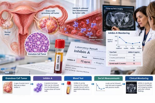
An inhibin A blood test can help evaluate and monitor ovarian granulosa cell tumors, a rare group of sex cord-stromal tumors that can produce reproductive hormones. Inhibin A is made from an alpha subunit paired with a beta-A subunit and normally participates in regulation of follicle-stimulating hormone. A granulosa cell tumor may release excess inhibin A into the bloodstream, so a high result can support the diagnosis when the clinical and imaging findings fit. The test is more useful for monitoring when inhibin A was elevated before treatment and then falls after surgery. A later rise can raise concern for persistent or recurrent disease, although imaging and clinical assessment are still required. Inhibin A is generally less sensitive than inhibin B for adult-type granulosa cell tumors, and normal values vary with reproductive status, menstrual cycle, pregnancy, and assay. It should not be used as a general ovarian cancer screening test or interpreted as a cancer diagnosis by itself.
- Inhibin A is a hormone produced by granulosa cells and can be elevated in ovarian granulosa cell tumors.
- A high inhibin A result supports, but does not prove, a granulosa cell tumor; the result must be interpreted with imaging and pathology.
- Inhibin B is usually a more sensitive serum marker than inhibin A for adult granulosa cell tumors, so both or AMH may be considered.
- Reference ranges depend on menopausal status, menstrual-cycle phase, pregnancy, and the laboratory assay; there is no universal tumor cutoff.
- For monitoring, the most informative comparison is often the patient’s own baseline value and serial trend using the same assay.
Table of Contents
- What Inhibin A Is
- Why Inhibin A Is Tested
- Understanding Inhibin A Results
- Inhibin A Versus Inhibin B and AMH
- Using Inhibin A for Monitoring
- Limitations and Noncancer Causes
- What Happens After an Abnormal Test
What Inhibin A Is
Inhibins are hormones produced mainly by ovarian granulosa cells. They help regulate the pituitary hormone FSH, which in turn influences ovarian follicle development.
There are two major dimeric forms:
- Inhibin A, made of an alpha subunit and beta-A subunit.
- Inhibin B, made of an alpha subunit and beta-B subunit.
The two hormones have different patterns during the menstrual cycle. Inhibin A tends to be more prominent in the luteal phase, while inhibin B is closely related to developing follicles. After menopause, ovarian production of both becomes very low.
Granulosa cell tumors arise from the same type of ovarian cells that normally produce inhibins. Many of these tumors retain hormone-secreting functions, which is why serum inhibin measurements can act as tumor markers.
Adult-type granulosa cell tumor is the most common form. It often contains a characteristic FOXL2 mutation and can produce estrogen, inhibins, and anti-Müllerian hormone. The tumors are usually slow growing, but an important feature is their ability to recur many years or even decades after the original diagnosis.
Inhibin A is therefore most useful in a specific clinical context: a known or suspected granulosa cell tumor. It is not a marker for all ovarian cancers. High-grade serous ovarian cancers are more often evaluated with markers such as CA-125, while germ cell tumors use markers such as AFP, beta-hCG, and LDH.
It is also important to distinguish a serum inhibin A test from inhibin immunohistochemistry. Pathologists may stain tumor tissue for inhibin-alpha to help identify sex cord-stromal differentiation. Tissue staining and blood measurement answer different questions and should not be treated as the same test.
Why Inhibin A Is Tested
A clinician may order inhibin A during the workup of an ovarian mass when age, symptoms, imaging, or hormone effects raise suspicion for a sex cord-stromal tumor.
Granulosa cell tumors can produce estrogen. Premenopausal patients may present with irregular bleeding, heavy bleeding, or amenorrhea. Postmenopausal patients may develop unexpected uterine bleeding. In children, hormone production can cause signs of precocious puberty. These hormonal clues can lead clinicians to consider inhibin testing.
The blood test can serve three main purposes:
- Support the initial evaluation. A clearly elevated value in the appropriate setting makes a granulosa cell tumor more plausible, although pathology is required for diagnosis.
- Establish a baseline. If inhibin A is high before surgery, the pretreatment value becomes a useful personal reference.
- Monitor disease over time. Falling levels after complete tumor removal can support treatment response, while a later sustained rise can trigger evaluation for recurrence.
The test is usually a simple venous blood draw. Fasting is not generally required. The clinician should know whether the patient is premenopausal, postmenopausal, pregnant, or using hormonal medications, because these factors can affect interpretation.
Timing is especially important when the purpose is to establish a baseline. Ideally, the pretreatment level is measured before the tumor is removed, because a postoperative value cannot show whether inhibin A was a useful marker for that tumor in the first place. If no baseline was obtained, later monitoring is still possible, but a normal value after surgery is harder to interpret.
The clinical history also matters because granulosa cell tumors may coexist with estrogen-related changes in the uterus. Excess estrogen can stimulate the endometrium, leading to thickening, hyperplasia, or occasionally a separate endometrial malignancy. Abnormal uterine bleeding therefore deserves direct evaluation rather than being attributed only to the ovarian marker result.
When a granulosa cell tumor is suspected, clinicians may also order inhibin B and AMH. Using more than one marker can be helpful because individual tumors do not all secrete the same hormones.
Understanding Inhibin A Results
There is no single inhibin A number that means ovarian cancer. The result must be compared with the reference interval from the performing laboratory.
In premenopausal patients, inhibin A changes across the menstrual cycle. A concentration that is expected in one phase may be unusual in another. Pregnancy can also produce high inhibin A because placental tissue contributes to circulating levels; inhibin A is used in a completely different context as part of some prenatal screening tests.
After menopause, inhibin A is normally very low or undetectable on many assays. For that reason, a clearly measurable or rising level can be more suspicious in a postmenopausal patient, especially if there is a known granulosa cell tumor history.
A high result can mean:
- active granulosa cell tumor that secretes inhibin A;
- residual tumor after treatment;
- recurrent disease in a patient whose tumor previously produced the marker;
- normal reproductive or pregnancy-related production, depending on the patient;
- less commonly, another tumor or biological source capable of producing related inhibin molecules.
A normal result does not exclude a granulosa cell tumor. In a large single-center cohort, inhibin A was elevated in about two-thirds of patients at diagnosis and was less sensitive than inhibin B. That means a substantial minority of confirmed tumors did not produce enough inhibin A to cross the test threshold.
For monitoring, one isolated borderline value is less informative than a repeatable trend. A small change can reflect assay variation, timing in the menstrual cycle, or biological fluctuation. A consistent rise, especially from a previously low post-treatment baseline, deserves clinical attention.
Inhibin A Versus Inhibin B and AMH
For adult granulosa cell tumors, inhibin B is generally the preferred inhibin marker because evidence shows greater sensitivity.
A frequently cited cohort comparing the two found inhibin A elevated in 67% of patients at diagnosis versus 89% for inhibin B. At recurrence, the corresponding proportions were 58% and 85%. In that study, both markers were highly specific in the control group, but inhibin B more often reflected active disease.
That does not make inhibin A useless. If a particular patient’s tumor produces inhibin A strongly, it can be an excellent personalized marker. Tumor-marker usefulness is partly determined by the biology of the individual cancer.
AMH is another granulosa-cell hormone used for diagnosis and follow-up. Some patients have both inhibin B and AMH measured because the combination may improve detection of active disease compared with either marker alone.
| Marker | Typical role in granulosa cell tumors | Important limitation |
|---|---|---|
| Inhibin A | Supportive marker and serial monitoring if elevated at baseline | Less sensitive than inhibin B in adult-type tumors; cycle and pregnancy affect levels |
| Inhibin B | Common serum marker for diagnosis and recurrence monitoring | Not elevated in every tumor; assay-specific cutoffs |
| AMH | Useful marker of granulosa-cell tumor activity and follow-up | Premenopausal ovarian reserve also affects concentration |
A clinician may choose one marker or follow several. The best marker is usually one that was clearly abnormal before treatment, normalized after successful therapy, and can be measured reproducibly over time.
Using Inhibin A for Monitoring
Granulosa cell tumors require long-term follow-up because late recurrence is a defining clinical concern. Serum markers can be especially useful when they begin to rise before symptoms or a mass becomes obvious.
After surgery, a previously high inhibin A concentration should usually decline if the hormone-producing tumor has been removed. The speed of decline depends on the starting level, assay, and remaining tumor burden. Persistent elevation may raise concern for residual disease, but the result must be interpreted with the operation, pathology, stage, and imaging.
During surveillance, clinicians may measure inhibin A at scheduled visits when it was informative at diagnosis. The frequency is individualized. Follow-up often becomes less frequent with time, but long-term surveillance remains important because recurrence can occur many years later.
A rising marker does not automatically mean treatment should start. The usual response is to confirm the trend and look for disease with history, examination, and imaging. A marker can provide an early warning, but imaging identifies where a recurrence is and whether it is measurable or operable.
The value of monitoring depends on consistency. Using the same laboratory and assay reduces variability. If the test platform changes, the old and new numbers may not be directly comparable.
A useful surveillance pattern is one in which the marker is clearly low during remission and rises reproducibly when disease becomes measurable. If inhibin A fluctuates without matching imaging or symptoms, the oncology team may rely more heavily on inhibin B, AMH, or radiologic follow-up. Marker performance can change over time as recurrent tumor clones evolve, so clinicians look for concordance rather than assuming the original pattern will always persist.
Long-term follow-up is particularly important for adult-type granulosa cell tumors because late relapse is well documented. A patient who has been disease free for many years should not assume that the risk has fallen to zero. The exact schedule varies by stage and treatment history, but continued clinical surveillance is a defining part of care.
Patients should also report new symptoms rather than waiting for the next blood test. Increasing abdominal or pelvic pain, bloating, a new mass sensation, abnormal uterine bleeding, early satiety, or unexplained changes in bowel or bladder function may justify earlier evaluation even if the previous inhibin A result was normal.
Limitations and Noncancer Causes
The main limitation of inhibin A is that it is not sufficiently sensitive or specific to diagnose a granulosa cell tumor by itself.
Normal reproductive physiology can raise the level
Premenopausal ovaries produce inhibin A normally. Menstrual-cycle timing therefore matters. Pregnancy can raise inhibin A substantially and changes the interpretation completely.
Not all granulosa cell tumors secrete inhibin A
A normal value cannot rule out disease. Inhibin B or AMH may be abnormal when inhibin A is not.
Other tumor types can show inhibin-related expression
Inhibin-alpha is widely used as a tissue marker for sex cord-stromal tumors, but expression can occur in other neoplasms. Serum findings must therefore be tied to the clinical diagnosis rather than used as a universal ovarian cancer marker.
Assays differ
Laboratories may use different antibody systems and reference intervals. Numeric results should not be compared across platforms without caution.
Hormone values do not replace pathology
The definitive diagnosis of a granulosa cell tumor depends on tissue morphology and, when needed, immunohistochemical and molecular findings. Adult-type tumors often carry a FOXL2 alteration, but no single blood marker substitutes for pathology review.
These limitations explain why inhibin A is best used as part of a focused sex cord-stromal tumor evaluation rather than a broad “ovarian cancer blood panel.”
What Happens After an Abnormal Test
If inhibin A is unexpectedly high, the clinician first considers whether the value can be explained by normal physiology. Menopausal status, menstrual timing, pregnancy, and previous values are reviewed. The laboratory reference range should be checked rather than using a cutoff from another institution.
When an ovarian mass is present, pelvic ultrasound or MRI can characterize the lesion. Granulosa cell tumors often appear as solid or mixed solid-cystic masses, but imaging is not diagnostic. Surgery provides tissue for definitive classification.
The pathology workup may include morphology, inhibin-alpha, calretinin, SF-1, FOXL2-related testing, and other stains depending on the differential diagnosis. The final diagnosis then determines staging and treatment.
If the patient has already been treated for a granulosa cell tumor and inhibin A rises, the next step is usually to confirm the increase and evaluate for recurrence rather than assuming the blood test alone proves relapse. Inhibin B, AMH, and imaging may add useful information.
A broader ovarian cancer biomarker profile is relevant mainly for epithelial tumors, whereas granulosa cell tumors have their own marker pattern. Knowing the histologic type prevents the wrong biomarker from being emphasized.
The key practical question is whether inhibin A has behaved as a reliable marker for this patient’s tumor. If it was never elevated before surgery, repeatedly measuring it after treatment may add little. If it was markedly elevated and normalized with treatment, serial testing can provide a useful signal during long-term surveillance.
Patients can ask for the actual numeric result and laboratory reference interval rather than only “high” or “normal.” Keeping a record of dates, values, menopausal status, and major treatment events makes trends easier to interpret over years. This is especially helpful if care moves between institutions or if the laboratory platform changes. A single unexpected value should generally be repeated or reconciled with the clinical picture before major decisions are made, particularly when the change is small and the patient has no other evidence of recurrence. Long-term trends are usually more informative than isolated minor fluctuations.
References
- Ovarian Sex Cord-Stromal Neoplasms: An Overview of Molecular Events and How to Correlate Morphology With Molecular Findings. 2025 (Review)
- Sex Cord-Stromal Tumors of the Ovary: An Update and Review. Part II – Pure Sex Cord and Sex Cord-Stromal Tumors. 2024 (Review)
- Immunohistochemical markers of prognosis in adult granulosa cell tumors of the ovary – a review. 2023 (Review)
- Adult-type granulosa cell tumor of the ovary 2022 (Review)
- Granulosa cell tumors of the ovary: the clinical value of serum inhibin A and B levels in a large single center cohort. 2007
Disclaimer
Inhibin A is a supportive tumor marker for granulosa cell tumors, not a stand-alone ovarian cancer test. Results vary with reproductive status, pregnancy, assay, and individual tumor biology, and a normal value does not exclude disease. Diagnosis and recurrence assessment require clinical evaluation, imaging, and pathology when appropriate.