Ki-67 is a protein found in cells that are actively dividing. In cervical dysplasia, pathologists can use Ki-67 staining to show abnormal cell proliferation, but Ki-67 by itself is not a stand-alone cervical cancer screening test or a single test that determines the CIN grade. Its most clinically established modern use is together with p16 in p16/Ki-67 dual-stain cytology, which identifies cervical cells that simultaneously show abnormal p16 expression and active proliferation. That combination is biologically unusual in normal cervical cells and can help triage people who test positive for high-risk HPV. A positive dual-stain result generally raises the risk of CIN3 or worse enough to support colposcopy in appropriate settings, while a negative result can allow 1-year follow-up for many HPV-positive patients. HPV 16 and HPV 18 remain important exceptions because current guidance recommends colposcopy even when dual stain is negative.
- Ki-67 is a cell-proliferation marker; more extensive staining can support the presence of a higher-grade cervical lesion but does not define CIN by itself.
- p16/Ki-67 dual stain is a cervical cytology triage test for many high-risk HPV-positive results, not a blood test.
- A dual-stain-positive result generally leads to colposcopy in guideline-supported HPV-positive screening pathways.
- A dual-stain-negative result can support 1-year HPV-based follow-up in many lower-risk situations, but HPV 16 or HPV 18 usually still requires colposcopy.
- CIN1, CIN2, and CIN3 are ultimately diagnosed from tissue morphology, with biomarkers used as supportive tools when appropriate.
Table of Contents
- What Ki-67 Measures
- Ki-67 in Cervical Biopsy Pathology
- p16/Ki-67 Dual-Stain Testing
- What Positive and Negative Results Mean
- How Ki-67 Relates to CIN Grades
- Limitations and Common Misunderstandings
- Follow-Up After Ki-67-Related Testing
What Ki-67 Measures
Ki-67 is a nuclear protein expressed during active phases of the cell cycle. Resting cells generally do not express it. Pathologists use an antibody against Ki-67 to visualize which cells in a sample are proliferating.
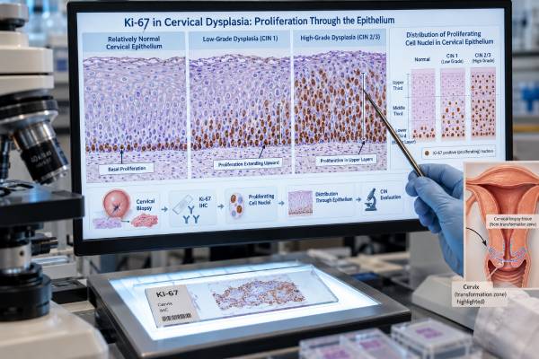
In normal squamous epithelium of the cervix, cell division is mainly confined to the basal and parabasal layers. As cells mature and move toward the surface, they stop dividing. Ki-67 staining should therefore be limited mainly to the lower portion of normal epithelium.
In cervical intraepithelial neoplasia, HPV-driven disruption of normal cell-cycle control can cause proliferating cells to extend upward into layers where they would not normally be dividing. A broader or more intense Ki-67 staining pattern can support the presence of dysplasia.
This biology makes Ki-67 useful, but it is not specific enough to diagnose cervical precancer on its own. Regeneration, inflammation, immature squamous metaplasia, and other benign processes can also increase cell proliferation.
Ki-67 should also not be confused with an HPV test. An HPV test detects carcinogenic viral types, while Ki-67 detects a host-cell proliferation state. The tests answer different questions.
The most important practical distinction is between Ki-67 immunohistochemistry on a biopsy and p16/Ki-67 dual-stain cytology. A biopsy test helps a pathologist interpret tissue architecture. Dual stain is performed on cervical cells collected for screening or triage and has specific clinical management recommendations.
Ki-67 in Cervical Biopsy Pathology
Cervical intraepithelial neoplasia is diagnosed primarily by the microscopic appearance and maturation pattern of the epithelium. Pathologists assess nuclear atypia, mitotic activity, and how far abnormal immature cells extend from the basal layer toward the epithelial surface.
Ki-67 can support this assessment. In low-grade lesions, proliferating cells are usually concentrated in the lower epithelial layers. In high-grade lesions, Ki-67-positive nuclei often extend into the middle or upper thirds.
However, there is no universal Ki-67 percentage cutoff that separates CIN1 from CIN2 or CIN3. A pathology report may describe the distribution rather than provide a numeric “Ki-67 score.”
The biomarker used more formally in difficult cervical biopsy classification is p16. Strong, continuous block-like p16 staining can support a transforming high-risk HPV effect in morphologically appropriate lesions. The p16 IHC test is especially relevant when morphology is equivocal or when distinction between a benign mimic and a high-grade lesion is difficult.
Ki-67 can add supportive information, but excessive reliance on proliferation staining can lead to overdiagnosis. Inflammation and tissue repair can produce Ki-67-positive cells without true high-grade precancer.
A biopsy diagnosis therefore integrates:
- tissue architecture and maturation;
- nuclear atypia and mitoses;
- the level of abnormal proliferation;
- p16 staining when indicated;
- clinical information such as HPV and cytology results.
The final CIN or squamous intraepithelial lesion diagnosis comes from this integrated pathology assessment, not from a single biomarker.
p16/Ki-67 Dual-Stain Testing
Dual stain is a different application of Ki-67. Instead of asking how far proliferating cells extend through a tissue layer, the test looks for the same cervical cell expressing both p16 and Ki-67.
This combination is meaningful because p16 overexpression reflects disruption of the retinoblastoma cell-cycle pathway by transforming HPV activity, while Ki-67 indicates active cell division. In normal physiology, a cell with strong p16-mediated cell-cycle arrest should not simultaneously be proliferating. Co-expression is therefore a marker of abnormal HPV-driven cell-cycle deregulation.
A commercial dual-stain test can be performed on cervical cytology material. The slide is examined for one or more cells showing the characteristic dual-positive pattern. The result is generally reported as positive or negative rather than as a percentage.
Current clinical guidance supports dual stain as a triage option for many people with a positive high-risk HPV test. It can reduce unnecessary colposcopies compared with referring every HPV-positive patient while still identifying many high-grade precancers earlier than cytology alone.
Dual stain is not the same as “two separate positive stains somewhere on the slide.” The key feature is co-expression within the same cell according to the assay’s interpretation criteria.
The test fits into a pathway that may also include extended HPV genotyping. If HPV 16 or HPV 18 is present, genotype risk generally takes priority and colposcopy is recommended even if dual stain is negative. For several other high-risk genotypes, a positive dual stain supports colposcopy and a negative result can support 1-year surveillance.
The test is interpreted on a slide by trained laboratory personnel. A positive result is based on the presence of qualifying cells with the expected p16 and Ki-67 staining pattern, not on whether the entire sample “looks strongly stained.” This matters because background staining, inflammatory cells, metaplastic cells, and technical artifacts can complicate interpretation. Quality-control rules and adequate cellularity are therefore part of a valid result.
Dual stain is particularly useful because it adds biological information after a positive HPV test. HPV tells the clinician that a carcinogenic virus is present; dual stain asks whether the sampled cervical cells are showing a pattern consistent with transforming cell-cycle disruption. It narrows the large HPV-positive population into groups with different near-term risks of CIN3+.
What Positive and Negative Results Mean
A dual-stain-positive result means at least one cervical epithelial cell meets the validated criteria for simultaneous p16 and Ki-67 expression. It does not mean cancer is present. It means the probability of clinically important cervical precancer is high enough to affect management.
In guideline-supported HPV-positive screening settings, dual-stain positivity generally leads to colposcopy. At colposcopy, suspicious areas can be biopsied to determine whether CIN2, CIN3, adenocarcinoma in situ, or invasive cancer is present.
A dual-stain-negative result means qualifying co-expressing cells were not identified. In many HPV-positive patients without HPV 16/18 or high-grade cytology, this lowers immediate CIN3+ risk enough to permit repeat HPV-based testing in 1 year.
Negative does not mean zero risk. Persistent HPV infection can later cause precancer, and the test can miss lesions because of sampling limitations or low numbers of abnormal cells.
The result also has to be read in context. A person with HSIL cytology, HPV 16, HPV 18, or a recent history of high-grade cervical disease is not managed the same way as someone with a first-time non-16/18 HPV-positive result and normal cytology.
For HPV-positive people, a specific HPV 16/18 result can therefore be as important as the dual-stain result.
Research meta-analyses generally show that p16/Ki-67 dual stain has high sensitivity for CIN2+ and CIN3+ with moderate specificity. Its main value is not perfect diagnosis; it is risk triage that helps direct colposcopy to the patients most likely to benefit.
How Ki-67 Relates to CIN Grades
CIN describes how much of the cervical squamous epithelium is replaced by abnormal immature cells.
CIN1
CIN1 is a low-grade HPV-associated lesion. Abnormal cells are mainly in the lower third of the epithelium, and maturation remains in the upper layers. Ki-67 staining may extend above the basal layer but is usually less extensive than in high-grade disease.
CIN1 often regresses without treatment, especially in younger patients. Management is usually observation rather than excision unless other risk factors change the plan.
CIN2
CIN2 is an intermediate category and can be difficult to reproduce between pathologists. Abnormal proliferation extends farther upward, and Ki-67 can be more extensive. p16 is often used to help determine whether a morphologically borderline lesion represents a transforming high-risk HPV lesion.
In selected patients, particularly those concerned about future pregnancy, carefully monitored observation of CIN2 may be an option. Others are treated.
CIN3
CIN3 is a high-grade precancer in which abnormal immature cells occupy most or all of the epithelial thickness. Ki-67-positive cells commonly extend into upper layers, and mitotic activity may be prominent.
CIN3 has a meaningful risk of progression to invasive cancer if untreated and is generally treated with an excisional procedure unless special circumstances apply.
The modern pathology terminology often groups p16-supported CIN2 and CIN3 as high-grade squamous intraepithelial lesion, or HSIL. The clinical goal is to identify the lesions that carry substantial cancer risk without overcalling transient low-grade HPV effects.
In practice, Ki-67 is most helpful when its distribution agrees with the morphology. A lesion that looks low grade and has proliferation largely restricted to the lower layers is biologically different from one with abnormal mitoses and proliferating cells high in the epithelium. If the stain and morphology conflict, the pathologist does not simply let the Ki-67 result “win.” The slide may be reviewed again, p16 may be added, deeper sections may be examined, or the diagnosis may remain qualified if the tissue is limited.
This is especially important in CIN2, which is less reproducible than clearly low-grade CIN1 or unequivocal CIN3. Biomarkers can improve consistency, but the diagnosis still depends on the full histologic pattern.
Limitations and Common Misunderstandings
A frequent misunderstanding is that “high Ki-67” means cervical cancer. It does not. Ki-67 indicates proliferation, and many benign or precancerous tissues contain proliferating cells.
Another misunderstanding is that a Ki-67 percentage from another cancer type can be applied to the cervix. In breast cancer and some neuroendocrine tumors, Ki-67 labeling indices may be reported numerically for grading or prognosis. Cervical dysplasia does not use those same cutoffs.
Sampling matters
A cytology specimen may not contain the abnormal cells even when a lesion is present. A tiny biopsy may also miss the most advanced area of a heterogeneous lesion.
Inflammation can increase proliferation
Reactive and reparative epithelium can show increased Ki-67. Morphology remains essential.
Dual stain does not replace colposcopy when genotype risk is high
HPV 16 and HPV 18 positivity generally still leads to colposcopy despite a negative dual-stain result under current recommendations.
Dual stain is not a treatment-response marker
The test is designed for screening triage, not for serial blood-like monitoring after treatment. Follow-up after CIN2+ treatment uses HPV-based surveillance according to risk-based guidelines.
A positive result does not specify lesion location
Dual stain shows that abnormal cells are present in the specimen but cannot show exactly where on the cervix the lesion is. Colposcopy and biopsy provide localization and diagnosis.
These limits help explain why the cervical cancer biomarker panel is best understood as a sequence of complementary tests rather than one combined yes-or-no cancer panel.
Follow-Up After Ki-67-Related Testing
The correct next step depends on whether Ki-67 was used on a biopsy or as part of dual-stain cytology.
If a pathology report mentions Ki-67 on a biopsy, the main result to follow is the final histologic diagnosis. Management is based on whether the tissue shows benign changes, LSIL/CIN1, HSIL/CIN2 or CIN3, adenocarcinoma in situ, or invasive cancer.
If a screening report gives a p16/Ki-67 dual-stain result, the next step follows HPV-based risk guidance. In many common pathways:
- dual-stain positive leads to colposcopy;
- dual-stain negative leads to repeat HPV-based testing in 1 year;
- HPV 16 or HPV 18 leads to colposcopy regardless of a negative dual stain;
- high-grade cytology can also override a reassuring triage result.
At the 1-year return, persistent HPV positivity or an abnormal triage result may lead to colposcopy. A negative follow-up test can lower risk and change the interval, depending on the prior history.
Patients who have already been treated for CIN2+ follow a longer surveillance pathway and should not assume that a negative dual stain returns them immediately to routine screening.
When reviewing a report, ask whether the term “Ki-67” refers to tissue IHC or p16/Ki-67 dual-stain cytology. That single distinction prevents many interpretation errors.
If the report comes from a biopsy, ask for the final diagnosis rather than focusing on the stain intensity. If it comes from screening cytology, ask whether the result was dual-stain positive or negative and which HPV genotype was present. The management pathway follows those validated categories, not a self-interpreted count of stained cells.
Cervical precancer usually causes no symptoms, which is why completing recommended follow-up matters. However, abnormal vaginal bleeding, bleeding after intercourse, persistent watery or bloody discharge, or pelvic pain should be evaluated diagnostically rather than waiting for a future screening test.
It is useful to keep the original HPV, cytology, dual-stain, colposcopy, and biopsy reports together. Risk-based management depends on sequence and history, and a future clinician may need to know whether a prior dual-stain-negative result was followed by persistent HPV, whether HPV 16/18 was present, and whether any high-grade lesion was ever treated. The history can change the meaning of a later “normal” test.
After treatment for a high-grade cervical lesion, Ki-67 or dual-stain results do not erase the need for the recommended post-treatment surveillance schedule. Prior CIN2, CIN3, or adenocarcinoma in situ changes future risk for years. Follow-up therefore relies on the complete history, usually including HPV-based testing at defined intervals, rather than on one reassuring biomarker result.
References
- Diagnostic accuracy of p16/Ki-67 dual-stain cytology for detecting CIN2+ and CIN3+ in HPV-positive women: A systematic review and meta-analysis 2026 (Systematic Review)
- P16/Ki67 dual staining versus cytology for identifying high-grade cervical intraepithelial neoplasia (CIN2+/CIN3+) in triage management of HR-HPV-positive women: a systematic review and meta-analysis 2026 (Systematic Review)
- Application of P16/Ki-67 dual-staining for the detection of high-grade cervical lesions in the triage of patients with minor abnormal cytology: A meta-analysis 2025 (Meta-Analysis)
- Recommendations for Use of p16/Ki67 Dual Stain for Management of Individuals Testing Positive for Human Papillomavirus. 2024 (Guideline)
- Enhancing Cervical Cancer Screening: Review of p16/Ki-67 Dual Staining as a Promising Triage Strategy 2024 (Review)
Disclaimer
Ki-67 is a proliferation biomarker used to support cervical pathology and, with p16, to triage some high-risk HPV-positive screening results. It does not diagnose cervical cancer by itself, and management depends on HPV genotype, cytology, biopsy findings, and prior history. Follow the colposcopy or surveillance plan recommended by your clinician rather than interpreting the stain in isolation.