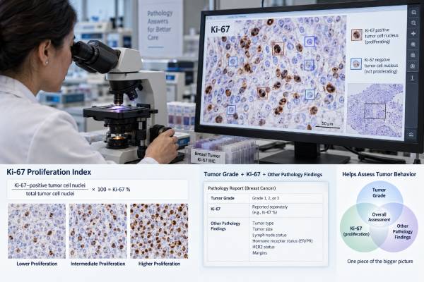
The Ki-67 breast cancer test estimates how actively tumor cells are dividing. A pathologist stains tumor tissue for the Ki-67 protein and reports the percentage of invasive cancer cell nuclei that are positive. A higher percentage generally means a larger fraction of the tumor is in the cell cycle and is associated, on average, with more rapid growth and less favorable prognosis. A lower percentage suggests slower proliferation.
Ki-67 is useful, but it is not a stand-alone cancer stage, grade, or treatment decision. Results can vary because different parts of a tumor may proliferate at different rates and because staining and scoring methods are not perfectly standardized between laboratories. This is why there is no single universal “normal” Ki-67 range for breast cancer. Very low and very high values are often more reproducible and informative than middle-range results. The score should be interpreted with ER, PR, HER2, tumor grade, stage, nodal status, and, when appropriate, genomic testing.
- Ki-67 is reported as the percentage of invasive tumor cell nuclei that stain positive; it is a proliferation index, not a blood level.
- Higher Ki-67 usually indicates faster tumor-cell proliferation, but there is no universal cutoff that separates “good” from “bad” breast cancer.
- International expert work has supported ≤5% and ≥30% as useful low and high prognostic cut points in selected stage I–II ER-positive/HER2-negative cancers when scoring is standardized.
- Ki-67 and tumor grade are related but different: grade evaluates several microscopic features, while Ki-67 measures proliferation specifically.
- In the United States, Ki-67 ≥20% is no longer required to determine eligibility for adjuvant abemaciclib in high-risk HR-positive/HER2-negative early breast cancer.
Table of Contents
- What the Ki-67 Test Measures
- How Ki-67 Testing Is Performed
- How to Interpret a Ki-67 Score
- Ki-67 vs Tumor Grade and Other Biomarkers
- What Ki-67 Means for Prognosis and Treatment
- Limitations, Variability, and Repeat Testing
- Questions to Ask About Your Ki-67 Result
What the Ki-67 Test Measures
Ki-67 measures the proportion of tumor cells that are actively cycling rather than resting. The Ki-67 protein is present during several phases of the cell cycle but is absent in resting cells. By staining a tumor sample for this protein, a pathologist can estimate how much of the cancer is proliferating at the time the tissue was collected.
The result is usually reported as a percentage. A Ki-67 score of 10%, for example, means that about 10% of the evaluated invasive tumor cell nuclei were positive under the scoring method used. It does not mean that the tumor is “10% cancer,” that 10% of the breast is affected, or that the cancer has a 10% chance of recurring.
Ki-67 is most often discussed in hormone receptor-positive, HER2-negative breast cancer, where proliferation contributes to prognosis and to the distinction between more indolent and more proliferative tumor biology. It may also be measured in other breast cancer subtypes, but its ability to direct treatment varies by setting.
The score belongs in a larger pathology picture. A complete breast cancer biomarker panel typically includes estrogen receptor, progesterone receptor, HER2, and other findings that answer different clinical questions. Ki-67 cannot substitute for any of them.
How Ki-67 Testing Is Performed
Ki-67 is an immunohistochemistry test performed on tumor tissue, not a routine blood test. Tissue may come from a core needle biopsy or a surgical specimen. The laboratory applies an antibody that binds the Ki-67 protein, creating visible nuclear staining in proliferating tumor cells.
A pathologist then evaluates the stained invasive cancer. Depending on the laboratory, scoring may use a standardized visual method, digital image analysis, or another validated approach. The reported value reflects the fraction of positive tumor cell nuclei among the cells counted or assessed.
Why specimen handling matters
Ki-67 is sensitive to pre-analytic and analytic conditions. Time before fixation, fixation duration, tissue processing, antibody clone, staining platform, and scoring technique can all influence the result. Quality control is therefore important if the percentage will be used in a clinical decision.
Tumors can also be heterogeneous. One region may have a dense “hot spot” of proliferating cells while another has relatively little Ki-67 staining. A scoring method that focuses only on the hottest area can produce a different percentage from one that samples the tumor more broadly. Modern recommendations emphasize standardized approaches to reduce this source of variation.
Turnaround and preparation
Patients do not usually need to fast, stop medications, or prepare specifically for Ki-67 testing. The test is performed on tissue already obtained for diagnosis or surgery. Turnaround depends on the pathology laboratory and whether the stain is performed routinely or sent to another center.
If the report does not include Ki-67, that does not necessarily mean anything was missed. Some guidelines and treatment decisions do not require it, and practice differs between institutions and countries.
How to Interpret a Ki-67 Score
A Ki-67 percentage should be interpreted as a continuum, with the greatest confidence at clearly low or clearly high values. There is no universal normal range and no single cutoff that applies to every breast cancer subtype or treatment decision.
The International Ki67 in Breast Cancer Working Group concluded that, when standardized scoring is used, values of 5% or less and 30% or more can help estimate prognosis in selected stage I or II, ER-positive/HER2-negative cancers with anatomically favorable features. Values between those extremes are harder to use as a stand-alone decision tool because interobserver and interlaboratory variation can be clinically meaningful.
| Ki-67 result | General interpretation | Important limitation |
|---|---|---|
| ≤5% | Very low proliferation under standardized scoring | Useful prognostically only in the appropriate clinical setting; does not guarantee low recurrence risk |
| 6%–29% | Intermediate proliferation | Cutoffs are less reproducible; other pathology and genomic information often matters more |
| ≥30% | High proliferation under standardized scoring | Does not by itself determine chemotherapy or targeted therapy |
You may also encounter a 20% cutoff in older reports, clinical trials, or subtype discussions. That threshold has been used for several purposes, including historical treatment eligibility and attempts to separate lower- from higher-proliferation hormone receptor-positive tumors. It should not be treated as a universal biological dividing line.
For example, a Ki-67 of 19% is not inherently a different disease from a Ki-67 of 21%. Small differences can fall within normal scoring variability, especially if different specimens or laboratories are involved. The entire report should be reviewed before drawing a treatment conclusion from a number near any cutoff.
Ki-67 vs Tumor Grade and Other Biomarkers
Ki-67 and tumor grade both relate to tumor aggressiveness, but they measure different things and should not be used interchangeably. Histologic grade is a composite microscopic assessment. In the commonly used Nottingham system, the pathologist scores tubule formation, nuclear pleomorphism, and mitotic activity, then combines those components into grade 1, 2, or 3.
Ki-67 focuses specifically on proliferation. A high-grade tumor often has a high Ki-67 score, but discordant cases occur. A grade 2 tumor may have a relatively low or high Ki-67, and the significance depends on the rest of the tumor profile.
ER and PR address hormone sensitivity rather than proliferation. The estrogen receptor test helps identify cancers likely to benefit from endocrine therapy, while the progesterone receptor test adds additional hormone-receptor information. HER2 testing identifies HER2 overexpression or amplification and, increasingly, lower HER2 expression that may affect antibody-drug conjugate options.
Ki-67 is not a genomic recurrence score
Ki-67 also should not be confused with multigene tests. A genomic expression assay measures the activity of multiple genes and may have prospective evidence for estimating recurrence risk or chemotherapy benefit in specific early-stage populations. Ki-67 is one immunohistochemical marker.
Studies have shown that Ki-67 and the Oncotype DX Recurrence Score do not agree closely enough to be treated as substitutes. A low Ki-67 may make a low genomic score more likely in some populations, but it cannot reliably predict an individual genomic result.
What Ki-67 Means for Prognosis and Treatment
Higher Ki-67 is generally associated with a higher risk of recurrence and less favorable outcomes, but its treatment value is much narrower than its prognostic association. Prognosis and prediction are different concepts: a prognostic marker relates to likely outcome, while a predictive marker tells whether a specific treatment is more likely to help.
In selected early-stage ER-positive/HER2-negative cancers, a very low or very high Ki-67 can add information when genomic assays are unavailable. The 2022 ASCO biomarker guideline allows Ki-67 combined with other parameters, or an IHC4 score, to help guide adjuvant treatment decisions in certain postmenopausal patients when genomic testing is not accessible. It is not recommended as a universal substitute for validated genomic assays.
The former 20% abemaciclib requirement
Ki-67 gained unusual visibility after the original U.S. adjuvant abemaciclib indication included a Ki-67 threshold of at least 20% for a high-risk HR-positive/HER2-negative early breast cancer population. That created the impression that 20% was a universal treatment cutoff.
In March 2023, the FDA expanded the indication and removed the Ki-67 testing requirement. High risk for the current U.S. indication is defined by nodal involvement plus specified clinical-pathologic features, not by a mandatory Ki-67 result. As a result, an older report stating that Ki-67 is required for abemaciclib eligibility may no longer reflect current U.S. labeling.
Ki-67 can still be informative in research, neoadjuvant endocrine therapy, prognostic models, and some non-U.S. clinical frameworks. Its role depends on the exact question being asked.
How the meaning changes by disease setting
In a newly diagnosed small, node-negative, strongly ER-positive/HER2-negative tumor, Ki-67 may help describe whether the cancer looks biologically quiet or highly proliferative. If the value falls in the middle, however, the result often does not settle whether chemotherapy is worthwhile. A validated genomic assay may provide stronger evidence when the patient meets criteria for that type of testing.
In neoadjuvant endocrine therapy, researchers and some specialist teams may compare Ki-67 before treatment with Ki-67 after a short period of endocrine therapy. A marked fall can show that proliferation is being suppressed. This is a different use from interpreting one baseline Ki-67 percentage, and the timing and protocol need to be standardized for the result to be meaningful. Ki-67 can also contribute to the Preoperative Endocrine Prognostic Index, or PEPI, in selected research-informed approaches after neoadjuvant endocrine treatment.
In HER2-positive or triple-negative breast cancer, Ki-67 is often high, but the score generally has less value for choosing systemic therapy because receptor status, stage, response to neoadjuvant treatment, and other validated biomarkers drive decisions more directly. A high Ki-67 should not be used to reclassify a tumor into another subtype.
In metastatic breast cancer, treatment selection usually depends on current ER, PR, HER2, actionable mutations, prior therapies, sites of disease, symptoms, and response history. A Ki-67 percentage from the original primary tumor may still describe its baseline biology, but it rarely functions as the decisive biomarker for a specific metastatic drug.
This setting-dependent use explains why two patients with the same Ki-67 percentage can receive very different recommendations. The number is meaningful only after the clinical question is defined.
Does high Ki-67 mean chemotherapy will definitely work better?
No. Rapidly dividing tumors can be more sensitive to some cytotoxic therapies, but Ki-67 alone is not reliable enough to predict individual chemotherapy benefit across breast cancer. Stage, nodal status, tumor size, grade, receptor status, genomic assays, age or menopausal status, and patient health can all be more important.
Limitations, Variability, and Repeat Testing
The main limitation of Ki-67 is reproducibility: the same tumor can receive meaningfully different percentages depending on specimen, laboratory methods, and scoring approach. Standardization has improved, but middle-range values remain challenging.
Common sources of variation include:
- differences in fixation and tissue processing;
- different antibody reagents or staining platforms;
- tumor heterogeneity and hot spots;
- visual estimation versus formal counting;
- which tumor areas are selected for scoring;
- interobserver differences between pathologists;
- biopsy versus surgical specimen differences.
A score should therefore not be interpreted with false precision. If one report shows 18% and another shows 23%, the five-point difference may not represent true biologic progression. The specimens may have been taken at different times, from different parts of the tumor, or processed in different laboratories.
Repeat testing is most useful when the result would genuinely change management and when there is a reason to question the original sample or method. Repeating Ki-67 simply to chase a preferred side of a cutoff can create more confusion rather than more certainty.
Digital pathology and validated image-analysis methods may improve reproducibility by applying consistent cell-counting rules. Recent guidance also emphasizes calibrated visual methods when digital tools are not available. Even with better technology, Ki-67 remains one part of a multiparameter assessment rather than a stand-alone score.
Questions to Ask About Your Ki-67 Result
The most useful next step is to ask how the Ki-67 result changes the interpretation of your specific tumor—not whether the percentage is simply “high” or “low.” A clinically meaningful explanation should connect the number to receptor status, stage, grade, and the treatment decision under discussion.
Questions that can clarify the result include:
- What was the exact Ki-67 percentage, and how was it scored?
- Was the result from the diagnostic biopsy or the surgical specimen?
- Does the laboratory use a standardized or validated scoring method?
- Is my tumor ER-positive and HER2-negative, where Ki-67 may have more established prognostic use?
- How does Ki-67 compare with the tumor grade and mitotic rate?
- Is a genomic recurrence test more appropriate for the chemotherapy question being considered?
- Is this Ki-67 result actually required for any treatment option in my current setting?
- Would repeating the test change management, or would it mainly add another potentially variable number?
A Ki-67 result can be helpful when used with discipline. Very low and very high values may strengthen a broader assessment of tumor biology, while intermediate values often need more context. The safest interpretation is one that treats the percentage as evidence about proliferation, not as a complete prognosis or automatic treatment rule.
If a treatment recommendation appears to hinge on one percentage point, ask whether the same decision would be made if the score were modestly higher or lower. That question can reveal whether Ki-67 is being used as supportive evidence or as an overly rigid cutoff. In many cases, the more reliable decision comes from combining clinical risk, pathology, and validated predictive tests.
When Ki-67 and other features point in different directions, the disagreement is clinically informative rather than a reason to average the numbers. A low Ki-67 does not erase a high tumor grade, and a high Ki-67 does not override receptor status, nodal involvement, tumor size, or a validated genomic assay. Treatment decisions should use the complete clinicopathologic picture and the evidence that applies to that specific setting.
References
- UK recommendations for Ki-67 immunohistochemical staining and interpretation in breast cancer 2026 (Guideline)
- Biomarkers for Adjuvant Endocrine and Chemotherapy in Early-Stage Breast Cancer: ASCO Guideline Update 2022 (Guideline)
- FDA expands early breast cancer indication for abemaciclib with endocrine therapy 2023
- Biomarkers in breast cancer 2024: an updated consensus statement by the Spanish Society of Medical Oncology and the Spanish Society of Pathology 2024 (Position Statement)
- The Ki67 dilemma: investigating prognostic cut-offs and reproducibility for automated Ki67 scoring in breast cancer 2024
- Assessment of Ki67 in Breast Cancer: Updated Recommendations From the International Ki67 in Breast Cancer Working Group 2021 (Position Statement)
Disclaimer
This article is for general education and does not replace interpretation by a breast pathologist or oncology team. Ki-67 methods and treatment uses vary by laboratory, country, and clinical setting, so a percentage should be interpreted with the complete pathology report and current treatment guidance. Do not change cancer treatment based on a Ki-67 number alone.