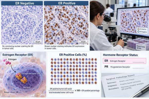
An estrogen receptor (ER) test shows whether breast cancer cells contain estrogen receptor protein and how many tumor-cell nuclei stain for it. Pathologists usually perform the test by immunohistochemistry (IHC) on a biopsy or surgical specimen. The result matters because ER-positive cancers may respond to endocrine, or hormone-blocking, treatments such as tamoxifen, aromatase inhibitors, fulvestrant, and newer estrogen-receptor degraders. Under the current ASCO/CAP standard, invasive breast cancers with at least 1% of tumor nuclei staining for ER are considered ER-positive. Tumors with 1% to 10% staining are reported as ER Low Positive because their biology and expected endocrine sensitivity can differ from cancers with stronger, more widespread ER expression. An ER result should therefore be read as more than a simple positive-or-negative label. The percentage, staining intensity, progesterone receptor result, HER2 status, tumor grade, stage, treatment setting, and quality controls all help determine what the result means for an individual patient.
- ER IHC measures estrogen receptor protein in tumor-cell nuclei; it is not a blood estrogen test.
- At least 1% ER-positive tumor nuclei is classified as ER-positive under current ASCO/CAP guidance.
- ER staining of 1%–10% is reported as ER Low Positive and needs careful clinical interpretation.
- ER-positive status supports possible benefit from endocrine therapy, but the amount of benefit depends on the whole clinical picture.
- ER status can change between a primary tumor and a recurrence, so repeat biopsy and retesting may matter in metastatic disease.
Table of Contents
- What the Estrogen Receptor Test Measures
- How ER IHC Is Scored and Reported
- What an ER-Positive Result Means for Treatment
- ER Low Positive: What 1%–10% Staining Means
- What an ER-Negative Result Means
- Why ER Results Can Change Between Samples
- How to Read an ER Pathology Report
What the Estrogen Receptor Test Measures
The ER test measures estrogen receptor protein inside the nuclei of breast cancer cells. Estrogen is a normal hormone that can activate this receptor. In many breast cancers, that signaling helps tumor cells grow. When the receptor is present, treatments that lower estrogen or block/degrade the receptor can interfere with an important growth pathway.
The standard clinical method is immunohistochemistry. A pathologist applies an antibody that binds to ER protein on a thin tissue section. After staining, the pathologist evaluates the invasive cancer cells and reports the proportion with nuclear staining. The result may also include staining intensity, although the percentage of positive nuclei is central to the positive/negative classification.
This is different from measuring estrogen in the blood. A person can have low circulating estrogen and still have an ER-positive tumor, or have normal hormone levels and an ER-negative tumor. The test describes a property of the cancer, not the patient’s overall hormone level.
ER is usually interpreted alongside the progesterone receptor (PR) test and HER2. Together, these markers define major treatment-relevant groups such as hormone receptor-positive/HER2-negative, hormone receptor-positive/HER2-positive, and triple-negative breast cancer. ER is the strongest established tissue marker for predicting benefit from endocrine therapy.
The test is performed on invasive breast cancer and is also commonly reported for ductal carcinoma in situ because endocrine therapy may be considered after local treatment in selected ER-positive DCIS. The clinical implications, however, are not identical to those for invasive disease.
How ER IHC Is Scored and Reported
The most important number on an ER report is the percentage of invasive tumor-cell nuclei that show specific staining. A result of 90%, for example, means about 90% of the evaluated invasive cancer cells had detectable nuclear ER staining. It does not mean that the tumor is “90% fueled by estrogen,” and it is not a percentage chance that endocrine therapy will work.
Under the ASCO/CAP guideline, 0% nuclear staining, or less than 1%, is interpreted as ER-negative when controls are adequate. Results from 1% through 100% are ER-positive. The 1%–10% range receives the special designation ER Low Positive because clinical evidence for endocrine sensitivity has historically been less certain and these cancers often have molecular and pathologic features closer to ER-negative disease.
Some pathology reports also give an Allred score or another semiquantitative score that combines proportion and intensity. Those systems can provide additional description, but the current clinical threshold for ER positivity is based on the percentage of positive tumor nuclei using a validated assay.
Quality controls are essential near the cutoff. Normal breast epithelium or separate control tissue should stain as expected. The 2020 ASCO/CAP update specifically recommends laboratory procedures to confirm or adjudicate low-positive and negative results because fixation, antibody performance, tissue handling, and interpretation can matter most when staining is weak or sparse.
A report may use words such as strong, moderate, or weak. Strong staining over most cells generally supports robust receptor expression, but intensity alone should not replace the percentage and the final interpretation. If the pathology wording is unclear, ask for the exact ER percentage and whether the case was classified as ER Low Positive.
What an ER-Positive Result Means for Treatment
An ER-positive result means endocrine therapy is potentially useful because the tumor expresses the molecular target those treatments act on. In early-stage disease, endocrine therapy can reduce the risk of recurrence and death in appropriately selected patients. The exact drug and duration depend on menopausal status, age, recurrence risk, side effects, prior therapy, and whether ovarian function is active.
Common strategies include tamoxifen, aromatase inhibitors, ovarian suppression plus endocrine therapy in selected premenopausal patients, and sequential approaches over several years. ER positivity does not by itself determine whether chemotherapy is needed. Tumor size, nodal status, grade, HER2 status, age, comorbidities, and sometimes a genomic recurrence assay all contribute to that decision. In suitable ER-positive/HER2-negative early breast cancers, tests such as Oncotype DX or EndoPredict can add prognostic information.
In metastatic disease, endocrine therapy is often a treatment backbone for ER-positive/HER2-negative cancer, commonly combined with a targeted drug such as a CDK4/6 inhibitor. Later treatment may depend on acquired tumor mutations, prior drug exposure, pace of disease, and organ involvement.
ER positivity is therefore a predictive biomarker, but not a guarantee. Some ER-positive cancers are intrinsically less endocrine-sensitive, and others acquire resistance over time. A cancer can remain ER-positive while developing an ESR1 mutation that reduces sensitivity to aromatase-inhibitor therapy. That distinction explains why a receptor-positive stain and endocrine resistance can coexist.
ER Low Positive: What 1%–10% Staining Means
ER Low Positive breast cancer occupies an important gray zone. These tumors meet the technical definition of ER-positive because 1%–10% of invasive tumor nuclei stain, but many behave more like ER-negative cancers than like tumors with high ER expression. They are more likely to have high grade, higher proliferation, and basal-like molecular features. That is why the pathology report should flag the result rather than simply label it “positive.”
The practical question is whether endocrine therapy still helps. The evidence is imperfect because ER-low tumors are uncommon and have been underrepresented in randomized trials designed specifically around this subgroup. However, newer observational syntheses support considering endocrine therapy rather than assuming there is no benefit. A 2026 systematic review and meta-analysis of more than 10,000 ER-low cases found endocrine therapy was associated with better disease-free and overall survival. Because the underlying studies were largely observational, the finding strengthens but does not settle the question for every patient.
Treatment planning should also respect the tumor’s higher baseline risk and biology. Clinicians may discuss chemotherapy using factors that would be relevant for ER-negative disease rather than assuming a small amount of ER staining makes the tumor biologically similar to a strongly ER-positive, low-grade cancer.
Repeat or confirmatory pathology review may be especially valuable when a result sits just above or below the 1% threshold, when internal controls are weak, or when the rest of the pathology seems discordant. The goal is not to “move” the score into a preferred category but to make sure a small amount of staining is real and reproducible.
What an ER-Negative Result Means
An ER-negative result means fewer than 1% of invasive tumor-cell nuclei show specific ER staining, assuming the assay and controls are valid. Standard endocrine therapies generally are not expected to help an ER-negative invasive breast cancer because their main molecular target is absent or essentially absent.
ER-negative does not define the whole tumor. HER2 may still be positive, creating a HER2-driven cancer with effective HER2-targeted options. If HER2 is also negative and PR is negative, the tumor is classified as triple-negative breast cancer, for which chemotherapy, immunotherapy in selected settings, antibody-drug conjugates, and germline or tumor biomarkers can guide treatment.
A negative result deserves a quality check when it conflicts with the morphology or prior specimens. Preanalytic problems such as delayed fixation, inadequate formalin exposure, decalcification of a bone biopsy, or very small amounts of viable tumor can reduce staining. ASCO/CAP guidance emphasizes appropriate controls and standard operating procedures for low and negative results.
The result can also differ between sites or over time because breast cancer evolves. A primary tumor may be ER-positive while a later metastasis is ER-negative, or the reverse can occur. Technical variation and tumor heterogeneity both contribute. When safely feasible and clinically useful, biopsy of recurrent or metastatic disease can therefore provide current receptor information.
An ER-negative result should not be “overruled” by a blood hormone level or by symptoms associated with estrogen. The relevant question is whether the cancer cells express receptor protein on a validated tissue test.
Why ER Results Can Change Between Samples
Breast cancers are made of evolving cell populations rather than one perfectly uniform clone. Treatment can suppress some populations and allow others to expand. As a result, receptor status in a recurrence may differ from the original diagnosis. Sampling can also matter because different regions of a large tumor, or different metastatic sites, may not have identical biology.
Technical factors add another layer. ER IHC depends on prompt and appropriate fixation, a validated antibody and platform, adequate controls, and correct identification of invasive tumor cells. Tiny biopsies, crushed tissue, scant viable tumor, or decalcified bone specimens may be harder to evaluate. None of these limitations automatically invalidates a result, but they can justify a repeat test when the answer would change treatment.
A change from strongly ER-positive to completely ER-negative can have major therapeutic implications. A small change from 95% to 80% usually does not. The clinically important issue is whether the category and expected endocrine sensitivity have changed, not whether two laboratories produced exactly the same percentage.
In metastatic breast cancer, current tissue biomarkers are interpreted together with molecular testing when indicated. For example, an ER-positive recurrence may undergo plasma or tissue sequencing to identify acquired resistance markers. Conversely, if a recurrence is now ER-negative, clinicians reassess the entire treatment framework rather than assuming the original hormone-receptor strategy still applies.
When two reports disagree, ask whether they evaluated the same specimen, whether one sample was a bone metastasis, whether controls were adequate, and whether expert pathology review is warranted. Discordance is a clinical problem to resolve, not a reason to average two percentages.
How to Read an ER Pathology Report
Read the ER line in a fixed order. First, confirm that the specimen contains invasive breast cancer and identify whether the result applies to the invasive component or to in situ disease. Second, find the exact percentage of tumor nuclei that stain. Third, note the final category: negative, ER Low Positive, or positive. Fourth, look for comments about staining intensity, controls, limited tissue, or testing difficulties.
Then place ER beside PR and HER2 status. A tumor described as ER 95%, PR 80%, HER2 0 has a very different treatment profile from ER 5%, PR 0%, HER2 0, even though both technically meet the ER-positive threshold. The numbers do not directly dictate treatment, but they describe different biological contexts.
Do not use the ER percentage as a recurrence-risk percentage. A result of ER 80% does not mean an 80% recurrence risk, an 80% response rate, or an 80% chance of cure. Likewise, the result cannot tell whether microscopic metastatic disease is present after surgery.
Useful questions for the oncology team include: Is this clearly ER-positive or ER-low? Is endocrine therapy recommended, and for how long? Does the tumor’s grade or genomic risk suggest chemotherapy should also be considered? Was HER2 assessed on the same specimen? If this is metastatic disease, should receptor testing or sequencing be repeated on a current site?
The best interpretation is treatment-linked and context-specific. ER is one of the most powerful biomarkers in breast cancer, but it works as part of a profile rather than as a stand-alone forecast.
Practical examples of ER result wording
An ER 95%, strong result is a classic high-expression pattern. It supports meaningful estrogen-receptor signaling and usually makes endocrine therapy an important part of treatment when the clinical setting is appropriate. It still does not tell whether chemotherapy is needed or whether the cancer will recur; those questions require stage, grade, HER2, nodal status, age, and sometimes genomic testing.
An ER 5%, weak, ER Low Positive result is different. It technically meets the positive threshold, so endocrine therapy may still be considered, but clinicians should not assume the tumor behaves like a strongly ER-positive cancer. PR status, grade, proliferation, chemotherapy sensitivity, and other features deserve particular attention. The patient may receive treatment strategies influenced by both hormone-receptor-positive and ER-negative biology.
An ER 0% result means endocrine therapy is generally not expected to help, provided the specimen is adequate and controls are valid. If the tumor is also PR-negative and HER2-negative, it fits the triple-negative category. If HER2 is positive, HER2-directed therapy remains available despite the absent ER target.
A borderline result deserves careful pathology context. If a tiny biopsy shows 1% staining but the surgical specimen later shows 0%, the team may review fixation, tumor heterogeneity, and which specimen better represents the invasive cancer. The numbers should not simply be averaged. Likewise, a metastatic biopsy can supersede an old receptor result when it shows convincing biological evolution and the current treatment decision depends on the new phenotype.
These examples show why the percentage is best treated as descriptive pathology data rather than a direct treatment-benefit percentage.
ER should also be separated from estrogen exposure. Menopause, hormone-replacement history, body weight, ovarian suppression, and blood estradiol levels can affect the hormonal environment, but none changes the pathology definition of ER status. The tissue test asks whether the cancer contains the receptor. A patient receiving an aromatase inhibitor can therefore still have a tumor that is strongly ER-positive even though circulating estrogen is intentionally very low.
When endocrine treatment fails, the next question is usually why the tumor has become resistant, not whether the original ER percentage was “wrong.” Resistance can arise through ESR1 mutations, pathway activation, cell-cycle changes, or loss of receptor dependence. That is why metastatic treatment decisions increasingly combine current receptor testing with targeted molecular testing when it can change therapy.
For patients receiving neoadjuvant therapy before surgery, receptor testing may be available from both the pretreatment biopsy and the residual tumor. If the results differ, the oncology and pathology teams consider treatment effects, sampling, and assay quality before deciding which phenotype should guide further therapy. A small change in percentage is usually less important than a true shift across a clinically meaningful category. Keeping the original and post-treatment reports together is useful because both may explain why certain therapies were chosen and why the plan later changed.
References
- Endocrine therapy in early-stage estrogen Receptor-Low breast cancer: a systematic review and Meta-Analysis 2026 (Systematic Review)
- Estrogen receptor-low positive breast cancer: Historical perspective and recent advancements 2025 (Review)
- The “lows”: Update on ER-low and HER2-low breast cancer 2024 (Review)
- Early breast cancer: ESMO Clinical Practice Guideline for diagnosis, treatment and follow-up 2024 (Guideline)
- Estrogen and Progesterone Receptor Testing in Breast Cancer: ASCO/CAP Guideline Update 2020 (Guideline)
Disclaimer
This article provides general information about estrogen receptor testing in breast cancer and does not interpret an individual pathology report. ER results should be reviewed with the treating oncology and pathology teams, especially for ER-low, discordant, limited, or metastatic samples where the testing context can change management.