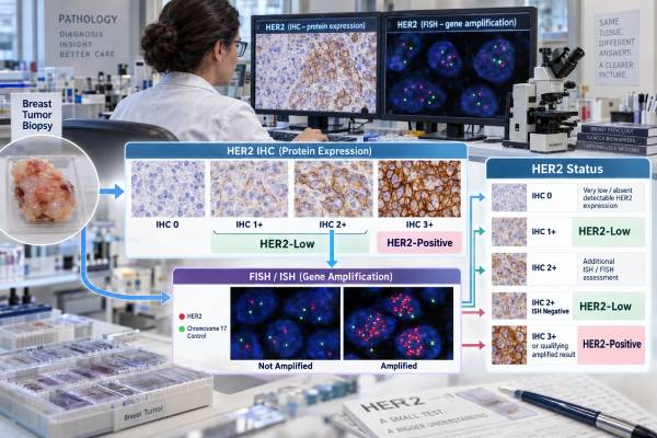
A HER2 breast cancer test determines how much HER2 protein a tumor expresses and, when needed, whether the HER2 gene is amplified. The result is central to breast cancer treatment because classic HER2-positive tumors can respond to HER2-targeted drugs such as trastuzumab, pertuzumab, and several antibody-drug conjugates. Testing usually starts with immunohistochemistry (IHC), scored 0, 1+, 2+, or 3+. An IHC 3+ result is HER2-positive, while IHC 2+ is equivocal and usually needs in situ hybridization such as FISH. IHC 0 or 1+ is not HER2-positive under the standard ASCO/CAP classification. However, low levels of HER2 protein now matter for some antibody-drug conjugates: HER2-low generally means IHC 1+ or IHC 2+/ISH-negative, and a 2025 FDA indication also uses HER2-ultralow, defined for that treatment context as IHC 0 with membrane staining. These newer treatment categories should not be confused with classic HER2 gene amplification.
- HER2 IHC measures protein expression; FISH or another ISH method measures HER2 gene amplification.
- IHC 3+ is HER2-positive, while IHC 2+ normally requires reflex ISH to determine final HER2 status.
- HER2-low generally means IHC 1+ or IHC 2+/ISH-negative; it is not the same as classic HER2-positive disease.
- For a 2025 FDA-approved metastatic HR-positive indication, HER2-ultralow means IHC 0 with detectable membrane staining.
- HER2 can differ between the original tumor and a recurrence, so repeat testing may be important when treatment decisions depend on current status.
Table of Contents
- What HER2 Testing Measures
- How HER2 IHC Scores 0, 1+, 2+, and 3+
- How FISH and Other ISH Tests Determine HER2 Status
- What HER2-Positive, Equivocal, and Negative Results Mean
- HER2-Low and HER2-Ultralow: New Treatment Categories
- Why HER2 Results Can Be Discordant or Change
- How to Read a HER2 Report Step by Step
What HER2 Testing Measures
HER2, also called ERBB2, is a gene that encodes a growth-signaling receptor on the cell surface. Some breast cancers contain extra copies of the HER2 gene. That amplification can drive production of large amounts of HER2 protein and create a cancer that depends heavily on HER2 signaling. Those tumors are called HER2-positive when they meet established testing criteria.
There are two main ways to evaluate this biology. Immunohistochemistry (IHC) stains the HER2 protein on tumor-cell membranes. In situ hybridization (ISH) counts HER2 gene copies in tumor-cell nuclei; fluorescence in situ hybridization, or FISH, is one common form. The tests answer related but not identical questions, which is why they are sometimes used together.
HER2 status is tested on invasive breast cancer because it predicts benefit from HER2-directed therapy. It is interpreted with ER and PR as part of the core breast cancer biomarker profile. A HER2-positive tumor may also be ER-positive or ER-negative; HER2 positivity does not imply a particular hormone-receptor status.
The newer terms HER2-low and HER2-ultralow describe lower protein-expression ranges that can matter for certain antibody-drug conjugates. They do not mean the HER2 gene is necessarily amplified. In fact, HER2-low cancers are, by definition, not HER2-positive under the classic amplification/overexpression framework.
This distinction is clinically important. Trastuzumab-based regimens for classic HER2-positive disease and antibody-drug conjugates used in HER2-low or ultralow settings may share a target but rely on different eligibility criteria and different evidence.
How HER2 IHC Scores 0, 1+, 2+, and 3+
HER2 IHC scoring is based on the intensity, completeness, and proportion of membrane staining in invasive tumor cells. Pathologists examine a properly prepared tissue section and compare the staining pattern with guideline-defined criteria.
An IHC 3+ result shows uniform, intense, complete circumferential membrane staining in more than 10% of invasive tumor cells and is considered HER2-positive. An IHC 2+ result is equivocal. In the usual pattern, more than 10% of cells show weak to moderate complete membrane staining. The tumor then needs reflex ISH unless a valid result is already available.
An IHC 1+ result shows faint or barely perceptible, incomplete membrane staining in more than 10% of tumor cells. IHC 0 includes no staining or only faint, incomplete membrane staining in 10% or fewer tumor cells under the traditional scoring framework. Both 0 and 1+ are HER2-negative for classic HER2-positive treatment decisions.
The difference between 0 and 1+ used to have little therapeutic consequence, so assays and training were optimized mainly to separate clearly positive from non-positive disease. Antibody-drug conjugates changed that. Pathologists now need greater consistency at the lower end of expression because 0, 1+, and 2+/ISH-negative results can affect eligibility for specific metastatic treatments.
For a focused explanation of these staining patterns, the HER2 IHC test should be read as a protein-expression result, not as a direct gene-copy measurement.
How FISH and Other ISH Tests Determine HER2 Status
FISH uses fluorescent DNA probes to count HER2 signals in tumor-cell nuclei, usually together with a reference probe for the chromosome 17 centromeric region, commonly called CEP17. The laboratory calculates an average HER2 copy number per cell and a HER2/CEP17 ratio. Those values place the specimen into an ASCO/CAP ISH group.
The simplest result is Group 1: HER2/CEP17 ratio at least 2.0 and average HER2 copy number at least 4.0 signals per cell. This is HER2-positive. Group 5, with a ratio below 2.0 and average HER2 copy number below 4.0, is HER2-negative. Groups 2, 3, and 4 require integration with the IHC result and, in selected circumstances, a blinded recount by another observer.
That integrated approach prevents a common mistake: calling a tumor positive or negative from the ratio alone. Unusual chromosome 17 patterns can create a high ratio despite a low absolute HER2 copy number, or a high HER2 copy number without a ratio of 2.0. The 2018 framework, reaffirmed by the 2023 update, uses IHC to help resolve these uncommon patterns.
Most patients encounter FISH after an IHC 2+ result. If ISH demonstrates amplification according to the integrated criteria, the tumor is HER2-positive. If it does not, the final tumor is HER2-negative for classic HER2-positive therapy, even though its IHC 2+ expression may place it in the HER2-low category.
The HER2 FISH result is therefore best read together with the IHC score and the laboratory’s final integrated interpretation.
What HER2-Positive, Equivocal, and Negative Results Mean
A HER2-positive result identifies a tumor with HER2 protein overexpression or qualifying gene amplification. In early-stage and metastatic breast cancer, this can open highly effective HER2-directed treatment pathways. The exact regimen depends on stage, tumor size, nodal status, prior therapy, residual disease after neoadjuvant treatment, and other clinical factors.
An equivocal IHC result means IHC 2+, not that the final cancer is “half positive.” It is a request for another layer of testing. The final status is usually resolved by ISH. Once the integrated workup is complete, the tumor should generally be assigned a final HER2-positive or HER2-negative status for the classic treatment framework.
A HER2-negative result means the tumor does not meet the criteria for HER2 overexpression/amplification. That category now contains biologically and therapeutically different lower-expression groups. IHC 0, IHC 1+, and IHC 2+/ISH-negative tumors are all non-amplified/non-overexpressed, yet some can qualify for antibody-drug conjugates in specific advanced-disease settings.
Do not use the HER2 score as a prognosis percentage. IHC 3+ does not mean the cancer is “three times worse,” and a HER2/CEP17 ratio does not measure how much cancer is present. These values classify a molecular target.
The implications also depend on timing. HER2-positive early breast cancer may be treated with curative-intent HER2-targeted therapy. HER2-low metastatic disease may become eligible for a different drug after specified prior treatment. The same laboratory words can therefore lead to different actions depending on stage and treatment history.
HER2-Low and HER2-Ultralow: New Treatment Categories
HER2-low is generally defined as HER2 IHC 1+ or IHC 2+ with a negative ISH result. These tumors are not HER2-positive by ASCO/CAP criteria. The term became clinically important because trastuzumab deruxtecan, an antibody-drug conjugate, can deliver chemotherapy into cells with lower HER2 expression and can also affect nearby tumor cells through a bystander effect.
HER2-ultralow is even more treatment-specific. In January 2025, the FDA approved trastuzumab deruxtecan for adults with unresectable or metastatic HR-positive breast cancer that is HER2-low or HER2-ultralow after progression on one or more endocrine therapies in the metastatic setting. For that indication, HER2-ultralow is IHC 0 with membrane staining as determined by an FDA-approved test.
This creates an apparent language conflict: a pathology report can say “IHC 0” while the treatment framework still cares whether faint membrane staining is detectable. The point is not that IHC 0 has become classic HER2-positive. It has not. Rather, a specific drug indication recognizes a very low expression subgroup inside the historical IHC 0 range.
ASCO/CAP’s 2023 guideline update did not create a new biologic HER2-low category as a replacement for the standard positive/negative interpretation. Laboratories should continue to report the actual IHC score and ISH result accurately. Clinicians can then map those results to current drug labels.
For readers comparing reports, the separate HER2-low classification is best understood as a treatment-selection layer added on top of conventional HER2 testing, not a new form of HER2 amplification.
Why HER2 Results Can Be Discordant or Change
HER2 testing is highly standardized, but discordance still occurs. One reason is tumor heterogeneity: different areas of the same tumor or different metastatic sites can contain cell populations with different HER2 expression or amplification. Another is true biological evolution under treatment pressure.
Technical factors also matter. Cold ischemia time, formalin fixation, decalcification, tissue age, assay platform, staining performance, and interpretation can affect IHC. FISH can be influenced by nuclear truncation, signal overlap, chromosome 17 abnormalities, and which invasive area is counted. This is why laboratories use validated protocols and pathologists correlate the molecular signals with tumor morphology.
A core biopsy and later surgical specimen may occasionally disagree. The ASCO/CAP guideline allows retesting on the excision specimen in selected cases when the initial negative result does not fit the tumor’s features or when technical concerns exist. In metastatic disease, a new biopsy can be especially useful because the treatment decision often depends on the current phenotype, not only the original diagnosis.
Discordance at the low end is becoming more visible because the difference between IHC 0 and 1+ can affect drug eligibility. Interobserver and inter-assay variability are greater in this range than for a classic, strongly positive 3+ tumor. When a low-expression result will determine access to an antibody-drug conjugate, review of an appropriate recent specimen with a validated companion diagnostic may be important.
The practical response to conflicting reports is not to average them. Ask which specimen is newest, whether it came from primary or metastatic disease, whether it was decalcified, and whether the laboratory issued a final integrated HER2 interpretation.
How to Read a HER2 Report Step by Step
Start with the IHC score. If it is 3+, the tumor is HER2-positive without needing routine confirmatory FISH. If it is 2+, look for the reflex ISH result and final integrated interpretation. If it is 0 or 1+, the tumor is not classic HER2-positive, but the exact low-expression wording may matter for an antibody-drug conjugate in advanced disease.
If FISH or another ISH method was performed, look for the average HER2 copy number, HER2/CEP17 ratio, ASCO/CAP ISH group if reported, the paired IHC score, and the final status. Do not interpret an unusual ratio without the integrated group rules.
Then place HER2 beside ER and PR. An ER-positive/HER2-positive cancer may receive both endocrine and HER2-directed strategies. An ER-negative/HER2-positive cancer still has a powerful HER2 target. An ER-positive/HER2-low metastatic cancer may become eligible for a lower-expression antibody-drug conjugate after the required prior therapy. The treatment context determines what part of the report matters most.
Ask whether the specimen is from the original diagnosis or a current recurrence. If metastatic treatment is being planned from an old primary-tumor report, ask whether a contemporary biopsy is feasible and useful. Receptor conversion does not happen in every patient, but it is common enough to matter.
Finally, distinguish status from score. The score is a laboratory observation. The final status is the guideline-based interpretation. Treatment eligibility may add another label such as HER2-low or ultralow. Keeping those layers separate makes a complicated HER2 report much easier to use.
Common HER2 report scenarios
A report that reads IHC 3+ is straightforward: the invasive cancer is HER2-positive under standard criteria. Routine FISH confirmation is generally unnecessary, and the oncology team considers HER2-directed therapy according to stage and treatment history. The score does not indicate how advanced the cancer is; a small stage I tumor and a metastatic tumor can both be IHC 3+.
A report that reads IHC 2+, FISH amplified is also HER2-positive. The IHC alone was equivocal, but gene amplification resolves the status. By contrast, IHC 2+, FISH not amplified is HER2-negative under the classic ASCO/CAP framework and falls into the HER2-low expression range. This is one of the clearest examples of why “2+” should never be read as a final positive result.
A report that reads IHC 1+ is non-amplified/non-overexpressed in the classic framework but can be clinically actionable in advanced disease under appropriate antibody-drug conjugate indications. A report that reads IHC 0 with membrane staining may meet the HER2-ultralow definition used in the 2025 FDA HR-positive metastatic indication, provided the required approved test and treatment conditions are met. IHC 0 with no membrane staining does not meet that ultralow definition.
Finally, an older primary-tumor result and a new metastatic result may not match. When the current treatment hinges on HER2 expression, clinicians may prioritize a recent, technically adequate biopsy while reviewing the old slides for context. The correct interpretation is not the highest score ever recorded; it is the best validated result for the disease that is being treated now.
A final practical issue is which specimen should guide a lower-expression treatment decision. HER2 expression can shift after therapy, and low scores are particularly sensitive to sampling and technical factors. When an advanced-disease drug requires a specific HER2 expression level, clinicians may review a recent metastatic specimen, prior archival tissue, or both. The goal is not to search indefinitely for the highest score, but to use an adequate specimen and the companion-diagnostic framework relevant to the drug.
Patients should also distinguish a pathology label from a drug label. “HER2-negative” remains correct for IHC 1+ and IHC 2+/ISH-negative under the classic ASCO/CAP overexpression/amplification definition, even when those same results qualify as HER2-low for a particular therapy. Both statements can be true because they answer different clinical questions.
This distinction helps prevent both undertreatment of truly HER2-positive disease and overinterpretation of low expression as gene amplification.
HER2 testing is also relevant after neoadjuvant therapy. Residual invasive cancer can have different HER2 expression from the pretreatment biopsy, and the amount of residual disease influences adjuvant treatment choices in classic HER2-positive cancer. In metastatic disease, prior HER2-targeted exposure can further shape tumor evolution. For that reason, the most informative report is not always the first report in the chart. Clinicians use the complete sequence of pathology results to understand both the original driver and the current therapeutic target.
References
- Human Epidermal Growth Factor Receptor 2 Testing in Breast Cancer: ASCO-College of American Pathologists Guideline Update 2023 (Guideline)
- HER2 Testing In Breast Cancer – 2023 Guideline Update – CAP 2023 (Guideline)
- Best pathology practices for HER2 testing interpretation, including HER2-low and ultralow, in the era of HER2-directed antibody drug conjugates (ADCs), aligned with the 2018 CAP/ASCO HER2 guidelines 2025 (Review)
- FDA approves fam-trastuzumab deruxtecan-nxki for unresectable or metastatic HR-positive, HER2-low or HER2-ultralow breast cancer | FDA 2025
- UK recommendations for HER2 assessment in breast cancer: an update 2023 (Guideline)
Disclaimer
This article explains HER2 testing in general and does not determine whether a specific patient qualifies for a HER2-directed drug. HER2 IHC and ISH results should be interpreted by the pathology and oncology teams using the tested specimen, current guideline criteria, disease setting, prior therapy, and the applicable drug label or companion diagnostic.