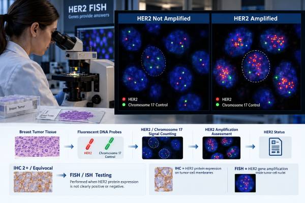
A HER2 FISH test examines breast cancer cells for extra copies of the HER2 gene, also called ERBB2. It is most often used when HER2 immunohistochemistry (IHC) is equivocal at 2+, although some laboratories use another validated in situ hybridization method instead of fluorescence. FISH does not simply return a positive or negative number. Dual-probe testing usually reports an average HER2 copy number per tumor cell and a HER2-to-CEP17 ratio, then combines those values with the IHC result for uncommon signal patterns. Under the ASCO/CAP framework reaffirmed in 2023, the five ISH groups are interpreted differently, and Groups 2, 3, and 4 cannot be classified from the ratio alone. A final HER2-positive result can make a patient eligible for established HER2-targeted treatments, while a non-amplified result may still be clinically relevant if IHC shows low HER2 expression. Understanding the report therefore requires the FISH numbers, the matching IHC score, and the laboratory’s final integrated HER2 status.
- HER2 FISH measures HER2 gene copy number and usually the HER2/CEP17 ratio in invasive breast cancer cells.
- FISH is commonly ordered after an equivocal HER2 IHC 2+ result.
- ISH Group 1 is HER2-positive and Group 5 is HER2-negative; Groups 2–4 require integration with IHC.
- A high HER2/CEP17 ratio alone does not always mean HER2-positive disease when average HER2 copy number is low.
- A non-amplified FISH result can still pair with IHC 1+ or 2+ to define HER2-low expression for certain advanced-disease treatments.
Table of Contents
- What the HER2 FISH Test Measures
- When HER2 FISH Is Ordered
- How to Read the HER2/CEP17 Ratio and Copy Number
- The Five ASCO/CAP HER2 ISH Groups
- What Positive and Negative FISH Results Mean
- Why FISH Results Can Be Difficult to Interpret
- Questions to Ask About a HER2 FISH Report
What the HER2 FISH Test Measures
FISH stands for fluorescence in situ hybridization. The test uses fluorescent DNA probes that bind to specific locations inside the nuclei of cancer cells. In breast cancer, one probe targets the HER2 gene and a second usually targets the centromeric region of chromosome 17, called CEP17. A technologist or pathologist counts signals in a defined number of invasive tumor-cell nuclei.
The laboratory calculates two core measurements: the average HER2 copy number per cell and the HER2/CEP17 ratio. Gene amplification means the cancer has gained extra HER2 copies beyond what is expected from the reference chromosome signal. Amplification can drive production of excess HER2 protein and create a strong therapeutic target.
FISH is different from HER2 IHC. IHC measures protein on the tumor-cell membrane. FISH measures gene copies in the nucleus. The tests correlate well in many cancers, especially at clear extremes, but unusual patterns and tumor heterogeneity can produce disagreement. Modern guidelines therefore integrate the tests rather than assuming one number always overrides the other.
FISH is not a blood test and does not measure circulating HER2. It is performed on fixed tumor tissue from a core biopsy, surgical specimen, or metastatic biopsy. The tissue must contain enough viable invasive cancer for accurate signal counting.
The final purpose is treatment classification. HER2 amplification identifies a group that may benefit from established HER2-directed therapy. A non-amplified result can also matter because an IHC 2+/ISH-negative tumor belongs to the HER2-low expression range used by some antibody-drug conjugate indications.
When HER2 FISH Is Ordered
The most common reason for FISH is an equivocal IHC 2+ result. IHC 3+ is already HER2-positive under standard criteria and usually does not need confirmatory FISH. IHC 0 or 1+ is negative for classic HER2 overexpression, so routine FISH is generally unnecessary unless there is a specific discordance, technical concern, unusual morphology, or another reason to question the initial result.
A laboratory may use FISH as its preferred ISH method, while others use bright-field dual in situ hybridization, silver ISH, or chromogenic ISH. The 2023 ASCO/CAP update continues to recognize validated ISH approaches. What matters clinically is that the assay meets quality standards and the result is interpreted using the appropriate algorithm.
FISH can also be repeated when an earlier result is technically inadequate or when a new specimen is clinically important. A core biopsy that is negative but has suspicious pathologic features can sometimes be retested on the excision specimen. In metastatic disease, a new biopsy may be tested because HER2 biology can change over time or differ between sites.
The test should be performed on invasive tumor, not accidentally on adjacent ductal carcinoma in situ. DCIS can have different HER2 characteristics, and counting the wrong cell population could misclassify the invasive cancer. Pathologist-guided selection of the area for analysis is therefore part of the test.
FISH is also not ordered simply because a patient wants a more precise numeric score. In a clearly IHC 3+ tumor, the extra test usually adds little. In a clearly IHC 0 tumor, FISH cannot determine HER2-low versus ultralow expression because those are protein-expression categories. The test is most useful when gene amplification is the unresolved question.
How to Read the HER2/CEP17 Ratio and Copy Number
A dual-probe HER2 FISH report usually gives the average number of HER2 signals per tumor cell, the average number of CEP17 signals, and the ratio of the two. It may also report how many cells were counted, whether a second observer performed a recount, and the final ASCO/CAP ISH group.
The ratio helps account for the number of chromosome 17 reference signals. For example, if a cell population has many HER2 signals but also many CEP17 signals, the ratio may stay below 2.0. Conversely, a low CEP17 count can produce a ratio of 2.0 or higher even when the average HER2 copy number is below 4.0. That is why neither value should be interpreted alone.
The simplest amplified pattern is ratio at least 2.0 with average HER2 copy number at least 4.0. The simplest non-amplified pattern is ratio below 2.0 with average HER2 copy number below 4.0. Values that fall between those patterns are assigned to Groups 2, 3, or 4 and require IHC integration.
The numbers are not percentages. A HER2/CEP17 ratio of 2.5 does not mean 250% HER2 expression, and an average copy number of 6 does not mean every tumor cell contains exactly six copies. The values are averages from the cells counted in the selected tumor area.
If a report only shows “amplified” or “not amplified,” the final conclusion may be enough for treatment, but the underlying values can clarify unusual cases. For a complete HER2 interpretation, pair FISH with the IHC score and final pathology comment.
The Five ASCO/CAP HER2 ISH Groups
The five-group system prevents rare signal patterns from being oversimplified. Group 1 has a HER2/CEP17 ratio of at least 2.0 and an average HER2 copy number of at least 4.0 signals per cell. This is HER2-positive.
Group 2 has a ratio of at least 2.0 but an average HER2 copy number below 4.0. This unusual pattern is not automatically positive. If IHC is 3+, it is HER2-positive. If IHC is 2+, a second observer recounts the ISH; if the pattern remains Group 2, the final result is HER2-negative with an explanatory comment. IHC 0 or 1+ is HER2-negative.
Group 3 has a ratio below 2.0 and average HER2 copy number of at least 6.0. IHC 3+ supports HER2-positive status. With IHC 2+, a blinded recount is performed; if the result remains Group 3, it is considered HER2-positive. IHC 0 or 1+ leads to a negative interpretation.
Group 4 has a ratio below 2.0 and average HER2 copy number at least 4.0 but below 6.0. IHC 3+ makes the case positive. With IHC 2+, recount is performed; if it remains Group 4, the case is HER2-negative. IHC 0 or 1+ is negative.
Group 5 has a ratio below 2.0 and average HER2 copy number below 4.0 and is HER2-negative. These integrated rules were introduced to reduce ambiguous classifications and avoid treating uncommon chromosome 17 patterns as unequivocal amplification without supporting protein expression.
What Positive and Negative FISH Results Mean
A final HER2-positive FISH/ISH interpretation means the tumor has qualifying HER2 gene amplification under the integrated guideline criteria. In the right disease setting, that result can support treatment with drugs that target HER2. In early breast cancer, HER2-targeted therapy may be part of neoadjuvant or adjuvant curative-intent treatment. In metastatic disease, several HER2-directed regimens are available, with selection based on prior treatment and clinical circumstances.
A final HER2-negative FISH result means the tumor does not meet the amplification criteria. It does not mean there is absolutely no HER2 protein. An IHC 2+/FISH-negative cancer often expresses enough membrane protein to be called HER2-low, which can matter for specific antibody-drug conjugates. FISH therefore answers the amplification question, while IHC supplies the lower-expression category.
A FISH result also does not predict the exact degree of benefit from treatment. A high ratio is not a dose-response scale for trastuzumab, and a low ratio is not a measure of recurrence risk. Treatment response depends on stage, other tumor biology, prior therapy, resistance mechanisms, and the particular drug.
If IHC and FISH appear contradictory, the laboratory should apply the integrated algorithm and may review the slides, recount cells, or test another specimen. True discordant cases exist, but technical issues and tumor heterogeneity must be considered first.
For patients, the most useful line on the report is often the final integrated statement: HER2-positive or HER2-negative, followed by the IHC score. Those two pieces usually explain both classic HER2 targeting and newer lower-expression treatment possibilities.
Why FISH Results Can Be Difficult to Interpret
FISH is quantitative, but it is not immune to biological and technical complexity. Tumor nuclei are cut into thin tissue sections, so some nuclei are incomplete. Fluorescent signals can overlap, split, or fade. The analyst must distinguish invasive cancer cells from non-neoplastic cells and choose representative tumor areas.
Chromosome 17 biology can complicate the ratio. Changes affecting the CEP17 region may alter the denominator without representing true whole-chromosome gain or loss. This is one reason older approaches that relied heavily on the ratio produced some counterintuitive classifications. The current integrated method uses both ratio and absolute HER2 copy number, plus IHC for Groups 2–4.
Heterogeneity is another challenge. A tumor may contain one amplified area and one non-amplified area. Guidelines provide approaches for evaluating a distinct amplified population when it represents a meaningful fraction of invasive cells. A metastatic lesion may also differ from the primary tumor because cancer evolves under treatment pressure.
Specimen quality matters. Decalcified bone metastases can be difficult for both IHC and ISH, depending on the decalcification method. Small biopsies can contain too little invasive tumor for confident analysis. If a result is technically unsuccessful, the report should not be forced into a positive or negative category; testing another block or specimen may be more reliable.
Recent studies continue to show that repeat testing can uncover discordance, especially in borderline or previously treated disease. The decision to retest should be linked to whether a different result would change therapy, not to a desire for repeated numbers without a clinical question.
Questions to Ask About a HER2 FISH Report
First ask why FISH was performed. If the answer was an IHC 2+ result, confirm that the FISH report is from the same tumor specimen and that the laboratory issued a final integrated HER2 status. If the test was performed on a recurrence, note whether it differs from the original tumor.
Next, identify the HER2/CEP17 ratio and average HER2 copy number. Ask which ASCO/CAP group the result fits if the report does not state it. For Groups 2, 3, and 4, ask how the concurrent IHC score affected the conclusion and whether a required recount was performed.
If the final result is negative, check the IHC score. IHC 2+/ISH-negative is not classic HER2-positive, but it falls within HER2-low breast cancer terminology and can matter for specific metastatic indications. An IHC 0 result requires different low-expression interpretation and cannot be converted to “HER2-low” from FISH alone.
If the report conflicts with older records, ask whether the difference could reflect a new specimen, treatment-related evolution, tumor heterogeneity, decalcification, or another technical factor. Pathology review may be more useful than assuming one report is wrong.
Finally, ask what treatment decision the result changes now. A laboratory result has its greatest value when connected to a specific choice: adding HER2-directed therapy, confirming a non-amplified lower-expression category, or deciding whether a new biopsy is needed. That question keeps the technical details tied to practical care.
Worked examples of common FISH patterns
Suppose a tumor has an average HER2 copy number of 8.0 signals per cell and a HER2/CEP17 ratio of 3.1. Both the ratio and copy number are high, placing it in Group 1. The final interpretation is HER2-positive. Now consider a tumor with an average HER2 copy number of 2.8 but a ratio of 2.3. The ratio is high, yet the absolute copy number is low, so this is Group 2 rather than an automatic positive result. Its final status depends on the concurrent IHC algorithm.
A third tumor might have a ratio of 1.7 and an average HER2 copy number of 6.5. That is Group 3: the ratio is below 2.0 but the copy number is high. IHC becomes essential. If IHC is 3+, the case is positive; if IHC is 2+, a recount is performed and a persistent Group 3 pattern is interpreted as positive; if IHC is 0 or 1+, it is negative under the integrated rules.
Group 4 illustrates the borderline zone: a ratio below 2.0 with 4.0 to fewer than 6.0 HER2 copies. A persistent Group 4 result paired with IHC 2+ is negative, while IHC 3+ supports positivity. Group 5, ratio below 2.0 with fewer than 4.0 copies, is negative.
These examples show why patients should not compare one FISH number with a single internet cutoff. The pair of values, ISH group, IHC score, and final integrated interpretation are the clinically meaningful unit.
FISH also cannot determine HER2-ultralow status. That category depends on very low membrane protein staining by IHC, not on gene amplification. A patient with a negative FISH result still needs the IHC score to know whether the tumor is IHC 2+/ISH-negative, IHC 1+, or IHC 0. Those lower-expression distinctions have different treatment implications from classic amplification.
When a report is older, it is worth asking whether the current disease has been retested. The answer does not always require a new biopsy, but metastatic treatment may justify reassessing HER2 when a safe, informative specimen is available and the result could open or close a treatment option.
The report may use the broader term ISH rather than FISH. That is not necessarily a different clinical question. Bright-field methods can visualize HER2 and chromosome 17 signals with chromogenic or silver detection instead of fluorescence, and validated methods can be interpreted under the same guideline framework. Patients should focus on whether the assay was validated, what ratio and copy-number pattern was found when dual probes were used, and what final HER2 status the laboratory assigned. The method name matters less than accurate performance and guideline-based interpretation.
References
- Human Epidermal Growth Factor Receptor 2 Testing in Breast Cancer: ASCO-College of American Pathologists Guideline Update 2023 (Guideline)
- UK recommendations for HER2 assessment in breast cancer: an update 2023 (Guideline)
- FISH Still a Very Important Tool to Monitor Breast Cancer: Review of Recent Literature, Alternative Methods, and Proposed Techniques. 2024 (Review)
- Breast Cancer Biopsies With Group 2 and Group 4 Human Epidermal Growth Factor Receptor 2 Fluorescence In Situ Hybridization Results Should Have Repeated Testing on Excision 2025
- Breast Cancers With Discordant Human Epidermal Growth Factor Receptor 2 Fluorescence In Situ Hybridization-Positive (Group 1) and Immunohistochemistry-Negative (0/1+) Results Following American Society of Clinical Oncology/College of American Pathologists Guideline 2026
Disclaimer
This article provides general information about HER2 FISH testing and does not interpret an individual pathology report. Borderline or uncommon ISH patterns must be integrated with HER2 IHC and reviewed by qualified pathology and oncology professionals before treatment decisions are made.