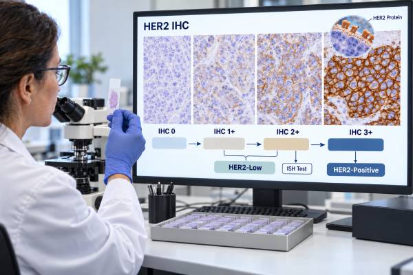
A HER2 IHC test measures HER2 protein on the surface of breast cancer cells and reports a score of 0, 1+, 2+, or 3+. The score helps determine whether the tumor is HER2-positive and whether additional gene-amplification testing is needed. Under the ASCO/CAP framework, IHC 3+ is HER2-positive, IHC 2+ is equivocal and normally requires reflex in situ hybridization, and IHC 0 or 1+ is negative for classic HER2 overexpression. Lower scores now have added treatment relevance. HER2-low generally means IHC 1+ or IHC 2+ with a negative ISH result. In a specific FDA-approved metastatic hormone receptor-positive setting, HER2-ultralow means IHC 0 with detectable membrane staining. That does not turn IHC 0 into classic HER2-positive disease; it identifies a very low expression range used for a particular antibody-drug conjugate indication. The most useful interpretation combines the exact IHC pattern, any reflex FISH/ISH result, disease stage, specimen timing, and current treatment criteria.
- HER2 IHC measures membrane HER2 protein in invasive tumor cells and is usually the first HER2 test performed.
- IHC 3+ is HER2-positive; IHC 2+ is equivocal and usually needs reflex FISH or another ISH test.
- IHC 1+ and IHC 2+/ISH-negative are HER2-low, not classic HER2-positive.
- IHC 0 includes a range from no staining to very faint limited staining; membrane staining can matter for a HER2-ultralow drug indication.
- Low-end HER2 scoring is technically challenging, so specimen quality and validated pathology interpretation are especially important.
Table of Contents
- What the HER2 IHC Test Measures
- What HER2 IHC Scores 0, 1+, 2+, and 3+ Mean
- What Happens After an IHC 2+ Result
- HER2-Low and HER2-Ultralow on IHC
- Why HER2 IHC Results Can Vary Between Samples
- What the IHC Result Means for Treatment
- How to Read a HER2 IHC Report
What the HER2 IHC Test Measures
Immunohistochemistry uses an antibody to detect HER2 protein in a thin section of tumor tissue. After staining, a pathologist examines the membranes of invasive breast cancer cells. HER2 is a cell-surface receptor, so the location, completeness, and intensity of membrane staining matter.
The test does not count HER2 genes. Gene amplification is assessed with FISH or another in situ hybridization method. IHC and ISH are connected because amplification often causes strong protein overexpression, but they are not interchangeable measurements.
HER2 IHC is normally performed on a core biopsy or surgical specimen at diagnosis of invasive breast cancer. It may also be repeated on recurrent or metastatic tissue because biomarker status can change. The result is considered alongside the estrogen receptor test and PR to define the main clinical subtype and treatment pathway.
A pathologist scores only the appropriate invasive component. Ductal carcinoma in situ can show strong HER2 staining even when nearby invasive cancer does not, so morphology matters. Proper fixation and validated staining procedures are also essential because underfixation, overprocessing, decalcification, or poor controls can alter membrane staining.
The familiar 0-to-3+ scale is not a linear measurement of tumor aggressiveness. It is a categorical interpretation designed to predict whether the tumor has HER2 overexpression and whether another assay is needed. More recently, the lower end of the same scale also helps identify patients who may benefit from antibody-drug conjugates despite lacking classic HER2-positive disease.
What HER2 IHC Scores 0, 1+, 2+, and 3+ Mean
An IHC 3+ result shows complete, intense circumferential membrane staining in more than 10% of invasive tumor cells. This is HER2-positive under ASCO/CAP criteria and generally does not need routine confirmatory ISH.
An IHC 2+ result is equivocal. The typical pattern is weak to moderate complete membrane staining in more than 10% of invasive cells. Some unusual strong but incomplete patterns can also fall into the equivocal category depending on morphology and guideline criteria. The next step is usually reflex ISH to determine whether the HER2 gene is amplified.
An IHC 1+ result shows faint or barely perceptible incomplete membrane staining in more than 10% of invasive cells. It is negative for classic HER2 overexpression but belongs to the HER2-low range used for certain advanced breast cancer treatments.
An IHC 0 result historically includes either no staining or faint, incomplete membrane staining in 10% or fewer invasive cells. This distinction within score 0 has become clinically important because the 2025 FDA indication for trastuzumab deruxtecan in HR-positive metastatic breast cancer defines HER2-ultralow as IHC 0 with membrane staining using an approved test.
These criteria show why the score is more than a simple intensity grade. A pathologist must consider what fraction of cells stain, whether the membrane pattern is complete, and how strong the staining is. The full HER2 testing pathway then combines IHC with ISH when required.
What Happens After an IHC 2+ Result
IHC 2+ is a testing waypoint, not a final biological diagnosis. The tissue usually undergoes reflex in situ hybridization to ask whether the HER2 gene is amplified. FISH is a common method, although validated bright-field ISH techniques can also be used.
If ISH shows qualifying amplification, the tumor is HER2-positive. If ISH is negative, the tumor is not HER2-positive under the classic framework. An IHC 2+/ISH-negative tumor, however, is categorized as HER2-low for treatment contexts that use that term.
Dual-probe ISH results may include an average HER2 copy number and a HER2/CEP17 ratio. Straightforward amplified and non-amplified patterns are easy to resolve. Less common ASCO/CAP Groups 2, 3, and 4 require integration with IHC and, in selected cases, a recount. This is why a patient should rely on the final integrated interpretation rather than trying to classify the cancer from one ratio.
If there is too little invasive tumor for successful ISH, the laboratory may test another block or specimen. A technically failed result is not the same as a negative result. Likewise, testing should not accidentally use a strongly staining in situ component instead of the invasive cancer.
The separate HER2 FISH report can look highly numerical, but its purpose is simple: resolve whether an equivocal protein-expression pattern represents true HER2 amplification. Once that question is answered, clinicians can apply the correct treatment pathway.
HER2-Low and HER2-Ultralow on IHC
HER2-low breast cancer is defined for current therapeutic use as IHC 1+ or IHC 2+/ISH-negative. These tumors do not have the overexpression or amplification required for classic HER2-positive status. The term became important after antibody-drug conjugates showed benefit in cancers with lower HER2 expression.
HER2-ultralow extends treatment attention into part of the IHC 0 range. In January 2025, the FDA approved trastuzumab deruxtecan for adults with unresectable or metastatic HR-positive breast cancer that is HER2-low or HER2-ultralow after progression on one or more endocrine therapies in the metastatic setting. In that indication, ultralow means IHC 0 with membrane staining as determined by an FDA-approved test.
This should not be interpreted as a new universal HER2-positive category. ASCO/CAP continues to use the established HER2-positive versus HER2-negative framework for overexpression/amplification. Low and ultralow are additional treatment-selection descriptors that depend on the drug, disease setting, and regulatory label.
Scoring at the low end is harder than recognizing a classic 3+ tumor. Very faint membrane staining can be affected by fixation, assay platform, slide age, background, and observer interpretation. Pathology training and appropriate controls are therefore increasingly important.
For a patient with metastatic disease, the most useful question is not “Am I a little HER2-positive?” It is: What is the exact IHC score, is ISH negative if IHC is 2+, and does that result meet the current criteria for a specific therapy? The HER2-low category is a treatment-relevant expression state, not a synonym for amplification.
Why HER2 IHC Results Can Vary Between Samples
HER2 IHC is sensitive to both tumor biology and specimen handling. A tumor can be heterogeneous, meaning one region contains more HER2-expressing cells than another. A small core biopsy may sample a different area from a later excision. Metastatic sites can also evolve after years of treatment and no longer match the primary tumor exactly.
Preanalytic variables can alter staining. Tissue should be placed in fixative promptly and processed according to validated laboratory procedures. Bone specimens may require decalcification, which can reduce antigen detection depending on the method. Old unstained slides can also lose antigenicity over time. Laboratories use controls to detect major assay failures, but borderline low-expression differences remain challenging.
Interpretation contributes additional variability. Distinguishing no staining from very faint incomplete staining, or deciding whether more than 10% of cells meet a criterion, can be difficult. That was less clinically consequential when IHC 0 and 1+ led to the same treatment category. It matters more now that lower expression can determine antibody-drug conjugate eligibility.
A repeat test is not automatically better. Testing another specimen is most useful when the sample is newer, technically stronger, or more relevant to current disease. If two high-quality specimens disagree, biological heterogeneity may be real.
When a treatment hinges on low or ultralow expression, ask whether the reported result came from an appropriate specimen and a validated assay. A pathology review can sometimes clarify borderline scoring, but it should not be used simply to search repeatedly for a preferred result.
What the IHC Result Means for Treatment
For IHC 3+ HER2-positive breast cancer, HER2-directed therapy is a major treatment component. In early disease, drugs such as trastuzumab and pertuzumab may be used with chemotherapy before or after surgery depending on stage and risk. Residual disease after neoadjuvant treatment can lead to an antibody-drug conjugate strategy. In metastatic HER2-positive disease, several sequential HER2-targeted regimens are available.
For IHC 2+, treatment cannot be chosen from IHC alone. ISH must resolve amplification. If positive, the tumor follows the HER2-positive pathway. If negative, it is HER2-low rather than classic HER2-positive.
For IHC 1+ and selected IHC 0 with membrane staining, the treatment relevance comes from newer antibody-drug conjugate evidence, not from classic trastuzumab sensitivity. In the 2025 FDA HR-positive metastatic indication, trastuzumab deruxtecan improved progression-free survival compared with physician’s-choice chemotherapy in the overall HER2-low/ultralow study population after endocrine therapy progression.
The exact drug indication matters. A lower HER2 score does not automatically justify a HER2-directed drug at initial diagnosis, and eligibility can depend on hormone-receptor status, metastatic setting, prior endocrine therapy or chemotherapy, and the companion diagnostic used.
IHC also does not determine prognosis by itself. HER2-positive breast cancer historically had aggressive biology, but modern targeted therapy has dramatically changed outcomes. The result is most useful as a treatment-selection biomarker, interpreted with stage and the rest of the tumor profile rather than as a stand-alone estimate of survival.
How to Read a HER2 IHC Report
Start with the numeric score and the final interpretation. 3+ should read as HER2-positive. 2+ should prompt you to find the reflex ISH result. 1+ is HER2-negative for classic amplification/overexpression but HER2-low for relevant treatment settings. 0 is also classically negative, yet the report may now distinguish whether any membrane staining is present because of ultralow eligibility.
Check whether the score applies to invasive carcinoma. If both invasive cancer and ductal carcinoma in situ are present, the clinically decisive score for systemic treatment is the invasive component. If the specimen is a metastasis, note the site and whether decalcification was used.
For IHC 2+, do not stop at the words “equivocal.” The final status should come from the integrated IHC/ISH workup. For IHC 0 or 1+ in metastatic disease, ask whether the exact low-expression distinction is relevant to the current treatment line and whether a recent specimen should be reviewed.
Compare HER2 with ER and PR. A hormone receptor-positive/HER2-positive tumor can use both endocrine and HER2 strategies. A hormone receptor-positive/HER2-low tumor follows a different sequence of therapies. A triple-negative tumor with HER2-low expression may have different antibody-drug conjugate options depending on prior treatment and current approvals.
Finally, remember that the score is not a percentage. IHC 1+ does not mean 25% HER2, and 3+ does not mean a threefold risk. It is a pathology category whose meaning comes from validated treatment evidence.
Practical scoring examples and common pitfalls
Imagine three invasive breast cancers. In the first, more than 10% of cells show crisp, complete, very dark circumferential membrane staining. That is the classic IHC 3+ pattern and is HER2-positive. In the second, more than 10% show complete but weak-to-moderate membrane staining. That is IHC 2+, which is equivocal until ISH resolves amplification. In the third, more than 10% show only faint, incomplete membrane staining. That is IHC 1+, which is not classic HER2-positive but is within the HER2-low range.
The most difficult distinction may be between IHC 0 and 1+. If faint incomplete staining is seen in only a small proportion of cells, the case remains score 0 under the traditional scheme. Yet whether any membrane staining is present can matter for the HER2-ultralow category in the specific 2025 FDA indication. That makes careful observation at the low end clinically meaningful without changing the fundamental 0-to-3+ scoring system.
Several pitfalls can distort interpretation. Strong staining in ductal carcinoma in situ should not be scored as though it were the invasive cancer. Cytoplasmic background should not be mistaken for membrane positivity. Edge artifact, crush, poor fixation, and decalcification can create misleading weak or uneven staining. Very old tissue blocks may also behave differently from fresh metastatic biopsies.
Pathologists address these problems by reviewing tumor morphology, controls, staining distribution, and specimen quality. When a borderline score will determine treatment eligibility, a recent, well-fixed specimen tested with the appropriate validated assay can be more informative than repeatedly restaining a poor-quality block.
For patients, the practical rule is to ask for the exact IHC score and the final status, then ask whether lower expression changes eligibility for a specific drug in the current disease setting. That is more useful than trying to interpret staining intensity in isolation.
The percentage of stained cells should not be confused with the IHC score itself. A tumor can show faint incomplete staining in 80% of cells and still be IHC 1+, while a tumor with intense complete staining in more than 10% can be IHC 3+. The score combines pattern and intensity rather than converting directly from a single percentage.
Low-expression categories also may change as assays, companion diagnostics, and drug labels evolve. A report created before HER2-low treatment existed may contain enough descriptive information to reinterpret the old specimen, but sometimes a pathologist must review the actual slide or repeat IHC. For current metastatic decisions, the oncology team should use the most appropriate validated test rather than relying only on a historical label such as “HER2-negative.”
This distinction helps prevent both undertreatment of truly HER2-positive disease and overinterpretation of low expression as gene amplification.
If a report says only “HER2 negative” without the numeric IHC score, obtaining the original pathology details can now matter. A historical negative result could represent IHC 0, IHC 1+, or IHC 2+/ISH-negative, and those distinctions can affect later metastatic treatment. The need is greatest when a lower-expression antibody-drug conjugate is being considered. The pathology laboratory may be able to provide the original score, review stored slides, or test an appropriate newer specimen rather than relying on an incomplete summary in the medical record.
References
- Human Epidermal Growth Factor Receptor 2 Testing in Breast Cancer: ASCO-College of American Pathologists Guideline Update 2023 (Guideline)
- HER2 Testing In Breast Cancer – 2023 Guideline Update – CAP 2023 (Guideline)
- Best pathology practices for HER2 testing interpretation, including HER2-low and ultralow, in the era of HER2-directed antibody drug conjugates (ADCs), aligned with the 2018 CAP/ASCO HER2 guidelines 2025 (Review)
- FDA approves fam-trastuzumab deruxtecan-nxki for unresectable or metastatic HR-positive, HER2-low or HER2-ultralow breast cancer | FDA 2025
- UK recommendations for HER2 assessment in breast cancer: an update 2023 (Guideline)
Disclaimer
This article provides general information about HER2 IHC testing and cannot determine treatment eligibility for an individual patient. Low and ultralow scoring can be technically sensitive, so the pathology result should be reviewed with the oncology team using the specific specimen, any ISH result, current drug indications, and approved companion diagnostic requirements.