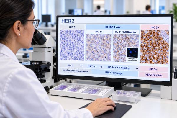
A HER2-low breast cancer test is not a separate laboratory test. It is a way of interpreting the amount of HER2 protein seen on a standard HER2 immunohistochemistry (IHC) test. In current clinical use, HER2-low usually means an IHC score of 1+ or an IHC score of 2+ with a negative in situ hybridization (ISH) result. These tumors are still classified as HER2-negative under the traditional HER2 testing system because they do not show the high protein expression or gene amplification that defines HER2-positive breast cancer.
The distinction matters because newer antibody-drug conjugates can work in some cancers with low HER2 expression. As a result, an exact IHC score that once had little treatment significance may now affect therapy choices in advanced disease. Interpretation is not always simple, however. Low-level staining can vary between samples, laboratories, and tumor sites, and newer terms such as HER2-ultralow have added another layer to reporting.
- HER2-low usually means IHC 1+ or IHC 2+/ISH-negative breast cancer; it is not the same as HER2-positive disease.
- IHC 2+ is not enough by itself to call a tumor HER2-low; ISH is needed to determine whether HER2 is amplified.
- HER2 IHC 0, 1+, 2+, and 3+ describe increasing membrane staining, but the traditional final interpretation remains HER2-negative or HER2-positive.
- Retesting may matter in metastatic disease because HER2 expression can differ between the original tumor and a later biopsy.
- Treatment decisions depend on stage, hormone-receptor status, prior therapy, the exact HER2 result, and current drug indications—not on the words “HER2-low” alone.
Table of Contents
- What HER2-Low Means
- How HER2 Testing Works
- Interpreting IHC Scores and ISH Results
- HER2-Low vs HER2-Positive, HER2-Zero, and HER2-Ultralow
- When HER2 Retesting Matters
- How HER2-Low Results Can Affect Treatment
- Limitations, Common Questions, and Next Steps
What HER2-Low Means
HER2-low describes breast cancers with detectable but not conventionally positive HER2 protein expression. The usual definition is HER2 IHC 1+ or IHC 2+ with no HER2 gene amplification by ISH. The term became important after clinical trials showed that some antibody-drug conjugates could benefit patients whose tumors fell into this range.
HER2 stands for human epidermal growth factor receptor 2. The HER2 protein sits on the surface of cells and helps transmit growth signals. Some breast cancers make very large amounts of HER2 because the ERBB2 gene is amplified. Those cancers are HER2-positive and have long been treated with HER2-directed therapies.
HER2-low tumors are different. They do not have enough HER2 overexpression or amplification to meet the standard definition of HER2-positive disease. Historically, IHC 0, IHC 1+, and IHC 2+/ISH-negative cancers were grouped together as HER2-negative. That traditional classification still applies, but the exact low-end IHC score can now have treatment implications in advanced breast cancer.
This is why a report may contain both statements that seem contradictory at first, such as “HER2 negative” and “IHC 1+.” The first is the formal interpretation for HER2 overexpression or amplification; the second gives the semiquantitative staining score. A patient can therefore have a HER2-negative cancer that falls within the clinical shorthand of HER2-low.
It is also important not to treat HER2-low as a fixed biologic subtype in the same way as classic HER2-positive disease. HER2 expression at low levels can vary within a tumor and can change over time. Hormone-receptor biology often remains more important for the overall identity and first-line treatment strategy of an estrogen-receptor-positive cancer. A broader breast cancer biomarker panel places HER2 alongside ER, PR, proliferation markers, and selected genomic tests rather than interpreting it in isolation.
How HER2 Testing Works
HER2 testing usually starts with immunohistochemistry on tumor tissue. IHC uses an antibody stain to show how much HER2 protein is present on the membranes of invasive breast cancer cells. A pathologist examines the staining pattern, intensity, and percentage of tumor cells and assigns a score of 0, 1+, 2+, or 3+.
The tissue usually comes from a core needle biopsy, surgical specimen, or biopsy of a metastatic site. The quality of tissue handling matters because delayed fixation, inadequate fixation, damaged tissue, and other pre-analytic problems can affect staining. Laboratories use validated assays, controls, and scoring procedures to reduce variability.
For readers who want the test mechanics in more detail, the HER2 IHC test focuses specifically on protein-expression scoring. The practical point is that IHC is a visual, semiquantitative test rather than a direct count of HER2 molecules on every cell. Distinguishing very faint staining from no staining can therefore be more difficult than distinguishing strong 3+ staining from clearly negative results.
When ISH or FISH is added
An IHC score of 2+ is considered equivocal for conventional HER2 status. The laboratory then uses an in situ hybridization test to evaluate the ERBB2 gene. Fluorescence in situ hybridization, commonly called FISH, is one type of ISH.
ISH asks a different question from IHC: instead of looking at protein on the cell surface, it assesses whether the tumor has extra copies or amplification of the HER2 gene. The HER2 FISH test is therefore especially important when IHC staining falls into the 2+ range.
If an IHC 2+ tumor is ISH-positive, it is HER2-positive rather than HER2-low. If it is ISH-negative, it is conventionally HER2-negative and fits the usual HER2-low definition.
Interpreting IHC Scores and ISH Results
The exact IHC pattern—not just the number on the report—determines the HER2 score. Current ASCO/CAP scoring criteria focus on membrane staining in invasive tumor cells.
| Result | Typical staining pattern | Traditional HER2 interpretation | HER2-low relevance |
|---|---|---|---|
| IHC 0 | No staining, or faint/incomplete membrane staining in 10% or fewer tumor cells | Negative | Not HER2-low by the original clinical-trial definition; some IHC 0 tumors with membrane staining may meet newer HER2-ultralow criteria for specific indications |
| IHC 1+ | Faint or barely perceptible incomplete membrane staining in more than 10% of tumor cells | Negative | Usually considered HER2-low |
| IHC 2+ | Weak-to-moderate complete membrane staining in more than 10% of tumor cells | Equivocal until ISH is completed | HER2-low only if ISH is negative |
| IHC 3+ | Complete, intense circumferential membrane staining in more than 10% of tumor cells | Positive | Not HER2-low |
The most easily misunderstood result is IHC 2+. It does not automatically mean HER2-positive and does not automatically mean HER2-low. The ISH result decides which path the interpretation follows. A 2+/ISH-positive tumor is HER2-positive. A 2+/ISH-negative tumor is HER2-negative for overexpression/amplification and falls into the HER2-low group used in major antibody-drug conjugate trials.
The IHC 0-versus-1+ boundary has also become more clinically important. Both scores are negative under the traditional HER2 system, but some drug indications distinguish them. Because the difference can be a small amount of faint membrane staining, pathologists may review borderline cases at higher magnification, use appropriate low-expression controls, or seek a second review when the distinction could affect treatment.
A complete HER2 breast cancer test interpretation should therefore consider the IHC score, any required ISH result, tissue quality, and the clinical setting rather than relying on a single label.
HER2-Low vs HER2-Positive, HER2-Zero, and HER2-Ultralow
HER2-low sits within the traditionally HER2-negative range, while HER2-positive disease remains a separate category defined by strong protein overexpression or gene amplification. Newer treatment research has made the lower end of the HER2 spectrum more detailed, but the terminology can be confusing.
HER2-positive
HER2-positive breast cancer generally means IHC 3+ or a result showing HER2 gene amplification by ISH under guideline criteria. These cancers have enough HER2 overexpression or amplification to qualify for established HER2-positive treatment pathways.
HER2-low
HER2-low generally means IHC 1+ or IHC 2+/ISH-negative. It was a trial-eligibility category that became clinically important because trastuzumab deruxtecan showed benefit in this population. ASCO/CAP has emphasized, however, that HER2-low is not a newly validated pathologic category that replaces the standard positive/negative interpretation.
HER2 IHC 0 and HER2-ultralow
IHC 0 historically meant either no membrane staining or faint/incomplete membrane staining in 10% or fewer invasive tumor cells. The newer term HER2-ultralow has been used for a subset of IHC 0 tumors that show some membrane staining, even though they do not meet the 1+ threshold.
In January 2025, the U.S. Food and Drug Administration expanded a trastuzumab deruxtecan indication to include certain patients with hormone receptor-positive, unresectable or metastatic HER2-ultralow breast cancer after progression on endocrine therapy in the metastatic setting. This makes it important to preserve the actual staining information rather than treating all IHC 0 results as biologically identical for every treatment question.
Terminology may continue to evolve as assays become more sensitive and more clinical trials test lower levels of HER2 expression. For now, readers should distinguish a pathology classification from a drug-eligibility definition. A treatment label can use “HER2-low” or “HER2-ultralow” without changing the core ASCO/CAP HER2-positive versus HER2-negative framework.
When HER2 Retesting Matters
Retesting can be useful when breast cancer recurs or becomes metastatic because HER2 expression may differ from the original tumor. The change may reflect true tumor evolution, differences between tumor sites, sampling, or the difficulty of reproducing low-level IHC scores.
A clinician may consider a new biopsy when it is safe and clinically useful, especially if the result could change systemic treatment. In advanced disease, the most relevant tissue may be a metastatic lesion rather than an old primary-tumor block from years earlier. A previously documented IHC 1+ or 2+/ISH-negative result may also be relevant for some treatment decisions, depending on the drug indication and the patient’s history.
Reasons a HER2 result may warrant review or repeat testing include:
- the report says IHC 2+ but does not show a final ISH result;
- the original sample was technically limited or very small;
- pathology and tumor features appear strongly discordant;
- recurrent or metastatic disease has developed and a new biopsy is available;
- the distinction between IHC 0, 1+, and low-level membrane staining could affect current treatment eligibility;
- different specimens have produced different HER2 scores.
A change from IHC 0 to 1+, or from 1+ to 0, does not necessarily mean the cancer has fundamentally transformed. Low HER2 expression is especially vulnerable to sampling and interpretive variability. The finding should be placed alongside hormone-receptor results and the rest of the tumor profile. For example, an ER-positive metastatic cancer may also need reassessment of endocrine-resistance markers as part of a hormone receptor-positive breast cancer biomarker panel.
How HER2-Low Results Can Affect Treatment
The main clinical importance of HER2-low testing is that it can identify some patients with unresectable or metastatic breast cancer who may be candidates for a HER2-directed antibody-drug conjugate despite being conventionally HER2-negative. It does not mean every person with IHC 1+ or 2+/ISH-negative disease should receive the same treatment.
Trastuzumab deruxtecan is an antibody-drug conjugate. The antibody component binds HER2, helping deliver a potent chemotherapy payload to HER2-expressing tumor cells. This mechanism differs from simply blocking HER2 signaling, which helps explain why lower HER2 expression can still be clinically useful as a drug-delivery target.
The phase 3 DESTINY-Breast04 trial established an important role for trastuzumab deruxtecan in previously treated HER2-low metastatic breast cancer. The trial defined HER2-low as IHC 1+ or IHC 2+/ISH-negative and showed longer progression-free and overall survival than physician’s-choice chemotherapy in the studied population. Later evidence and regulatory decisions expanded use in hormone receptor-positive metastatic disease to an earlier point after endocrine therapy and included HER2-ultralow tumors with membrane staining under a specific approved testing framework.
That does not make the HER2 score a stand-alone treatment prescription. Decisions can depend on:
- whether disease is early-stage, unresectable, recurrent, or metastatic;
- whether the tumor is ER- or PR-positive;
- previous endocrine therapy, chemotherapy, and targeted therapy;
- the current HER2 IHC and ISH results;
- other actionable biomarkers;
- organ function, prior toxicities, and overall health;
- the exact current indication and companion-diagnostic requirements for a drug.
Hormone receptor-negative cancers require their own treatment context. In triple-negative breast cancer biomarker testing, for example, HER2 is interpreted together with ER, PR, PD-L1, germline BRCA-related risk, and other findings that may open different treatment pathways.
Trastuzumab deruxtecan can cause serious adverse effects, including interstitial lung disease or pneumonitis, so eligibility is only one part of the decision. New cough, shortness of breath, fever, or other respiratory symptoms during treatment require prompt medical assessment. The prescribing oncology team also monitors blood counts, heart function when indicated, nausea, and other treatment-related effects.
Limitations, Common Questions, and Next Steps
The biggest limitation of HER2-low testing is that standard HER2 IHC assays were originally optimized to identify HER2 overexpression, not to make ultra-precise measurements at the very bottom of the expression range. A result near the 0-versus-1+ boundary therefore carries more uncertainty than the neat numeric labels may suggest.
Does HER2-low mean the cancer is less aggressive?
Not necessarily. HER2-low is not a simple prognosis score. Outcome depends on stage, hormone-receptor status, tumor grade, disease burden, prior treatment, and many other factors. The term is currently more useful for identifying potential treatment options than for predicting how aggressive an individual cancer will be.
Can HER2-low become HER2-positive or HER2-zero?
Different samples from the same person can produce different HER2 results. Some differences may represent biologic change, while others reflect tumor heterogeneity, specimen handling, sampling, or scoring variability. A new result should be interpreted in context rather than assumed to represent a permanent switch.
Is a blood test used to diagnose HER2-low status?
Routine HER2-low classification is tissue based. IHC evaluates HER2 protein in tumor cells, and ISH evaluates HER2 gene status when needed. Blood-based genomic tests may identify ERBB2 alterations in selected settings, but they do not replace the validated tissue IHC scoring system used to define HER2-low expression for current breast cancer treatment decisions.
What should you ask about a HER2 report?
Useful questions include:
- What was the exact IHC score: 0, 1+, 2+, or 3+?
- If the score was 2+, was ISH or FISH performed, and what was the final result?
- Was the test performed on the original breast tumor or a metastatic biopsy?
- Is there another tumor sample worth testing if the result is borderline or old?
- Does the exact HER2 result affect treatment options at the current stage of disease?
- Does hormone-receptor status or another biomarker take priority for the next treatment decision?
The most useful report is one that preserves the exact IHC score and any ISH result rather than reducing everything to a single “positive” or “negative” word. As HER2-directed drug indications continue to evolve, those details can become clinically relevant later.
One more practical point is that a HER2-low result is not a blood level that can be trended. It is a tissue-based description of protein staining on a particular specimen. If treatment depends on the distinction between IHC 0, 1+, and 2+/ISH-negative, the pathology report and available tissue should be reviewed rather than trying to infer the category from an older “HER2-negative” summary.
References
- HER2 Testing in Breast Cancer – 2023 Guideline Update 2023 (Guideline)
- Trastuzumab Deruxtecan in Previously Treated HER2-Low Advanced Breast Cancer 2022 (RCT)
- FDA approves fam-trastuzumab deruxtecan-nxki for unresectable or metastatic HR-positive, HER2-low or HER2-ultralow breast cancer 2025
- US Food and Drug Administration Approval Summary: Trastuzumab Deruxtecan for the Treatment of Adult Patients With Hormone Receptor-Positive, Unresectable or Metastatic Human Epidermal Growth Factor Receptor 2-Low or Human Epidermal Growth Factor Receptor 2-Ultralow Breast Cancer 2025 (Review)
- Trastuzumab deruxtecan in HER2-low metastatic breast cancer: long-term survival analysis of the randomized, phase 3 DESTINY-Breast04 trial 2025 (RCT)
Disclaimer
This article explains HER2-low breast cancer testing for general education and does not replace interpretation by a pathologist or oncology team. HER2 terminology, companion diagnostic requirements, and treatment indications can change, so treatment decisions should use the complete pathology report, disease setting, and current prescribing guidance. Seek prompt medical care for urgent symptoms or serious treatment side effects.