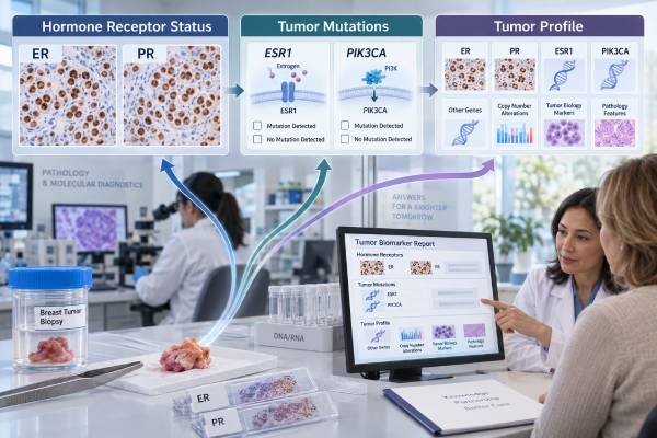
A hormone receptor-positive breast cancer biomarker panel is not one universal laboratory panel. It is the combined set of pathology and molecular tests used to define the tumor’s hormone-receptor biology, HER2 status, prognosis, and potentially actionable mutations. The foundation is estrogen receptor (ER) and progesterone receptor (PR) testing on tumor tissue. In advanced disease, molecular testing may add ESR1, PIK3CA, and other genes when the results can guide a treatment choice.
These markers answer different questions. ER and PR show whether tumor cells use hormone signaling. ESR1 mutations can reveal acquired resistance to some endocrine therapies. PIK3CA mutations identify abnormal PI3K pathway signaling and can make certain targeted treatments relevant. HER2 must also be known because it changes how the cancer is classified and treated. The best “panel” therefore depends on whether the cancer is newly diagnosed, early-stage, recurrent, or metastatic and on which treatment decision needs to be made now.
- ER positivity is the key marker that predicts potential benefit from endocrine therapy; PR adds useful biologic and prognostic context.
- ESR1 testing is most useful in advanced ER-positive/HER2-negative disease, especially after exposure to aromatase inhibitor therapy.
- PIK3CA mutations are common actionable alterations in HR-positive/HER2-negative advanced breast cancer and can be tested in tissue or circulating tumor DNA.
- A negative plasma mutation result may not rule out a tumor mutation because low levels of circulating tumor DNA can reduce sensitivity.
- The panel should be matched to the clinical decision: early-stage prognosis testing is different from metastatic resistance and targeted-therapy testing.
Table of Contents
- What a Hormone Receptor-Positive Biomarker Panel Measures
- ER and PR: The Core Hormone Receptor Tests
- ESR1 Mutation Testing and Endocrine Resistance
- PIK3CA Testing and the PI3K Pathway
- Building the Full Tumor Profile
- When and How Testing Is Done
- Interpreting Results and Planning Next Steps
What a Hormone Receptor-Positive Biomarker Panel Measures
A hormone receptor-positive breast cancer panel combines markers of tumor identity with markers that may predict response or resistance to treatment. It should not be thought of as a single fixed blood test or a standard bundle that every patient needs at every stage.
The first question is whether the invasive breast cancer expresses estrogen receptor or progesterone receptor. A tumor is generally described as hormone receptor-positive when ER and/or PR is positive, although ER status is the main predictor of benefit from endocrine therapy. HER2 is tested separately but is essential because HR-positive/HER2-positive and HR-positive/HER2-negative cancers follow different treatment pathways.
Later questions depend on the disease setting. In early-stage HR-positive/HER2-negative cancer, the main concern may be recurrence risk and whether chemotherapy adds enough benefit to justify its toxicity. In that setting, selected genomic expression assays may be useful. In metastatic disease, the focus shifts toward mutations that can drive resistance or create a targeted treatment opportunity, including ESR1 and PIK3CA.
A practical tumor profile may therefore contain several layers:
- ER and PR protein expression by immunohistochemistry;
- HER2 protein expression and, when required, gene amplification testing;
- tumor grade and proliferation information;
- a genomic recurrence assay in selected early-stage cancers;
- somatic mutation testing in advanced disease;
- germline testing when inherited cancer risk is relevant;
- repeat biomarker testing on recurrent or metastatic tissue when results may have changed.
The broader breast cancer biomarker panel explains how these categories fit together across breast cancer subtypes.
ER and PR: The Core Hormone Receptor Tests
ER and PR are usually measured by immunohistochemistry on tumor tissue, and the percentage of tumor cell nuclei that stain is reported. These are foundational tests because they help determine whether endocrine therapy is likely to be useful.
Estrogen receptor
ER is the most important hormone receptor marker for predicting endocrine sensitivity. Under ASCO/CAP guidance, invasive breast cancers with 1% to 100% of tumor nuclei staining for ER are considered ER-positive. Tumors with 1% to 10% ER staining are reported as ER Low Positive because evidence for endocrine benefit is less robust than for tumors with stronger or more extensive ER expression.
An estrogen receptor test report may include the percentage of positive cells, staining intensity, and sometimes a combined scoring system. The percentage alone does not tell the whole story, but it is clinically central.
Progesterone receptor
PR is regulated partly by estrogen signaling, so its presence can support evidence of an active hormone-receptor pathway. PR is not as important as ER for deciding whether endocrine therapy should be offered, but it can add prognostic and biologic information.
A progesterone receptor test is also performed by IHC. Discordant patterns can occur. For example, an ER-positive/PR-negative tumor is still hormone receptor-positive, but loss of PR may be associated with different tumor biology from a strongly ER-positive/PR-positive cancer.
ER and PR are tissue markers. They are not the same as blood estrogen or progesterone levels, and hormone blood tests cannot substitute for tumor receptor testing.
ESR1 Mutation Testing and Endocrine Resistance
ESR1 mutations are mainly resistance biomarkers in advanced ER-positive/HER2-negative breast cancer, not routine markers for a newly diagnosed untreated early-stage tumor. ESR1 is the gene that encodes estrogen receptor alpha. Certain acquired mutations in its ligand-binding domain can activate estrogen-receptor signaling even when estrogen production is suppressed.
These mutations often emerge under the selective pressure of aromatase inhibitor therapy. They are uncommon in treatment-naive primary tumors but become more frequent in metastatic disease after endocrine treatment. That makes timing important: a test performed years earlier may not reflect the tumor’s current resistance biology.
Testing is often done with circulating tumor DNA from plasma because a blood sample can capture DNA shed from multiple metastatic sites and can be repeated more easily than a tissue biopsy. Tumor tissue can also be tested with next-generation sequencing.
The ESR1 mutation test is clinically actionable because several estrogen-receptor-directed treatments now have U.S. indications tied to ESR1 mutation status. As of 2026, approved options include oral estrogen-receptor antagonists or degraders for selected patients whose advanced disease has progressed after endocrine therapy. A September 2026 accelerated approval also introduced a strategy in which an ESR1 mutation detected in circulating tumor DNA during aromatase inhibitor plus CDK4/6 inhibitor therapy can trigger a treatment switch before radiographic progression in a defined setting.
That development makes serial molecular monitoring more relevant, but it does not mean every patient needs ESR1 blood testing at every visit. Testing frequency, assay choice, and treatment timing should follow the specific therapy being considered and current oncology guidance.
A “no ESR1 mutation detected” result also needs context. If very little tumor DNA is circulating, the blood test may be negative even when a mutation exists somewhere in the cancer. The laboratory report, tumor burden, and whether tissue is available can influence whether another method is useful.
PIK3CA Testing and the PI3K Pathway
A pathogenic PIK3CA mutation can identify HR-positive/HER2-negative advanced breast cancer that may respond to therapies directed at the PI3K/AKT pathway. PIK3CA encodes the p110-alpha catalytic subunit of PI3K, a signaling protein involved in cell growth and survival.
PIK3CA mutations are among the most common genomic alterations in hormone receptor-positive breast cancer. Unlike ESR1 resistance mutations, PIK3CA mutations may be present in the primary cancer from the start and can remain detectable later. Common activating mutations occur at hotspots such as E542K, E545K, and H1047R, although approved assays can evaluate a broader defined set.
The PIK3CA mutation test can use either tumor tissue or circulating tumor DNA, depending on the assay and treatment being considered. ASCO guidance has emphasized an important limitation of plasma testing: if PIK3CA is not detected in ctDNA, testing available tumor tissue can identify some additional mutation-positive patients.
PIK3CA results can affect treatment in more than one way. Alpelisib plus endocrine therapy established an early mutation-directed strategy in advanced disease. Later approvals expanded PI3K/AKT-pathway targeting. In October 2024, the FDA approved inavolisib with palbociclib and fulvestrant for a defined group with endocrine-resistant, PIK3CA-mutated, HR-positive/HER2-negative advanced breast cancer. Capivasertib with fulvestrant is another pathway-targeted option for selected cancers with PIK3CA, AKT1, or PTEN alterations.
A positive mutation is not a guarantee that a drug will work, and it does not automatically determine treatment order. Prior therapies, pace of disease, diabetes risk, other mutations, symptoms, and drug toxicity all influence the final decision.
Building the Full Tumor Profile
ER, PR, ESR1, and PIK3CA answer only part of the treatment question; the full tumor profile also needs HER2 status and, in selected settings, other pathology, genomic, and hereditary information.
HER2 testing is required for invasive breast cancer because it separates cancers with HER2 overexpression or amplification from those without it and can identify lower levels of HER2 expression that may matter for antibody-drug conjugate therapy. A complete HER2 breast cancer test may include IHC and reflex ISH for an equivocal IHC 2+ result.
For early-stage HR-positive/HER2-negative disease, multigene expression assays can help estimate recurrence risk and chemotherapy benefit in appropriately selected patients. Tests such as Oncotype DX Breast Recurrence Score, MammaPrint, EndoPredict, Prosigna, or Breast Cancer Index are not interchangeable with mutation panels. They measure patterns of gene activity for prognosis or treatment decision support rather than simply looking for actionable DNA variants.
In advanced disease, a broader next-generation sequencing panel may include PIK3CA, ESR1, AKT1, PTEN, ERBB2, and other genes. Depending on the clinical picture, testing may also evaluate germline BRCA1/2 or other inherited predisposition genes because those findings can affect both treatment and family risk.
Pathology still matters. Tumor grade, histologic type, lymph-node involvement, stage, and sites of metastasis can be as important as molecular results. A mutation report should never be interpreted as if it replaces the diagnosis and stage.
| Marker or test type | Sample | Main question | Typical setting |
|---|---|---|---|
| ER | Tumor tissue | Is endocrine therapy likely to be useful? | Initial diagnosis and often recurrence |
| PR | Tumor tissue | What additional hormone-receptor biology is present? | Initial diagnosis and often recurrence |
| HER2 | Tumor tissue | Is HER2-directed treatment relevant? | Initial diagnosis and metastatic reassessment |
| ESR1 | Plasma ctDNA or tumor tissue | Has an endocrine-resistance mutation emerged? | Advanced ER-positive/HER2-negative disease |
| PIK3CA | Tumor tissue or plasma ctDNA | Is a PI3K-pathway targeted option relevant? | Advanced HR-positive/HER2-negative disease |
| Genomic recurrence assay | Primary tumor tissue | What is recurrence risk, and will chemotherapy likely add benefit? | Selected early-stage HR-positive/HER2-negative cancers |
When and How Testing Is Done
Testing should be timed to the treatment decision because some biomarkers are stable while others can emerge under therapy. At diagnosis, ER, PR, and HER2 are core pathology tests. Additional testing is selected rather than automatic.
Newly diagnosed early-stage disease
The initial biopsy or surgical specimen establishes ER, PR, HER2, histologic type, and grade. If the cancer is HR-positive/HER2-negative and clinical features make the value of chemotherapy uncertain, a validated genomic recurrence assay may be considered according to age, menopausal status, nodal involvement, and other factors.
Routine ESR1 mutation testing is generally not useful in an untreated early-stage tumor because the mutation usually develops later under endocrine selection pressure. PIK3CA testing also does not routinely guide standard adjuvant treatment for every early-stage HR-positive cancer.
Recurrent or metastatic disease
A biopsy of recurrent or metastatic cancer can confirm the diagnosis and allow reassessment of ER, PR, and HER2. Molecular testing may then look for actionable alterations such as PIK3CA and ESR1. Blood-based ctDNA can be especially useful when tissue is difficult to obtain or when repeated assessment is needed.
No special fasting or medication preparation is usually required for tissue biomarker testing. A plasma ctDNA test is typically a routine blood draw, but the assay’s pre-analytic instructions still matter. The timing relative to treatment and disease burden can affect how much tumor DNA is detectable.
Interpreting Results and Planning Next Steps
A biomarker result is useful only when it is linked to a specific clinical question. A long molecular report can contain many variants, but not every finding is actionable, validated, or relevant to breast cancer treatment.
When reviewing a panel, separate results into categories:
- Tumor-defining markers: ER, PR, and HER2 establish the main biologic classification.
- Predictive biomarkers: Certain PIK3CA or ESR1 findings may predict eligibility for a targeted treatment in a defined setting.
- Prognostic tests: Genomic recurrence scores can estimate future risk in selected early-stage disease but are not general mutation screens.
- Resistance markers: ESR1 can indicate evolving endocrine resistance and, in some settings, influence when or how endocrine therapy is changed.
- Hereditary findings: Germline variants answer inherited-risk questions and require different interpretation from tumor-only mutations.
Ask for the exact result rather than a simplified “positive” or “negative” summary. For ER and PR, that means the percentage and staining interpretation. For PIK3CA or ESR1, ask which mutation was found, what specimen was tested, whether the assay is validated for the contemplated drug, and whether a negative plasma result should be followed by tissue testing.
It is also reasonable to ask whether a result has changed treatment now. Some findings may become relevant later, while others should trigger an immediate discussion of options. In metastatic disease, the tumor profile can evolve, so testing is sometimes repeated at a meaningful treatment transition rather than treated as a one-time lifetime record.
Finally, biomarker-guided treatment still requires clinical judgment. Performance status, symptoms, disease location, prior responses, medication interactions, glucose control, reproductive considerations, and personal goals can all influence which technically eligible therapy is the best next choice.
Why a negative molecular result may need a second look
A negative result does not always mean the pathway or mutation is absent. With plasma ctDNA, the amount of tumor DNA in the bloodstream can be very low when disease burden is small, when metastases shed little DNA, or when treatment has recently suppressed tumor activity. The laboratory may report that no actionable alteration was detected even though the specimen simply did not contain enough tumor-derived DNA to reveal one.
This is especially important when a treatment decision depends on PIK3CA status. If a validated plasma assay does not detect a PIK3CA mutation and archived or newly obtained tumor tissue is available, tissue testing may identify an alteration that the blood test missed. The reverse issue can also occur: an older tissue block may not show a resistance mutation such as ESR1 that developed later under endocrine therapy, while a current plasma sample can capture it.
Ask whether the report includes a measure of tumor fraction, assay sensitivity, or another indication that the blood sample contained detectable tumor DNA. Also ask whether the tested specimen was collected before or after a major treatment change. These details can determine whether a negative result is considered reassuring, inconclusive, or worth repeating with a different specimen.
References
- Biomarkers for Adjuvant Endocrine and Chemotherapy in Early-Stage Breast Cancer: ASCO Guideline Update 2022 (Guideline)
- Biomarkers for Systemic Therapy in Metastatic Breast Cancer: ASCO Guideline Update 2022 (Guideline)
- Testing for ESR1 Mutations to Guide Therapy for Hormone Receptor-Positive, Human Epidermal Growth Factor Receptor 2-Negative Metastatic Breast Cancer: ASCO Guideline Rapid Recommendation Update 2023 (Guideline)
- FDA approves inavolisib with palbociclib and fulvestrant for endocrine-resistant, PIK3CA-mutated, HR-positive, HER2-negative, advanced breast cancer 2024
- FDA grants accelerated approval to camizestrant with a CDK4/6 inhibitor for ESR1-Mutated HR-positive, HER2-negative locally advanced or metastatic breast cancer 2026
- Estrogen and Progesterone Receptor Testing in Breast Cancer: ASCO/CAP Guideline Update 2020 (Guideline)
Disclaimer
This article is for general education and does not replace individualized pathology or oncology advice. Biomarker testing and drug indications change over time, and the most appropriate test depends on disease stage, prior treatment, specimen availability, and the therapy being considered. Discuss exact ER, PR, HER2, ESR1, and PIK3CA findings with the treating oncology team before making treatment decisions.