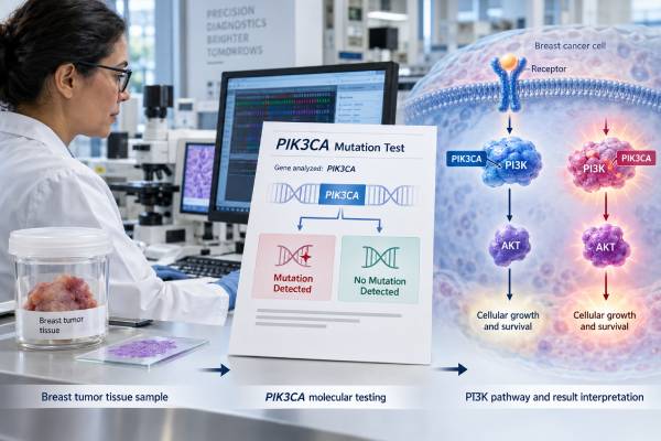
A PIK3CA mutation test looks for tumor DNA changes that activate the PI3K signaling pathway, one of the most commonly altered growth pathways in hormone receptor-positive, HER2-negative breast cancer. In advanced disease, a positive PIK3CA result can identify patients who may be eligible for PI3K- or AKT-pathway targeted treatments in specific clinical settings. Testing can be performed on tumor tissue or, for some indications, circulating tumor DNA from plasma. A positive result usually reflects a somatic mutation that developed in the cancer rather than an inherited family-risk variant. A negative blood-based result can be falsely negative when too little tumor DNA is circulating, so tumor-tissue testing may still be appropriate if available. PIK3CA status is not interpreted as a simple prognosis score and does not replace ER, PR, HER2, stage, or treatment history. The exact mutation, assay, disease setting, and intended drug all matter because different targeted therapies have different companion-diagnostic requirements and eligibility criteria.
- PIK3CA encodes the p110-alpha catalytic subunit of PI3K, a key cell-growth signaling protein.
- PIK3CA mutations are common in hormone receptor-positive, HER2-negative breast cancer and are usually acquired tumor changes.
- Testing may use tumor tissue or circulating tumor DNA, depending on the treatment decision and assay.
- A negative plasma result may need follow-up tissue testing because ctDNA can be below the detection limit.
- A positive result can be actionable, but drug choice depends on prior therapy, disease setting, and the exact approved indication.
Table of Contents
- What PIK3CA and the PI3K pathway do
- How PIK3CA testing is performed
- What positive and negative results mean
- How PIK3CA results can guide targeted treatment
- Common mutations and why assay coverage matters
- Limits, timing, and repeat testing
- Questions to ask after receiving a PIK3CA result
What PIK3CA and the PI3K pathway do
PIK3CA is the gene that encodes p110-alpha, the catalytic subunit of class I phosphatidylinositol 3-kinase, or PI3K. The PI3K-AKT-mTOR signaling pathway helps regulate cell growth, survival, metabolism, and proliferation. Normal cells switch the pathway on and off in response to growth signals. Certain PIK3CA mutations keep the pathway abnormally active, allowing cancer cells to grow or survive more efficiently.
PIK3CA is one of the most frequently mutated genes in breast cancer. Alterations are especially common in hormone receptor-positive, HER2-negative tumors, although they can occur in other breast cancer subtypes as well. The mutation is usually somatic, meaning it arose in the tumor after conception and is not present in every cell of the body.
That distinction matters. A tumor PIK3CA mutation does not normally mean children or siblings have inherited the same cancer risk. Germline hereditary testing for genes such as BRCA1, BRCA2, or PALB2 answers a different question. A breast cancer hereditary gene panel is designed to assess inherited susceptibility, while PIK3CA testing in breast oncology is generally performed to characterize the cancer and guide treatment.
Rare germline or mosaic PIK3CA variants can cause PIK3CA-related overgrowth spectrum disorders, but that is a different clinical context from the common somatic mutations found in breast tumors. A routine breast cancer PIK3CA-positive result should not be interpreted as an inherited syndrome without separate evidence.
PIK3CA also is not the same as AKT1 or PTEN. These genes act in the same signaling network, and some therapies are approved for tumors with alterations in any of the three. A report may therefore describe an “AKT-pathway alteration” panel that includes PIK3CA, AKT1, and PTEN rather than testing PIK3CA alone.
How PIK3CA testing is performed
PIK3CA mutations can be detected in tumor tissue or in circulating tumor DNA, abbreviated ctDNA, found in plasma. The best method depends on which specimen is available, how much disease is present, the assay’s validated use, and which treatment is being considered.
Tumor testing usually uses DNA from formalin-fixed, paraffin-embedded tissue taken during biopsy or surgery. The sample may come from the original breast tumor or a metastatic site. Next-generation sequencing, or NGS, can evaluate PIK3CA along with many other cancer genes. Some targeted assays test only a defined group of known hotspot mutations.
Plasma testing uses a blood sample to look for fragments of tumor DNA released into the circulation. It is often called a liquid biopsy. A breast cancer ctDNA test can be useful when a tissue biopsy is difficult, when archival tissue is limited, or when the current metastatic tumor may have evolved since the original diagnosis.
The major caution is sensitivity. Not every cancer releases enough DNA into plasma for a mutation to be detected. A positive ctDNA result is generally informative when the assay is validated, but a negative plasma result can mean either “no mutation” or “not enough tumor DNA to detect the mutation.” ASCO guidance therefore recommends testing tumor tissue, if available, when plasma testing is negative and PIK3CA status is still needed to determine eligibility for PI3K-targeted therapy.
A PIK3CA report should ideally identify:
- the specimen type and collection date;
- the assay or platform;
- the exact PIK3CA variant detected;
- whether the laboratory classifies it as pathogenic or activating;
- variant allele frequency when relevant;
- whether the assay is an FDA-approved companion diagnostic for the intended treatment; and
- other actionable alterations found on a broader panel.
Unlike germline testing, tumor sequencing can show many variants that are not treatment targets. The presence of a PIK3CA alteration therefore should be matched to a clinically validated drug indication rather than interpreted only from a generic “pathogenic” label.
What positive and negative results mean
A positive PIK3CA result means the assay found a mutation in the tumor DNA that meets its reporting criteria. In hormone receptor-positive, HER2-negative advanced breast cancer, that result can be predictive because several targeted treatment strategies depend on activation of the PI3K-AKT pathway.
“Positive,” however, does not mean the cancer is more advanced or that targeted therapy must be started immediately. Treatment still depends on whether the disease is early-stage or metastatic, whether it is endocrine-sensitive or endocrine-resistant, which therapies have already been used, and whether the person can safely receive the targeted agent.
A negative result must be interpreted according to the sample:
| Result situation | What it may mean | Possible next step |
|---|---|---|
| Negative high-quality tumor assay | No covered PIK3CA mutation detected in the tested tumor | Use other biomarkers and treatment options |
| Negative plasma ctDNA assay | No mutation detected, but low tumor DNA shedding can cause a false negative | Test tumor tissue if available and clinically needed |
| Mutation outside a limited hotspot panel | May be missed if the assay does not cover that region | Consider broader validated sequencing when relevant |
PIK3CA status is not a direct recurrence percentage. Although the pathway is biologically important and has been associated with tumor behavior in research, the main current use of testing is predictive: identifying a molecular target for therapy. A person should not interpret a positive mutation as proof of a poor prognosis or a negative result as proof of a better prognosis.
The result should also be distinguished from the hormone receptor-positive breast cancer biomarker panel. ER and PR establish hormone sensitivity, HER2 determines whether HER2-directed therapy is appropriate, and PIK3CA adds a later molecular treatment option in selected advanced disease.
How PIK3CA results can guide targeted treatment
PIK3CA became clinically important because drugs can inhibit the pathway it activates. The exact treatment pathway has expanded over time, so a PIK3CA-positive result should be interpreted using current drug indications rather than older rules alone.
Alpelisib is a PI3K-alpha inhibitor. It was the first drug approved specifically for PIK3CA-mutated, hormone receptor-positive, HER2-negative advanced breast cancer in combination with fulvestrant after progression on endocrine therapy. The SOLAR-1 trial showed a progression-free survival benefit in the PIK3CA-mutated group. Important toxicities include hyperglycemia, rash, diarrhea, and other metabolic or inflammatory effects, so baseline health and glucose control matter.
Capivasertib is an AKT inhibitor. In 2023, the FDA approved capivasertib with fulvestrant for HR-positive, HER2-negative locally advanced or metastatic breast cancer with one or more PIK3CA, AKT1, or PTEN alterations after progression on an endocrine-based regimen in the metastatic setting or recurrence on or within 12 months of completing adjuvant therapy. A PIK3CA mutation can therefore qualify as one of several pathway alterations for this regimen.
Inavolisib is a selective PI3K-alpha inhibitor that also promotes degradation of mutant p110-alpha. In 2024, the FDA approved inavolisib with palbociclib and fulvestrant for adults with endocrine-resistant, PIK3CA-mutated, HR-positive, HER2-negative locally advanced or metastatic breast cancer following recurrence on or after completing adjuvant endocrine therapy. In the INAVO120 trial, median progression-free survival was 15.0 months with inavolisib, palbociclib, and fulvestrant versus 7.3 months with placebo, palbociclib, and fulvestrant.
These approvals illustrate why a positive PIK3CA result does not point to one universal drug. The oncology team considers:
- whether the disease is HR-positive and HER2-negative;
- whether the cancer is locally advanced or metastatic;
- when recurrence occurred relative to adjuvant endocrine therapy;
- which endocrine and CDK4/6 treatments have already been used;
- whether the assay meets the companion-diagnostic requirement; and
- the toxicity profile and the person’s medical conditions.
For example, diabetes or difficult-to-control blood glucose can be especially relevant to PI3K inhibitors because hyperglycemia is a known toxicity. Capivasertib has a different adverse-effect pattern, including diarrhea and rash, and every regimen has its own monitoring requirements.
Treatment recommendations also change as new trial data and approvals appear. A result generated years ago may still be biologically valid, but the available drugs and sequencing strategy should be reviewed using current guidance.
Common mutations and why assay coverage matters
Many activating PIK3CA mutations cluster in hotspot regions of the gene. Common examples include E542K and E545K in the helical domain and H1047R in the kinase domain. Other pathogenic variants occur as well.
Older or highly targeted assays may test a limited set of mutations that were included in pivotal trials. Broader NGS panels can detect a wider spectrum. Research using comprehensive genomic profiling has shown that a meaningful minority of PIK3CA-mutated advanced breast cancers contain pathogenic alterations outside the classic set used in the original SOLAR-1 assay definition.
That does not mean every rare PIK3CA change is actionable. The laboratory must determine whether a variant is activating or clinically significant, and the drug’s approved companion-diagnostic framework matters. A broad report may contain variants of uncertain significance that should not be treated as equivalent to a known activating hotspot.
The exact notation can look technical. A result such as PIK3CA p.H1047R means the amino acid histidine at position 1047 has been replaced by arginine. Laboratories may also provide the DNA-level notation. Patients do not need to memorize the code, but retaining the full report is useful because future therapies may define eligibility by specific alterations.
Variant allele frequency, or VAF, is sometimes reported. In a tumor sample, VAF is influenced by tumor purity, copy-number changes, and whether the mutation is present in all cancer cells. In plasma, VAF is also influenced by how much ctDNA is being released. A low VAF does not automatically mean the mutation is unimportant, and a high VAF does not directly measure tumor burden across the whole body.
If the test was performed long ago on a small hotspot panel and treatment now depends on pathway status, ask whether that assay covered the mutations recognized by the current companion diagnostic. The answer may determine whether repeat or broader testing is worthwhile.
Limits, timing, and repeat testing
PIK3CA mutations often arise early in breast cancer development and can remain detectable through disease progression, but tumor evolution and sampling still matter. Testing may be done on the primary tumor or on metastatic tissue depending on availability and the current treatment question.
For early-stage breast cancer, routine PIK3CA testing generally does not have the same established role in choosing standard adjuvant therapy as it does in advanced HR-positive, HER2-negative disease. A mutation found incidentally on an early-stage panel should not be assumed to require a PI3K inhibitor outside an approved or studied setting.
In metastatic disease, testing becomes most useful when the result can change systemic therapy. A practical sequence may be to use available archival tissue, obtain a current biopsy when clinically appropriate, or use plasma ctDNA. If a validated plasma test is positive, a separate tissue confirmation is often unnecessary for that target. If plasma is negative, tissue can resolve whether the result reflects true wild-type disease or inadequate ctDNA shedding.
Repeat testing may be reasonable when:
- the original assay had limited PIK3CA coverage;
- the earlier sample was inadequate or failed quality control;
- a plasma result was negative but tissue is available;
- a new metastatic biopsy is being obtained for other clinical reasons; or
- a broader panel is now needed to evaluate multiple treatment targets at once.
Repeat testing solely to look for a different answer is not useful. The purpose should be to improve assay sensitivity, obtain current tumor information, or meet a treatment-selection requirement.
PIK3CA testing also does not replace ESR1 mutation testing. ESR1 mutations can emerge under aromatase-inhibitor pressure and guide endocrine treatment in advanced HR-positive disease, whereas PIK3CA identifies a different signaling pathway. Both may be relevant at the same time.
Questions to ask after receiving a PIK3CA result
A useful result is one that can be connected to a specific treatment decision. Ask the oncology team to interpret the mutation in the current disease setting rather than only reading the word “positive.”
Helpful questions include:
- Was PIK3CA tested in tumor tissue or plasma ctDNA?
- What exact mutation was found, and is it considered activating and actionable?
- If plasma was negative, is there enough tumor tissue to test directly?
- Is my current cancer HR-positive and HER2-negative?
- Which PIK3CA- or AKT-pathway treatment indication fits my previous therapy history?
- Does the assay I had meet the companion-diagnostic requirement for the proposed drug?
- What benefit was seen in patients like me in the relevant trial?
- What toxicities are most important for my health, especially blood-sugar, rash, or diarrhea risks?
- Were AKT1, PTEN, ESR1, BRCA1/2, or other actionable biomarkers evaluated at the same time?
- Is this mutation somatic, or is there any reason to consider separate germline genetic testing?
The central point is that PIK3CA is a treatment-selection biomarker, not a stand-alone diagnosis. A positive mutation can open targeted options, while a negative result—especially from plasma—must be judged in light of assay sensitivity. The most accurate interpretation combines the molecular result with receptor status, disease setting, treatment history, and the current approved therapy landscape.
The reported variant allele fraction can add laboratory context, but it is not a treatment-response percentage. A low fraction may reflect low tumor content, a small tumor-DNA contribution to plasma, or tumor heterogeneity. If a recognized actionable PIK3CA mutation is confidently detected by a validated assay, clinicians interpret the mutation itself together with the approved treatment criteria; they do not require a high allele fraction simply to call the result positive.
References
- Biomarkers for Systemic Therapy in Metastatic Breast Cancer: ASCO Guideline Update 2022 (Guideline)
- FDA approves inavolisib with palbociclib and fulvestrant for endocrine-resistant, PIK3CA-mutated, HR-positive, HER2-negative, advanced breast cancer 2024 (FDA)
- Inavolisib-Based Therapy in PIK3CA-Mutated Advanced Breast Cancer 2024 (Randomized Trial)
- FDA approves capivasertib with fulvestrant for breast cancer 2023 (FDA)
- Capivasertib in Hormone Receptor-Positive Advanced Breast Cancer 2023 (Randomized Trial)
- Biology and Targetability of the Extended Spectrum of PIK3CA Mutations Detected in Breast Carcinoma 2023
Disclaimer
This article is for general education and does not replace advice from a breast oncologist or molecular pathologist. PIK3CA-directed treatment depends on the exact mutation, assay, receptor status, stage, prior therapies, companion-diagnostic requirements, and individual health risks. A negative plasma ctDNA result should not automatically be treated as proof that the tumor is PIK3CA-wild type when tissue testing is feasible and the result would change treatment.