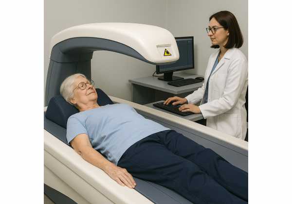
Healthy bones are central to healthy aging. A single wrist or hip fracture can trim years off independence, slow recovery from other illness, and restrict the activities that keep you fit. Dual-energy X-ray absorptiometry (DEXA or DXA) is the standard test to measure bone mineral density (BMD) and estimate fracture risk. Yet scan printouts can be confusing: T-scores, Z-scores, vertebrae excluded, and charts that don’t always agree from one site to another. This guide explains what a DEXA report actually shows, who benefits from scanning, how to prepare, and how to use results alongside clinical risk tools to make better decisions. If you’re building a broader tracking plan for long-term health, visit our curated hub for longevity biomarkers and practical tools. The aim is to spot risk early, track change correctly, and pair the right next step with your goals.
Table of Contents
- What DEXA Reports Show: T-Scores, Z-Scores, and Sites Scanned
- Who Should Consider Scanning and at What Age
- Before the Scan: Clothing, Calcium, and Recent Imaging
- Interpreting Risk in Context: FRAX and Clinical Factors
- Change Over Time: Least Significant Change (LSC) and Repeats
- Artifacts and Errors: Degenerative Changes, Implants, and Positioning
- Next Steps to Discuss with Your Clinician
What DEXA Reports Show: T-Scores, Z-Scores, and Sites Scanned
A DEXA report quantifies bone mineral density (BMD) in grams per square centimeter (g/cm²) and then compares your BMD to reference populations to estimate fracture risk.
Core numbers you’ll see
- BMD (g/cm²): The direct measurement at each skeletal site. This is the value used to monitor change over time.
- T-score: Your BMD compared with a young adult reference (typically women aged 20–29 from NHANES III). It tells how many standard deviations (SD) your BMD sits above or below the young-adult mean at that site.
- Normal: T ≥ −1.0
- Low bone mass (“osteopenia”): −1.0 > T > −2.5
- Osteoporosis: T ≤ −2.5
- Established (severe) osteoporosis: T ≤ −2.5 plus a fragility fracture
- Z-score: Your BMD compared with an age-, sex-, and ethnicity-matched reference population. Z is mainly used in premenopausal women, younger men, and anyone with suspected secondary causes. A Z ≤ −2.0 is often flagged as “below the expected range for age” and prompts a search for reversible contributors.
Sites that matter
- Lumbar spine (L1–L4): Sensitive to metabolic changes, but can be falsely elevated by osteophytes or aortic calcification. Reports may exclude vertebrae that are clearly abnormal or outliers; the remaining vertebrae are averaged for diagnosis.
- Total hip and femoral neck: Less affected by degenerative change and crucial for fracture prediction and FRAX calculations. If both hips are scanned, the lowest hip T-score determines diagnostic category, while mean total hip BMD is often used for monitoring.
- Forearm (33% radius): Used when hip/spine can’t be measured or are confounded (e.g., bilateral hip replacements, extensive spinal hardware), and in certain hyperparathyroid or severe obesity scenarios.
Additional tools sometimes included
- VFA (Vertebral Fracture Assessment): A lateral spine image to detect vertebral deformities that might be silent yet double future fracture risk.
- TBS (Trabecular Bone Score): A texture metric derived from the lumbar image that can refine risk estimates, especially when diabetes or degenerative changes complicate interpretation.
- Body composition add-ons: Some centers include whole-body lean/fat analysis; this doesn’t affect bone diagnosis but can contextualize training and nutrition.
Radiation and safety
Modern DEXA delivers a very low dose, often comparable to roughly a day of natural background radiation and typically lower than a chest X-ray. Pregnancy remains a reason to delay unless benefits clearly outweigh risks.
How to read it all together
Anchor your interpretation in one diagnostic site (lowest of spine or hip) and one monitoring strategy (usually mean total hip or the same evaluable vertebral set). Keep the BMD (g/cm²) values handy—they’re the numbers used to judge significant change later.
Who Should Consider Scanning and at What Age
Age-based screening
- Women aged 65 and older: Routine screening is recommended in most jurisdictions because fracture risk rises steeply with age and DEXA can identify those who benefit from therapy or targeted prevention.
- Postmenopausal women under 65 with risk factors: Scan if clinical risk suggests elevated 10-year fracture probability (family history of hip fracture, low body weight, prior fragility fracture, smoking, excessive alcohol, long-term glucocorticoid therapy, rheumatoid arthritis, or conditions linked to secondary osteoporosis).
What about men?
Evidence for universal screening in men is mixed. Still, many expert groups advise DEXA for men ≥70, and younger men with risk factors (hip or vertebral fracture after age 50, long-term steroids, androgen-deprivation therapy for prostate cancer, low BMI, heavy alcohol use, chronic kidney or liver disease, hypogonadism). If a man has a fragility fracture, scan to guide management regardless of age.
Therapy-related indications
Certain medications accelerate bone loss:
- Glucocorticoids (e.g., ≥5 mg prednisone daily for ≥3 months)
- Aromatase inhibitors (breast cancer)
- Androgen-deprivation therapy (prostate cancer)
- Certain antiepileptics, PPIs, some SSRIs, and thiazolidinediones may modestly increase risk—consider DEXA if other risk factors are present.
Fracture or imaging clues
- Prior low-trauma fractures (wrist, humerus, vertebral) after age 50
- Height loss (>4 cm lifetime or >2 cm in a year), kyphosis, or vertebral deformity on chest or spine imaging
- Bone microarchitecture concerns in diabetes, celiac disease, malabsorption, or hyperparathyroidism
Nutritional and endocrine considerations
Low calcium intake, low body weight, or malabsorption raise risk. Assess and address 25-hydroxyvitamin D and calcium intake as part of bone health conversations; see our focused guide on vitamin D testing for practical ranges and interpretation.
Bottom line
Screen those at highest absolute risk first. If you’re on therapies or have conditions that accelerate bone loss—or you’ve had a fragility fracture—don’t wait for a birthday to qualify. A well-timed baseline scan gives you a reference point for future decisions.
Before the Scan: Clothing, Calcium, and Recent Imaging
A clean, repeatable protocol improves scan quality and comparability.
What to wear (and what to remove)
- Choose lightweight, metal-free clothing: no zippers, snaps, underwire, or belts over the scanned regions.
- Remove keys, coins, phones, jewelry, and watches.
- If you have a body piercing over the spine or hip that cannot be removed, tell the technologist—artifacts can distort the region of interest.
Food, supplements, and meds
- You can eat normally, but many centers suggest skipping calcium supplements for 24 hours prior to the scan. Large calcium tablets are radiopaque and can appear as artifacts if still in the GI tract.
- Hydration is fine. Regular medications are usually continued; bring a list.
Recent imaging and procedures
- Barium studies (e.g., barium swallow/enema) and contrast from certain imaging can obscure anatomy. As a rule of thumb, schedule DEXA 1–2 weeks after barium procedures and at least a few days after nuclear medicine tests.
- If you’ve had spinal surgery (laminectomy, fusion, vertebral augmentation) or hip replacement, alert the technologist. These details guide which sites are valid and whether the forearm (33% radius) should be included.
Positioning details that matter
- Spine: You’ll lie supine with knees supported to flatten the lumbar curve; the technologist will center L1–L4 and set consistent region lines.
- Hip: Feet are placed in modest internal rotation to standardize femoral neck positioning. If both hips are scanned, diagnosis uses the lower T-score of femoral neck or total hip; monitoring often uses mean total hip BMD across visits.
- Forearm: The non-dominant arm is used unless contraindicated (e.g., prior fracture, hardware).
Safety and radiation
- DEXA uses very low radiation, commonly similar to a day of natural background exposure.
- Pregnant or possibly pregnant? Defer unless the clinical benefit is clear—discuss with your clinician.
If your center also offers whole-body DEXA for soft-tissue analysis, protocol differences (hand position, leg straps, artifact handling) can affect body composition outputs. For practical context on those workflows, see our guide to body composition DXA basics.
Bring prior results
- If you’ve scanned elsewhere, bring previous DEXA reports (including the machine brand/model if known). This helps decide whether the current exam is a new baseline or can be cautiously compared.
Interpreting Risk in Context: FRAX and Clinical Factors
A T-score is useful, but absolute fracture risk drives decisions. That’s where FRAX and clinical context come in.
FRAX in practice
FRAX estimates your 10-year probability of a major osteoporotic fracture (hip, clinical spine, forearm, humerus) and hip fracture using age, sex, BMI, selected clinical risks (parental hip fracture, smoking, glucocorticoids, rheumatoid arthritis, secondary osteoporosis, alcohol), and optionally femoral neck BMD.
- FRAX is country-specific; thresholds for “treat” versus “monitor” vary by health system. Some use fixed probabilities; others use age-dependent intervention thresholds.
- TBS-adjusted FRAX (when available) can refine risk in conditions like type 2 diabetes where T-scores may underestimate fragility risk.
When a high FRAX trumps a “borderline” T-score
- Example: A 72-year-old with a femoral neck T-score of −1.8, on glucocorticoids, with a parental hip fracture, might have a higher 10-year risk than a younger person with a lower T-score but fewer risk factors. In many systems, high absolute risk justifies treatment even without a T ≤ −2.5.
What FRAX does not capture well
- Falls history and balance impairments: A strong driver of fracture risk; layer in fall-prevention strategies regardless of FRAX.
- Dose-response steroid effects: FRAX assumes a “standard” glucocorticoid dose; higher chronic doses may warrant upward risk adjustment.
- Atypical femur fracture history, bone-active cancer therapies, or recent multiple fractures: These call for specialist input beyond FRAX outputs.
- Advanced chronic kidney disease (CKD): Mineral-bone disorders in CKD can uncouple BMD from true bone strength; management needs renal-aware expertise. To understand kidney risk workups in healthy aging, see our overview of kidney risk markers.
Reading the report with FRAX
- Confirm which hip metric your FRAX calculation used (femoral neck BMD is standard).
- Note whether TBS adjustment was applied and whether VFA identified vertebral fractures.
- Compare risk numbers with your local treatment thresholds (your clinician will align with national or specialty guidelines).
- Use the lowest T-score (hip or spine) to label diagnostic category but use absolute risk to choose action.
Key takeaway
Don’t treat a T-score in isolation. Combine BMD, FRAX (± TBS), fracture history, medications, and falls risk to decide whether to start therapy, intensify training and nutrition, or recheck later.
Change Over Time: Least Significant Change (LSC) and Repeats
Whether your plan is medication, targeted exercise, or “watchful improvement,” the question is the same: Did BMD truly change? The answer rests on precision and LSC.
Precision and LSC, demystified
- Precision error reflects how much repeated measurements on the same person vary due to positioning and analysis. Every DEXA facility should measure its own precision (not rely on manufacturer values).
- LSC (Least Significant Change) is the minimum change needed to be confident (usually 95% confidence) that a real biological change occurred, not just measurement noise.
- At 95% confidence, LSC ≈ 2.77 × precision SD.
- Typical minimum acceptable precision for a trained technologist is around 1.8–2.5% depending on site, giving LSCs near 5–7% (e.g., ~5.0% at total hip, ~5.3% at lumbar spine, ~6.9% at femoral neck). Your center will state its own numbers.
What that means for you
- If your total hip BMD rose +2% after a year, but the LSC is 5%, you can’t be sure you beat noise.
- If your spine BMD fell −6% and your center’s spine LSC is 5.3%, that’s a real decline and warrants action (check adherence, calcium/vitamin D status, medications, and consider therapy escalation if appropriate).
How often to recheck
- On stable therapy (e.g., oral bisphosphonate): commonly every 1–2 years until response is established, then less frequently.
- On anabolic therapy or after major changes (e.g., new aromatase inhibitor, high-dose steroids): consider earlier follow-up (e.g., 12 months) to ensure the trajectory is favorable.
- No therapy, low risk: recheck every 2–3 years, sooner if new risk factors arise or a fracture occurs.
Make comparisons valid
- Whenever possible, scan on the same machine, with the same technologist, and identical positioning. If you must change devices, ask the center whether they performed cross-calibration or whether you should treat the first scan on the new system as a new baseline.
- Reports should state your facility’s LSC and whether a change exceeds it. If that line is missing, request it.
For a broader picture of physical resilience while you track bone, consider pairing DEXA with simple functional strength tests (e.g., chair stands, grip strength, timed walk). Functional capacity complements BMD when setting realistic goals.
Bottom line
Trends only matter when they exceed LSC. Anchor follow-up intervals to your risk, therapy, and facility precision, and ensure each recheck is worth doing because it can change your plan.
Artifacts and Errors: Degenerative Changes, Implants, and Positioning
DEXA is only as accurate as the images and region lines allow. Recognize common pitfalls so you and your clinician can trust what you’re seeing.
Degenerative changes that inflate spine BMD
- Osteophytes, endplate sclerosis, and facet arthropathy can make lumbar vertebrae look denser than they are.
- Aortic calcification projects over the lumbar spine, artificially raising measurements.
- Good practice: Exclude clearly abnormal vertebrae or those >1.0 T-score different from neighbors; average the rest. If only one vertebra remains evaluable, shift diagnosis to the hip or forearm.
Fractures and vertebral anomalies
- Old compression fractures and vertebroplasty cement distort BMD. A VFA image can clarify deformities and strengthen risk assessment.
- Laminectomy or spinal hardware can change anatomy so much that lumbar analysis is unreliable; rely on hip and forearm.
Hip measurement errors
- Insufficient internal rotation shortens the femoral neck and can alter regions of interest. Consistent foot positioning devices help.
- Hip replacements: Do not analyze the prosthetic side; use the contralateral hip and forearm.
Obesity and body size
- Very high BMI can push scanners to weight limits or create field-of-view constraints. Some centers use offset scans or alternative sites; forearm becomes more important.
Labeling and line placement
- Mislabeling L1–L4, including ribs or iliac crest in the ROI, or failing to remove artifacts (buttons, snaps) can skew results. Experienced technologists follow checklists and include quality notes in the report.
Device changes and drift
- Switching machines or brands without cross-calibration breaks trend lines. Good centers track phantom scans weekly and post-service to ensure stability.
- If your report compares current images to outside studies, expect cautious language or a fresh baseline unless the devices are validated against each other.
If your center also offers whole-body scans for soft tissue, many of these artifact rules apply there, too—see our overview of whole-body DEXA nuances for positioning and quality checks that keep composition outputs trustworthy.
Key takeaway
When an individual site looks “too good to be true,” it might be. Use multiple sites, VFA/TBS when appropriate, and clinical judgment to separate artifact from reality.
Next Steps to Discuss with Your Clinician
A useful DEXA report does three things: it clarifies today’s risk, guides what to do next, and sets a clean baseline for future comparisons. Use the conversation prompts below to get value from your results.
1) Align on risk category and goals
- Confirm the diagnostic label (normal, low bone mass, osteoporosis) and the absolute 10-year risk (major osteoporotic and hip) from FRAX (± TBS).
- Review fracture history, falls risk, and any silent vertebral fractures on VFA.
2) Decide what to change now
- Lifestyle:
- Resistance training 2–3 days/week (major muscle groups; progress load safely).
- Impact or weight-bearing to tolerance (walking, stair climbing, brief hops for those cleared to do so).
- Balance work (single-leg stands, Tai Chi) and a fall-proofing audit at home (lighting, rugs, rails).
- Nutrition:
- Discuss total calcium intake (food plus supplements) and whether you’re meeting typical daily targets (often ~1,000–1,200 mg/day for older adults; individual needs vary).
- Review vitamin D status and plan maintenance or correction if low.
- Ensure adequate protein intake—many older adults do well around 1.0–1.2 g/kg/day, adapted to kidney function and clinical context.
- Medications:
- Discuss whether currently used medications affect bone (e.g., steroids, PPIs, certain SSRIs, antiandrogens, aromatase inhibitors) and whether alternatives or risk-mitigating strategies exist.
3) Consider pharmacotherapy when appropriate
- Treatment decisions should reflect absolute risk, patient preferences, and comorbidities. Options range from antiresorptives to anabolic agents, sometimes in sequence.
- If therapy is started, agree on monitoring intervals (commonly 1–2 years), adherence checks, and lab follow-up (e.g., calcium, vitamin D, renal function as indicated).
- Prior to certain antiresorptives, a dental evaluation can reduce rare jaw complications; your clinician will advise.
4) Set the follow-up plan now
- Choose the monitoring site (often mean total hip BMD) and ensure your report includes the facility’s LSC so future change can be judged correctly.
- Plan to return to the same device and bring this report to every follow-up.
5) Keep the big picture in view
Bone health supports mobility, independence, and metabolic well-being. Pair DEXA with strength, balance, and cardiorespiratory goals you can track monthly. This integrated view keeps the focus on what matters most: living well for longer.
References
- Screening for Osteoporosis to Prevent Fractures: US Preventive Services Task Force Recommendation Statement 2025 (Guideline)
- Official Positions 2023 – ISCD 2023 (Position Statement)
- Radiation protection of patients during DXA 2025 (Guidance)
- Clinical guideline for the prevention and treatment of osteoporosis 2024 (Guideline)
- Calculation Tool – FRAX – University of Sheffield 2025 (Tool)
Disclaimer
This article is for educational purposes and does not replace personalized medical advice. Bone health decisions—including when to scan, how to interpret results, and whether to start medication—should be made with a qualified clinician who knows your history, medications, and goals. Never ignore professional advice or delay seeking care because of information you read here.
If this guide helped you, please consider sharing it on Facebook, X (formerly Twitter), or any platform you prefer, and follow us for future updates. Your support helps us continue creating clear, evidence-guided resources.


