
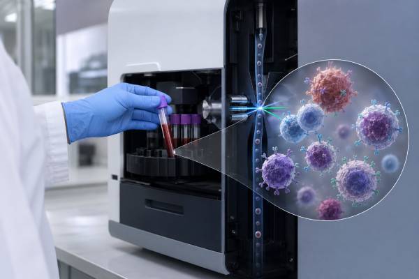
A lymphocyte subset panel uses flow cytometry to count major immune-cell populations in blood, usually CD3 T cells, CD4 helper T cells, CD8 cytotoxic T cells, CD19 or CD20 B cells, and CD16/CD56 natural killer cells. It commonly reports both percentages and absolute counts, along with the CD4/CD8 ratio. The panel helps evaluate suspected immune deficiency, monitor HIV, assess immune reconstitution after transplantation, and follow medicines that deplete or suppress selected lymphocytes. It describes how many circulating cells carry particular marker combinations; it does not prove that those cells function normally, produce effective antibodies, recognize a specific infection, or are active inside tissues. Results change with age, acute illness, stress, corticosteroids, biologic therapy, pregnancy, exercise, time of day, and the total lymphocyte count. A low percentage can coexist with a normal absolute count, and a normal percentage can conceal lymphopenia. Interpretation therefore focuses on the pattern across cell types, the patient’s age and clinical history, and related functional testing—not on a single flagged number or a universal “ideal” ratio.
- CD3 identifies most mature T cells; CD4 and CD8 subdivide major T-cell populations.
- CD19 or CD20 is used for B cells, while CD3-negative CD16/CD56-positive cells define most NK cells.
- Absolute counts and percentages answer different questions and should always be read together.
- Age-specific ranges are essential, especially in infants and young children.
- Normal cell numbers do not guarantee normal lymphocyte function.
Table of Contents
- What the Panel Counts
- How Flow Cytometry Produces the Results
- Why a Lymphocyte Subset Panel Is Ordered
- Absolute Counts, Percentages, and the CD4/CD8 Ratio
- Meaning of Low T-, B-, or NK-Cell Patterns
- Meaning of High Counts and Percentages
- Age, Treatment, and Timing Effects
- Follow-Up Tests and Practical Interpretation
What the Panel Counts
Lymphocytes are white blood cells with different roles in adaptive and innate immunity. A basic panel separates them by surface markers rather than by appearance under a microscope. The exact marker combinations should be stated on the report because no single marker identifies every population by itself.
CD3 is part of the T-cell receptor complex and marks most mature T cells. CD3-positive CD4-positive cells are commonly called helper T cells because they coordinate immune responses, support B-cell antibody production, activate macrophages, and maintain immune memory. CD3-positive CD8-positive cells include cytotoxic T cells that can kill infected or abnormal cells. Small populations may express both or neither CD4 and CD8, so CD4 plus CD8 does not always equal total CD3 exactly.
B cells are usually identified as CD19-positive and CD3-negative; some panels also use CD20. B cells can become plasma cells that secrete antibodies and memory cells that respond to future exposures. Mature plasma cells often lose CD19 or CD20, so a standard B-cell count is not a plasma-cell count and cannot measure antibody production directly.
Natural killer cells are typically CD3-negative and express CD16, CD56, or both. They recognize stressed, infected, or transformed cells without the same antigen-specific receptor rearrangement used by T and B cells. A basic count does not distinguish all NK subsets or evaluate killing, degranulation, or cytokine production.
Some expanded panels add naïve and memory T-cell markers such as CD45RA, CD45RO, CCR7, or CD27; recent thymic emigrant markers; activated T-cell markers; switched-memory B cells; transitional B cells; plasmablasts; or NK subsets. These additions answer more focused questions and require their own reference ranges, controls, and interpretation standards for each age group.
The panel measures cells circulating in peripheral blood. Many lymphocytes reside in lymph nodes, spleen, bone marrow, gut, skin, lungs, and other tissues. Temporary movement between blood and tissue can change the count without changing the body’s total lymphocyte pool. This is why a blood panel can look reassuring while an organ-specific immune process remains active, or look depleted during temporary trafficking without permanent cell loss. Tissue biopsy, imaging, microbiology, and organ-specific testing may be more informative when symptoms are localized.
How Flow Cytometry Produces the Results
Whole blood is mixed with fluorescently labeled antibodies that bind selected markers. Red blood cells are lysed or otherwise excluded, and leukocytes pass one at a time through a flow cytometer. Lasers excite the fluorescent tags, detectors record light scatter and marker intensity, and software groups cells through a process called gating.
CD45 expression and side scatter commonly define the lymphocyte region. Within that gate, combinations such as CD3/CD4, CD3/CD8, CD19/CD3, and CD16/CD56/CD3 distinguish the major populations. Good gating matters because monocytes, debris, dead cells, doublets, and abnormal cells can distort percentages if included. Laboratories also verify that marker patterns are biologically plausible and that enough events were collected for a reliable estimate. Rare populations require more cells than abundant populations; a tiny reported subset based on only a handful of events may have wide uncertainty even when the instrument produces a precise-looking number. Internal controls, compensation for overlapping fluorescent signals, and review by trained personnel are therefore part of the result, not merely technical details.
Absolute counts may be obtained with a single-platform method that uses counting beads or an internal standard. Another approach combines flow-cytometry percentages with an absolute lymphocyte count from a complete blood count. Differences in timing between the CBC and flow sample can affect a calculated absolute result, particularly when counts are changing quickly.
A result may list cells per microliter and percentage of total lymphocytes. The denominator must be understood. CD4 percentage is usually the fraction of lymphocytes or T cells depending on the report; naïve CD4 percentage may be the fraction of CD4 cells. Similar-looking percentages can therefore describe different calculations.
Specimen quality affects accuracy. Clotted blood, delayed transport, extreme temperature, cell degradation, or incorrect anticoagulant can alter marker expression and cell recovery. Laboratories set acceptance limits and may reject an unsuitable sample.
A subset panel is different from leukemia or lymphoma immunophenotyping. The latter uses broader marker combinations, morphology, clinical information, and hematopathologist interpretation to identify abnormal clonal populations. A routine enumeration panel should not be used to diagnose unexplained lymphocytosis or classify a malignancy.
Why a Lymphocyte Subset Panel Is Ordered
Suspected inborn error of immunity is a major indication. Severe, recurrent, unusual, or persistent infections; opportunistic organisms; chronic thrush; poor growth; chronic diarrhea; vaccine complications; autoimmunity; lymphoproliferation; or a family history of immune disease may prompt testing. In infants, an abnormal newborn screen for severe combined immunodeficiency requires urgent T-, B-, and NK-cell phenotyping.
The pattern can narrow the differential diagnosis. Very low T cells with preserved B cells and absent NK cells suggests a different group of defects from low T cells with preserved B and NK cells, or from absent B cells with normal T cells. These T-B-NK patterns guide genetic and functional testing but are not sufficient for a final diagnosis. In suspected severe combined immunodeficiency, clinicians often summarize the phenotype as T-negative, B-positive or B-negative, and NK-positive or NK-negative. That shorthand helps prioritize pathways such as cytokine-receptor signaling, V(D)J recombination, purine metabolism, or thymic development. However, maternal lymphocytes, partial or “leaky” defects, recent transfusion, and treatment can blur the classic categories, so urgent functional and genetic confirmation remains necessary.
HIV monitoring is another use. CD4 count and percentage estimate immune status, while viral load measures treatment control. Modern HIV care generally does not use the total panel at every visit when suppression is stable, but the appropriate schedule depends on current guidelines and the patient’s clinical situation.
Transplant teams follow immune reconstitution after hematopoietic cell transplantation, solid-organ transplantation, or cellular therapy. T, B, and NK cells recover on different timelines, and counts may relate to infection risk, vaccine planning, graft-versus-host disease, and medication adjustment. Donor chimerism and cell function add information that simple counts cannot provide.
Biologic drugs can selectively deplete cell populations. Anti-CD20 therapy can produce very low circulating B cells for months, while antithymocyte globulin, alemtuzumab, chemotherapy, JAK inhibitors, corticosteroids, and other treatments can alter T or NK cells. Monitoring should follow the medicine’s indication and protocol.
The panel may also be used in autoimmune disease, cancer treatment, unexplained lymphopenia, recurrent infections after chemotherapy, or before selected live vaccines in immunocompromised patients. It is not an appropriate general screening test for vague symptoms without a clinical reason.
Absolute Counts, Percentages, and the CD4/CD8 Ratio
Absolute count is the estimated number of cells in a volume of blood. Percentage is the share of the lymphocyte pool represented by that subset. They can move in different directions because the denominator changes.
Suppose the absolute CD4 count remains stable while CD8 cells expand during a viral infection. The CD4 percentage and CD4/CD8 ratio may fall even though CD4 cells were not lost. Conversely, a person with a very low total lymphocyte count can have a normal-looking CD4 percentage but an abnormally low absolute CD4 count.
The CD4/CD8 ratio divides the CD4 count or percentage by the corresponding CD8 value. A low ratio may result from low CD4, high CD8, or both. A high ratio may result from high CD4, low CD8, or both. The component values determine the meaning; the ratio alone cannot show which pattern is present.
Age changes the expected distribution. Infants and young children normally have higher absolute lymphocyte and CD4 counts than adults. Adult reference ranges should not be applied to a baby. Laboratories should report age-specific intervals, and pediatric immunologists may interpret values against developmental stage and vaccination history.
Absolute counts can fluctuate from day to day. Hydration, acute infection, exercise, circadian rhythm, stress hormones, and corticosteroids alter circulating cell traffic. A small isolated deviation in a clinically well person may be less important than a persistent pattern confirmed on repeat testing.
The report should also be checked for internal consistency. CD3 should roughly encompass the major CD4 and CD8 T-cell groups, while T, B, and NK percentages usually account for most lymphocytes. Minor gaps are expected, but large unexplained discrepancies may require review of gating, unusual populations, or specimen quality.
Meaning of Low T-, B-, or NK-Cell Patterns
Low total T cells can occur with severe combined immunodeficiency, thymic-development disorders, combined immune defects, advanced HIV, chemotherapy, transplantation, severe infection, malnutrition, protein loss, corticosteroids, and other immune-suppressing treatments. The urgency depends on age, degree, persistence, naïve T-cell numbers, and infection history.
A low CD4 count is associated with impaired helper function and infection risk in specific settings, especially HIV. Outside HIV, the same number may have different implications. Idiopathic CD4 lymphocytopenia is a diagnosis of exclusion requiring repeated low counts and evaluation for secondary causes.
Low CD8 cells can occur in combined immune defects, treatment effects, marrow disorders, or temporary redistribution. Because CD8 cells are important in antiviral and tumor surveillance, persistent severe reduction may prompt functional and genetic evaluation, but the clinical phenotype remains essential.
Low B cells suggest possibilities such as X-linked agammaglobulinemia, selected combined immunodeficiencies, anti-CD20 therapy, chemotherapy, or post-transplant suppression. Normal B-cell numbers do not guarantee normal immunoglobulin levels or vaccine responses. A B-cell count must be paired with antibody testing.
Low NK cells can occur in severe combined immunodeficiency patterns, GATA2 deficiency, selected genetic defects, marrow failure, viral illness, or treatment effects. A low count does not automatically prove impaired NK function, and a normal count does not rule out a degranulation or cytotoxicity defect.
Pancytopenia or broad lymphopenia points toward a different process from an isolated subset abnormality. Complete blood count, smear, marrow function, infection testing, nutrition, kidney and liver disease, and medication exposure help determine whether the problem is primary or secondary.
Severity and persistence matter more than the direction of a flag alone. A mildly low subset during influenza is not equivalent to an infant with nearly absent naïve T cells, persistent thrush, and poor growth. Similarly, a person receiving a planned B-cell-depleting medicine may have an expected low B-cell count but still need assessment for recurrent infection, falling immunoglobulins, or delayed recovery. Clinicians combine the numeric pattern with the organism history: recurrent encapsulated bacterial infections suggest an antibody problem, opportunistic viral or fungal infections raise concern for T-cell dysfunction, and severe herpesvirus disease can point toward cytotoxic T- or NK-cell defects.
Meaning of High Counts and Percentages
High lymphocyte subset counts often reflect reactive immune expansion. Viral infections can increase CD8 T cells, alter the CD4/CD8 ratio, and expand activated populations. Recovery from infection may leave changes for weeks or months. Symptoms and trends are more informative than a one-time flag.
High B-cell or T-cell counts can also occur with smoking, chronic infection, autoimmune disease, asplenia, medication effects, or clonal lymphoproliferative disorders. A routine panel cannot determine clonality. Persistent unexplained lymphocytosis requires a complete blood count, smear, and appropriate leukemia/lymphoma flow cytometry.
High NK-cell counts may be reactive or persistent. Large granular lymphocyte expansions can involve T cells or NK cells and may be associated with cytopenias, autoimmune disease, or clonal disorders. Classification needs morphology, receptor studies, molecular testing, and hematopathology.
A high percentage may simply reflect a low competing population. For example, B-cell percentage can look high because T cells are reduced while the absolute B-cell count remains normal. Similarly, an elevated CD4/CD8 ratio can result from low CD8 rather than excess CD4.
High counts do not prove “strong immunity.” Expanded cells can be exhausted, dysfunctional, treatment-related, or clonal. Function, diversity, and tissue behavior are not captured by quantity alone.
Age, Treatment, and Timing Effects
Age is the most important physiologic modifier. Newborns, children, adults, and older adults have different absolute counts and naïve-memory distributions. The thymus is most active early in life, so young children normally have many naïve T cells. With age, memory populations accumulate and thymic output declines.
Acute infection can redistribute lymphocytes. Early stress responses may lower circulating counts, while later antiviral responses can expand selected T cells. Sampling during fever may not reflect baseline. Severe infection can cause broad lymphopenia associated with illness severity.
Corticosteroids can rapidly lower circulating lymphocytes through redistribution and other effects. Chemotherapy and radiation suppress marrow and lymphocyte recovery. Anti-CD20 medicines can make CD19/CD20 B cells nearly absent; CD20-based detection may be especially affected by receptor occupancy, so laboratories often use CD19 for monitoring. Other drugs alter where lymphocytes circulate rather than simply destroying them. Sphingosine-1-phosphate receptor modulators can retain lymphocytes in lymphoid tissue and lower blood counts, while some kinase inhibitors or immune therapies shift specific compartments. After B-cell maturation antigen-directed or CD19-directed cellular therapy, the expected pattern may include prolonged B-cell aplasia, low immunoglobulins, or delayed reconstitution. The treatment date, target antigen, and anticipated recovery curve are therefore essential context.
After transplantation, NK cells may recover before T and B cells, while functional recovery and vaccine response lag behind numeric recovery. A normal count does not automatically mean it is safe to stop prophylaxis or receive a live vaccine. Decisions follow transplant protocols and the full immune assessment.
Pregnancy, strenuous exercise, smoking, sleep disruption, and time of day can shift counts modestly. Repeat testing should be performed under comparable conditions when a trend matters.
Follow-Up Tests and Practical Interpretation
A subset panel is a starting map. Follow-up depends on the pattern and clinical question. Basic companion tests include a complete blood count with differential, immunoglobulin G, A, and M, vaccine antibody titers, HIV testing when appropriate, and review of medicines and infection history.
T-cell function can be assessed with a lymphocyte proliferation test, cytokine production, activation markers, or receptor-signaling assays. B-cell function is evaluated through immunoglobulins, specific antibody responses, and expanded B-cell phenotyping. NK-cell evaluation may require a natural killer cell function test or degranulation assay.
Naïve and memory T-cell phenotyping is particularly useful in infants with suspected severe combined immunodeficiency because maternal or expanded memory-like cells can create a misleading total T-cell count. T-cell receptor excision circles, maternal engraftment studies, genetic testing, and proliferation complete the evaluation.
Persistent abnormal counts may lead to targeted gene panels, exome or genome sequencing, bone marrow studies, imaging, or hematopathology. Testing should be selected by phenotype rather than ordering every available immune marker. When lymphocytosis is the problem, the next test is often not another enumeration panel. A peripheral smear and diagnostic immunophenotyping can assess light-chain restriction in B cells, aberrant antigen expression, T-cell receptor patterns, or an expanded large-granular population. Molecular or cytogenetic studies may then establish clonality. This distinction matters because a reactive antiviral expansion and a lymphoid malignancy can both produce a high subset count, but they require very different evidence and management.
Repeat testing is reasonable when a result was obtained during acute illness, soon after corticosteroids, or with questionable specimen quality. The same laboratory and similar timing improve comparability. Repetition should not delay urgent management in an infant or severely ill patient.
The report is best read as a pattern: Which cells are truly low in absolute number? Are percentages distorted by another population? Does age explain the value? Is there a drug designed to deplete that marker? Are counts consistent with function and infection history? These questions prevent both overdiagnosis from mild flags and missed immune deficiency behind normal-looking percentages.
For practical comparison, keep the full report rather than recording only the CD4 number or ratio. Note the collection date, illness status, recent vaccines, steroid exposure, chemotherapy or biologic dosing, and whether the same laboratory and method were used. Trends are most useful when the clinical context is similar. A meaningful recovery may involve rising absolute counts, improving naïve or memory composition, normal immunoglobulins, protective vaccine responses, and fewer infections—not merely movement into a printed reference interval. Conversely, worsening symptoms or opportunistic infection can justify urgent evaluation even when several subset values remain within range.
References
- Diagnostic tests for primary immunodeficiency disorders: Classic and genetic testing 2024 (Review)
- Flow cytometry-based diagnostic approach for inborn errors of immunity: experience from a resource-limited setting 2024 (Research Article)
- Quantitative Lymphocyte Subsets: T, B, and Natural Killer Cells, Blood 2026 (Laboratory Test Guide)
- Lymphocyte Subset Panel 7 – Congenital Immunodeficiencies 2026 (Laboratory Test Guide)
- Primary Immunodeficiency Diseases – Immunoglobulin Disorders 2026 (Clinical Guidance)
- Severe Combined Immunodeficiency 2026 (Clinical Guidance)
Disclaimer
This article is for general education and does not diagnose an immune disorder, HIV, leukemia, or lymphoma. Lymphocyte subset results require age-specific ranges and interpretation with absolute counts, percentages, medicines, symptoms, and functional tests. Infants with an abnormal newborn screen or anyone with severe opportunistic infection need prompt specialist care.





