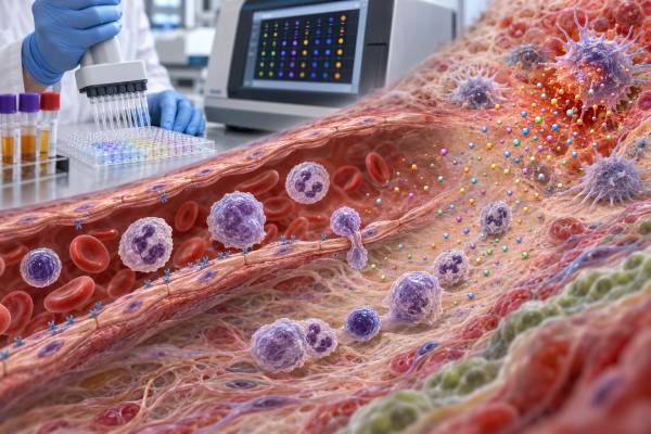
A chemokine panel measures several small immune-signaling proteins that help direct white blood cells toward infection, injury, tumors, or inflamed tissue. Unlike a routine blood count, it does not simply count immune cells. It looks for chemical “traffic signals” such as CXCL8, CXCL9, CXCL10, CCL2, CCL5, and related markers. These tests are used most often in research, specialty immunology, infectious disease, cancer, and selected inflammatory evaluations rather than routine screening. A high result can reflect active immune recruitment, but it does not identify one disease by itself. A low result may be normal, assay-related, treatment-related, or meaningful only in a carefully defined clinical setting. Interpretation depends on which chemokines were measured, the specimen type, timing, laboratory method, symptoms, medications, and other findings. The most useful question is usually not whether one value is “good” or “bad,” but whether the overall pattern fits the suspected immune process.
- Chemokines guide immune-cell movement; they are not the same as cell counts.
- Panels vary widely, so two laboratories may measure different markers.
- High levels usually indicate signaling activity, not a specific diagnosis.
- Serum, plasma, cerebrospinal fluid, and tissue results are not interchangeable.
- Trends and marker combinations are often more informative than one isolated value.
Table of Contents
- What a chemokine panel measures
- Major chemokines and what they signal
- Why the test may be ordered
- How the panel is performed
- How to interpret high, low, and normal results
- How doctors interpret chemokine patterns
- Limitations and sources of variation
- What usually happens after the result
What a chemokine panel measures
A chemokine panel measures a selected group of chemokines in a biological sample. Chemokines are signaling proteins released by immune cells, blood-vessel cells, tissue cells, and sometimes tumor cells. Their main job is chemotaxis: creating chemical gradients that attract particular cells to a location. A neutrophil, monocyte, T cell, eosinophil, or natural killer cell may detect a chemokine through a matching receptor and move toward the strongest signal.
The names can look confusing because two naming systems are still encountered. Modern names describe the arrangement of conserved cysteine amino acids. The major families are CC chemokines, such as CCL2 and CCL5, and CXC chemokines, such as CXCL8 and CXCL10. Less common families include CX3C and XC chemokines. The “L” means ligand. Receptors use “R,” as in CCR2 or CXCR3. Therefore, CCL2 can signal through CCR2, while CXCL9 and CXCL10 commonly signal through CXCR3.
Some markers also have older names. CXCL8 is widely known as interleukin-8, or IL-8. CCL2 is also called monocyte chemoattractant protein-1, or MCP-1. CCL5 is called RANTES in older literature. CXCL12 may be called stromal cell-derived factor-1, or SDF-1. A report may use one or both names.
There is no single universal chemokine panel. One laboratory may offer a small inflammation panel containing four or five targets. Another may use a multiplex platform that reports dozens. A cancer study, transplant program, or neuroinflammation laboratory may select a completely different group. The report should therefore be interpreted according to its exact analyte list, not according to the general label “chemokine panel.”
Chemokines overlap with cytokines, but the terms are not identical. Cytokine is a broad category covering many immune messengers, including interleukins, interferons, colony-stimulating factors, tumor necrosis factors, and chemokines. A cytokine panel may include chemokines, but it may also focus on proteins that activate, suppress, or mature immune cells rather than mainly directing their movement.
Most chemokine tests measure protein concentration, commonly in picograms per milliliter. They do not directly show whether the corresponding receptor is present, whether cells respond normally, or whether the protein is active inside tissue. A blood result is best understood as a partial snapshot of a larger signaling network.
Major chemokines and what they signal
The importance of a marker depends on the cells that produce it, the receptor it activates, and the clinical setting. Several chemokines appear frequently in specialty panels.
CXCL8 (IL-8) strongly attracts and activates neutrophils. It may rise during bacterial infection, tissue injury, acute inflammation, and some cancers. Because neutrophils can release additional inflammatory substances after arriving at a site, CXCL8 may participate in a self-amplifying response. A blood CXCL8 value should be compared with symptoms and, when appropriate, a neutrophil count. The two tests answer different questions: one measures a recruiting signal, while the other counts circulating cells.
CXCL9, CXCL10, and CXCL11 are often associated with interferon-driven immunity and the CXCR3 receptor. They help recruit activated T cells and natural killer cells. CXCL9 and CXCL10 can increase in viral and intracellular bacterial infections, autoimmune inflammation, transplant rejection, and other states with strong interferon signaling. CXCL10 is sometimes called interferon gamma-induced protein 10, or IP-10. These markers can support a biologic pattern, but they do not prove that interferon-gamma is the only cause. An interferon-gamma test, pathogen testing, tissue findings, and clinical context may be needed.
CCL2 (MCP-1) recruits monocytes and related myeloid cells, commonly through CCR2. It has been studied in metabolic disease, vascular inflammation, fibrosis, infection, autoimmune disorders, and cancer. Elevated circulating CCL2 may indicate monocyte-attracting activity, but it does not show where that activity is occurring.
CCL3 and CCL4, formerly called MIP-1 alpha and MIP-1 beta, participate in leukocyte recruitment and can be produced by macrophages, lymphocytes, and other cells. They may appear in panels designed to assess innate and T-cell activation. CCL5 (RANTES) attracts T cells, eosinophils, basophils, and other cells depending on the receptor environment. Platelets can release substantial CCL5, making sample handling especially important.
CCL11 (eotaxin-1) is linked to eosinophil recruitment through CCR3. It may be evaluated in allergy, asthma, eosinophilic disease, aging research, and selected inflammatory studies. A high CCL11 result is not equivalent to a high eosinophil count; the signal can rise before, during, or independently of an obvious blood eosinophilia. It may be compared with an eosinophil count, IgE, symptoms, and organ-specific testing.
CX3CL1 (fractalkine) is unusual because it can exist in a membrane-bound form that helps cells adhere and a soluble form that attracts cells expressing CX3CR1. It has been investigated in vascular disease, kidney disease, neurologic inflammation, autoimmune disorders, and cancer. Its meaning depends heavily on whether the test measures circulating soluble fractalkine or tissue expression.
CXCL12 (SDF-1) helps regulate stem-cell retention and trafficking in bone marrow, immune-cell migration, tissue repair, and blood-vessel biology. It is important physiologically, so an abnormal concentration is not automatically evidence of harmful inflammation. Its effects also vary with receptor expression, local concentration gradients, and tissue conditions.
No marker acts alone. Chemokine receptors can bind more than one ligand, and a ligand may interact with more than one receptor. Some receptors attract cells; atypical chemokine receptors may instead capture, transport, or clear chemokines. This redundancy protects the immune system but makes simple one-marker interpretations unreliable.
Why the test may be ordered
A chemokine panel is usually ordered to answer a focused question, not as a general wellness test. In clinical practice, its use is much narrower than common tests such as a complete blood count or C-reactive protein.
An immunologist or infectious disease specialist may use selected chemokines to investigate immune activation, monitor an unusual inflammatory syndrome, or support research-based classification. CXCL9 and CXCL10, for example, may be studied when an interferon-dominant process is suspected. In some infections, chemokine profiles are being evaluated as biomarkers of disease activity or treatment response, but many proposed uses remain investigational and are not stand-alone diagnostic standards.
In rheumatology and autoinflammatory medicine, panels may be used when conventional markers do not explain the pattern of illness or when a specialist is evaluating a pathway-targeted therapy. The result may help describe whether signaling appears neutrophil-dominant, monocyte-dominant, eosinophilic, or interferon-associated. It cannot replace diagnostic criteria, examination, imaging, or organ-specific tests.
Transplant and cellular-therapy programs may measure chemokines while evaluating rejection, graft-versus-host disease, immune-effector-cell toxicity, or response to immunosuppression. Here, timing matters greatly. A result before treatment, during fever, after steroid therapy, and after clinical recovery may represent four different biologic states.
Cancer centers and research programs may measure chemokines because tumors can recruit suppressive myeloid cells, T cells, or other leukocytes. Chemokines may also relate to angiogenesis, metastasis, and response to immunotherapy. However, most circulating chemokines are not validated cancer-screening tests. A high level should not be interpreted as proof of cancer, and a normal level cannot exclude it.
Neurology teams may analyze chemokines in cerebrospinal fluid rather than blood when investigating central nervous system infection or inflammation. The specimen compartment changes the question. A concentration in spinal fluid reflects signaling near the brain and spinal cord more directly than a blood value, although collection, blood contamination, and local laboratory standards still affect interpretation.
Researchers also use panels to discover biomarkers, divide patients into biologic subgroups, and examine response to vaccines or medications. A test offered by a research laboratory may not have the same validation, reference intervals, regulatory status, or clinical meaning as a routine hospital assay. Patients should ask whether the panel is a clinically validated test, a laboratory-developed test, or research-only measurement.
How the panel is performed
Most panels use blood collected from a vein. The ordering laboratory specifies whether serum or plasma is required and, for plasma, which anticoagulant tube to use. These details matter. Clotting activates platelets and other cells, so serum concentrations can differ substantially from plasma concentrations. A result from an EDTA-plasma assay should not be compared directly with a serum reference value.
Other samples may include cerebrospinal fluid, urine, saliva, bronchoalveolar lavage fluid, synovial fluid, or tissue-culture fluid. These specimens require their own validated methods. A “normal range” from blood cannot be transferred to spinal fluid or urine.
Many laboratories use bead-based multiplex immunoassays. Each bead set carries an antibody for a different chemokine, allowing many proteins to be measured from a small sample. Other platforms use electrochemiluminescence, microfluidics, proximity-extension methods, or single-analyte enzyme-linked immunosorbent assays. Multiplex testing is efficient, but combining many antibody reactions can introduce cross-reactivity, matrix effects, and differences in sensitivity.
The sample may need rapid processing, centrifugation, freezing, and transport on dry ice. Repeated freeze-thaw cycles can alter some proteins. Delayed separation of plasma from blood cells may allow cells to release or consume chemokines after collection. Vigorous exercise, acute stress, a recent infection, vaccination, smoking, meals, circadian rhythm, and medications may influence results. Preparation instructions are assay-specific; fasting is not automatically required unless the laboratory or clinician says so.
The report commonly includes a concentration, a laboratory interval, and flags such as high, low, or below detection. Some research reports provide only raw values or comparison with a study cohort. “Below the lower limit of quantification” does not necessarily mean that no chemokine is present. It means the assay cannot measure it accurately below that threshold.
Turnaround time may be longer than for routine blood tests because samples are sometimes batched. A laboratory may wait until enough specimens are available to run a full plate. Panels sent to a specialty laboratory may also require additional transport and review.
How to interpret high, low, and normal results
A chemokine result should first be checked against the reference information for that exact method, specimen, and laboratory. There is no universal normal range for a broad chemokine panel. Even when two reports use the same units, their antibodies, calibration materials, detection limits, and sample-processing rules may differ.
A high chemokine level usually means increased production, release, or reduced clearance. It may reflect infection, autoimmune activity, tissue injury, allergic inflammation, fibrosis, vascular disease, treatment effects, or cancer-related signaling. The list is broad because chemokines are part of normal immune responses as well as disease. A high CXCL10, for example, supports interferon-associated recruitment but does not distinguish viral infection from autoimmune inflammation on its own.
The degree of elevation can matter, but more is not always proportionally worse. Chemokines act locally in gradients. A modest blood increase can accompany intense tissue production, while a very high circulating level may partly reflect spillover from many sites. Receptor binding, protein degradation, kidney and liver clearance, and treatment can all change the measured concentration.
A low chemokine level is often difficult to interpret. Healthy people may have concentrations near or below an assay’s detection limit. Immunosuppressive drugs, corticosteroids, pathway-targeted biologic therapy, severe immune dysfunction, or timing after an inflammatory peak may reduce a value. Yet a low blood concentration can also occur while a chemokine is concentrated inside tissue. Unless a validated low threshold has a known clinical meaning for the specific application, “low” should not be labeled immune failure.
A normal result means only that the measured value falls within the laboratory’s comparison interval or decision range. It does not rule out localized inflammation, intermittent signaling, receptor abnormalities, or a disease driven by chemokines not included in the panel. It also does not show that immune cells function normally.
When several markers are abnormal, the direction and relationship may be more helpful than any one number. The clinician may ask whether related ligands rise together, whether a chemokine matches the expected cell count, and whether values change with symptoms. Repeating a panel can be useful only when the same specimen type, method, and similar collection conditions are used.
Reference intervals deserve special caution. Some specialty panels establish ranges from a small group of healthy donors. Age, sex, ancestry, pregnancy, obesity, and chronic conditions may be underrepresented. Pediatric values may differ from adult values. A statistical interval is not automatically a disease cutoff, and a result just outside it may reflect normal biologic variation.
How doctors interpret chemokine patterns
Pattern interpretation begins with the clinical question. The same set of numbers can mean different things in a person with fever after transplantation, chronic asthma, suspected tuberculosis, or a tumor receiving immunotherapy.
An interferon-associated pattern may include CXCL9, CXCL10, and sometimes CXCL11. These ligands can accompany activated T-cell and natural killer-cell recruitment. A clinician may compare them with symptoms, pathogen tests, autoantibodies, liver tests, ferritin, lymphocyte subsets, or an activated T-cell marker test. Concordant findings make the pattern more persuasive; discordance may indicate timing, treatment, or a different source of signaling.
A neutrophil-recruiting pattern may feature CXCL8 and related CXC chemokines. It can appear in acute bacterial inflammation, tissue damage, severe viral illness, lung inflammation, and malignancy. The next question is whether neutrophils are elevated, depleted, or trapped in tissue. A high recruiting signal with a low blood neutrophil count can occur in a very different situation from the same signal with marked neutrophilia.
A monocyte or macrophage-recruiting pattern may include CCL2, CCL3, CCL4, or CCL7. These markers may be examined alongside monocyte counts, ferritin, liver findings, tissue pathology, and macrophage-activation indicators. Because the same chemokines participate in repair and chronic disease, persistence and context matter more than a single elevation.
An eosinophilic or type 2 pattern may include CCL11, CCL17, CCL22, or CCL26, depending on the panel. The clinician may compare it with eosinophils, total IgE, allergen testing, lung function, skin findings, or tissue biopsy. A blood pattern can suggest cell recruitment without proving that eosinophils have infiltrated a particular organ.
A vascular or tissue-remodeling pattern may involve CX3CL1, CXCL12, CCL2, and other markers. These signals can be produced by endothelial cells, stromal cells, and injured organs. They may overlap with inflammation, fibrosis, and cancer biology, so they rarely point to one diagnosis.
Doctors also compare chemokines with broad markers. C-reactive protein reflects a liver response to inflammatory signaling, while chemokines describe selected communication pathways. A CRP test may be high when a chemokine panel is unremarkable, or vice versa. Flow cytometry answers another question by identifying immune-cell populations and activation markers. A flow cytometry immune panel can show which cells are present, while chemokines suggest where those cells may be called and which pathways may be active.
The strongest interpretation usually combines four layers: the patient’s symptoms and timing, conventional laboratory findings, cell populations or tissue evidence, and the chemokine pattern. A panel is most useful when it changes the probability of a diagnosis or guides a specific next step.
Limitations and sources of variation
Chemokine panels have substantial preanalytic, analytic, and biologic limitations. These do not make the tests useless, but they explain why results require expert interpretation.
Preanalytic variation happens before the assay begins. Serum versus plasma, tube material, anticoagulant, clotting time, processing delay, centrifugation, storage temperature, and freeze-thaw cycles can all change values. Platelet-derived chemokines are particularly sensitive to clotting and handling. Hemolysis or contamination by blood cells may distort a result.
Analytic variation comes from the assay. Different antibody pairs may recognize different molecular forms. A calibration standard may not behave exactly like the natural protein in a patient sample. One chemokine can interfere with measurement of another, especially in a large multiplex panel. Very high concentrations can occasionally exceed the assay range or create a nonlinear response. Laboratories use dilution and quality controls to reduce these problems, but cross-platform agreement may remain limited.
Biologic variation is often larger than expected. Chemokines can rise rapidly and fall within hours. The blood compartment may not reflect tissue gradients. Kidney or liver dysfunction may alter clearance. Age, pregnancy, body composition, sleep, exercise, infection exposure, and medications can shift baseline concentrations. Steroids and targeted immune drugs may suppress some signals while leaving others active.
Panels can also create false certainty through the sheer number of results. When 30 or 40 markers are measured, one or two may fall outside a statistical reference interval by chance. Looking for a disease explanation for every flagged value can lead to unnecessary tests. The ordering clinician should decide in advance which markers and patterns are clinically meaningful.
Another limitation is validation. A chemokine may be strongly associated with a disease in published research but still lack a standardized clinical cutoff. Association does not prove that the test diagnoses the disease, predicts an individual outcome, or improves treatment decisions. Study populations, assay platforms, and sample timing may differ from the patient’s situation.
Results from direct-to-consumer or research-use-only panels deserve extra caution. Ask whether the laboratory is accredited for clinical testing, whether the assay was validated for the specimen, and whether a specialist can interpret it. Repeating a surprising result with a validated method may be more useful than drawing conclusions from one broad panel.
What usually happens after the result
The next step depends on why the test was ordered. A mildly abnormal value in a stable person may lead only to review of symptoms, recent infections, medications, and collection conditions. A marked or coordinated abnormal pattern in a seriously ill patient may prompt urgent evaluation, but treatment is directed at the underlying condition rather than at the laboratory number alone.
The clinician may confirm conventional markers such as a complete blood count, metabolic panel, liver tests, C-reactive protein, erythrocyte sedimentation rate, ferritin, or cultures. Infection testing may include molecular assays, serology, imaging, or tissue sampling. Autoimmune evaluation may require autoantibodies, complement tests, urinalysis, and organ-specific studies. When abnormal cell populations are suspected, lymphocyte subsets or flow cytometry may be more directly actionable.
A repeat chemokine panel may be considered when the first result conflicts with the clinical picture, when a preanalytic problem is suspected, or when a validated trend is used to monitor therapy. The same laboratory and assay are preferable. The repeat should be timed consistently relative to treatment, fever, infusion, exercise, or other major influences.
Questions worth asking include:
- Which exact chemokines were measured, and why were they chosen?
- Is the test clinically validated for this condition or mainly investigational?
- Was the sample serum, plasma, or another fluid?
- Is the reported range appropriate for the person’s age and specimen?
- Which abnormal values form a meaningful pathway pattern?
- What other test or clinical finding confirms or contradicts that pattern?
- Will the result change diagnosis, treatment, or monitoring?
Seek prompt medical care for severe symptoms such as trouble breathing, confusion, fainting, chest pain, rapidly worsening weakness, persistent high fever, or signs of shock. A chemokine report should never delay assessment of an acutely ill person.
For most people, the value of a chemokine panel lies in careful integration rather than a single flagged number. The panel can map selected immune traffic signals, but it cannot by itself reveal the full location, cause, or consequence of inflammation. The most reliable interpretation connects the pattern to cells, organs, timing, and a focused clinical question.
References
- Atypical chemokine receptors in the immune system 2024 (Review)
- The role of CCL2/CCR2 axis in cancer and inflammation 2024 (Review)
- Interleukin-8: An evolving chemokine 2022 (Review)
- Fractalkine in Health and Disease 2024 (Review)
- Role of chemokine systems in cancer and inflammatory diseases 2022 (Review)
- Plasma chemokines CXCL10 and CXCL9 as potential biomarkers of drug-resistant and drug-sensitive tuberculosis 2023 (Clinical Study)
Disclaimer
This article is for general educational purposes and is not a diagnosis or a substitute for medical care. Chemokine assays, reference intervals, and clinical uses vary substantially, so results should be interpreted by the ordering clinician or an appropriate specialist in the context of symptoms, medications, specimen handling, and other tests. Seek urgent medical help for severe or rapidly worsening symptoms.