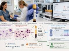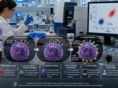
An eosinophil count measures a type of white blood cell involved in allergic inflammation, defense against some parasites, and several immune and blood disorders. It is usually reported as part of a complete blood count with differential. The most useful number is the absolute eosinophil count, or AEC, rather than the percentage alone. Mild elevation is common with asthma, allergic rhinitis, eczema, and medication reactions, but the result is not an allergy test and cannot identify a specific trigger. Higher or persistent counts may require evaluation for tissue-invasive parasites, eosinophilic lung or gastrointestinal disease, vasculitis, adrenal disease, immune disorders, or a hematologic condition. The urgency depends on the level, how long it has been present, symptoms, organ findings, medicines, travel, and prior results. A normal count does not exclude allergic disease or tissue eosinophilia, especially after corticosteroid treatment. The test is most useful as one part of a focused clinical assessment rather than as a stand-alone diagnosis.
- Use the absolute eosinophil count, not only the eosinophil percentage.
- A common adult threshold for eosinophilia is above about 500 cells/µL, but laboratory ranges vary.
- Counts of 1,500 cells/µL or higher are called hypereosinophilia and deserve closer evaluation, especially when persistent.
- Allergy and asthma often cause mild elevation, but marked eosinophilia should not automatically be blamed on atopy.
- Some drugs and tissue-invasive parasites can raise eosinophils, so exposure and travel history matter.
- Chest pain, breathlessness, weakness, neurologic symptoms, fever, or organ dysfunction with high eosinophils require prompt care.
Table of Contents
- What an Eosinophil Count Measures
- Eosinophil Ranges and Thresholds
- Allergy, Asthma, and Eosinophilic Inflammation
- Parasites, Drugs, and Other Reactive Causes
- When High Eosinophils Suggest Systemic Disease
- How Eosinophilia Is Evaluated
- Monitoring, Treatment, and Next Steps
What an Eosinophil Count Measures
Eosinophils are granulocytes made in the bone marrow. They circulate briefly in blood and then move into tissues, especially the gastrointestinal tract and sites of inflammation. Their granules contain eosinophil cationic protein, major basic protein, peroxidase, and other substances that can damage parasites but can also injure human tissue.
A complete blood count with differential reports eosinophils in two ways:
- Eosinophil percentage: the share of measured white blood cells that are eosinophils
- Absolute eosinophil count: the actual number of eosinophils in a volume of blood
The AEC is calculated by multiplying the total white blood cell count by the eosinophil percentage. For example, a white count of 10,000 cells/µL with 8% eosinophils gives an AEC of 800 cells/µL.
The percentage can mislead. If the total white count is low, a relatively high percentage may still produce a normal absolute count. If neutrophils are elevated during infection or corticosteroid use, the eosinophil percentage may look low even when the absolute number is clinically relevant. Interpretation should therefore start with the AEC.
Units may be written as cells/µL, cells/mm³, or ×10⁹/L. A value of 500 cells/µL equals 0.5 ×10⁹/L. Confusing these unit systems can create a thousand-fold error, so the complete report and reference range should be retained.
Counts vary during the day and can be higher in the early morning. Illness, stress hormones, pregnancy, and medicines can also change them. Oral or injected corticosteroids may lower blood eosinophils quickly by changing survival and movement. Inhaled corticosteroids can reduce type 2 airway inflammation and may lower counts over time.
The blood count does not measure eosinophils inside an organ. A patient can have tissue eosinophilia in the esophagus, lungs, skin, or nose with a normal AEC. Conversely, a high blood count does not prove that an organ is being damaged.
The test also differs from an eosinophil cationic protein test. The count estimates cell number, while ECP is intended to reflect release of a granule protein and is more sensitive to sample handling.
Eosinophil Ranges and Thresholds
Reference intervals vary with laboratory, age, population, and method. Many clinicians use an AEC above approximately 500 cells/µL as eosinophilia in adults, but the printed laboratory range takes priority.
A commonly used severity framework is:
| AEC | Description | General interpretation |
|---|---|---|
| Up to about 500 cells/µL | Usually within or near the reference range | Does not exclude allergy, asthma, or tissue eosinophilia |
| 500–1,500 cells/µL | Mild eosinophilia | Often reactive; review allergy, skin disease, medicines, infection, and trend |
| 1,500–5,000 cells/µL | Moderate eosinophilia or hypereosinophilia | Requires explanation, especially if persistent or symptomatic |
| Above 5,000 cells/µL | Severe or marked eosinophilia | Prompt evaluation for systemic, parasitic, inflammatory, or clonal disease |
These labels describe magnitude, not cause or danger. Mild eosinophilia can be important when accompanied by organ symptoms, while a temporary higher count can resolve after a short-lived exposure. The trend and clinical setting matter.
Hypereosinophilia is generally defined as an AEC of at least 1,500 cells/µL. Older definitions required persistence for six months before hypereosinophilic syndrome could be diagnosed. Modern practice does not wait that long when organ damage is possible. Repeated elevation over weeks, or a single marked elevation with compatible organ findings, can justify urgent investigation.
Hypereosinophilic syndrome requires more than a number. It refers to hypereosinophilia associated with organ damage or dysfunction attributable to eosinophils after appropriate evaluation. The heart, lungs, skin, nervous system, gastrointestinal tract, and blood vessels can be affected.
One result should be compared with previous counts. A stable AEC of 650 cells/µL for years in a person with eczema is different from a rise from 100 to 4,000 cells/µL after starting a new medication. The rate of change can point toward the cause and urgency.
The total white count and other cell lines also matter. An isolated eosinophil elevation with otherwise normal blood counts suggests a different range of possibilities from eosinophilia accompanied by anemia, low platelets, abnormal immature cells, or very high total leukocytes. Those additional findings can increase concern for marrow or systemic disease and may speed referral to the appropriate specialist for timely assessment before potentially preventable organ injury develops or progresses over time.
Laboratory flags should be read carefully. Some systems mark a percentage high even when the AEC is normal, while others flag a value that is only slightly above a conservative reference interval. The clinician should verify the arithmetic and units before labeling the patient. A repeat sample is often reasonable for an unexpected mild elevation, but it should not delay evaluation when the count is marked or symptoms suggest organ involvement.
Age matters, especially in infants and young children, whose reference intervals may differ. Pregnancy, smoking, altitude, and population factors may also affect distributions. Do not apply an adult internet cutoff to a child without the laboratory’s pediatric range.
Allergy, Asthma, and Eosinophilic Inflammation
Allergic conditions are among the most common causes of mild eosinophilia. Signals such as interleukin-5 promote eosinophil production and survival, while chemokines draw the cells into tissues.
Allergic rhinitis, asthma, and atopic dermatitis can raise the AEC, particularly during active disease or allergen exposure. However, the count cannot identify whether pollen, mites, pets, food, or another allergen is responsible. Specific IgE or skin testing is needed when a trigger must be established.
A normal count does not exclude allergic disease. Eosinophils may have moved from blood into tissue, the disease may not be eosinophil-dominant, or treatment may suppress the count. Many people with seasonal rhinitis have normal counts outside their active season.
In asthma, blood eosinophils are used as a marker of type 2 inflammation and future exacerbation risk at the population level. Higher values can support an eosinophilic phenotype and help select some biologic medicines in patients with severe uncontrolled disease. The relevant thresholds vary by treatment, clinical trial, regulator, and payer.
A blood eosinophil number does not diagnose asthma. Asthma requires variable respiratory symptoms and objective evidence of variable airflow limitation when possible. Spirometry, bronchodilator response, peak-flow variation, and clinical assessment remain essential.
Counts can fluctuate. Current asthma guidance notes that repeating eosinophils may be useful when severe asthma is being phenotyped, particularly if a previous sample was taken during oral corticosteroid treatment. A low value after steroids may not reflect the patient’s untreated biology.
The same count can have different meaning at different stages of care. In a person with newly diagnosed wheeze, it may support type 2 inflammation but cannot replace confirmation of variable airflow. In someone already taking high-dose inhaled therapy with repeated attacks, it may contribute to biologic selection. In a patient doing well, a mildly elevated value may not require treatment escalation. Clinical purpose must be defined before interpreting the threshold.
Asthma severity is also not proportional to the AEC. Some patients with severe attacks have low counts, while others with higher counts have few symptoms between episodes. Prior intensive-care treatment, frequent oral steroids, low lung function, poor adherence, smoke exposure, and uncontrolled comorbid disease are independent risk factors that should not be overshadowed by the biomarker.
Allergic bronchopulmonary aspergillosis is a specific condition in which asthma or cystic fibrosis coexists with immune reaction to Aspergillus and characteristic clinical findings. Eosinophilia can support the diagnosis, but total IgE, Aspergillus sensitization, imaging, and other criteria are needed.
Eosinophilic granulomatosis with polyangiitis can initially resemble difficult asthma with sinus disease and eosinophilia. Neuropathy, purpura, kidney findings, cardiac disease, constitutional symptoms, or lung infiltrates should prompt broader evaluation rather than simply escalating allergy treatment.
Food allergy can cause eosinophil involvement, but a routine AEC is not a diagnostic food allergy test. Eosinophilic esophagitis and other eosinophilic gastrointestinal diseases require organ-specific assessment, often including endoscopy and biopsy. Blood eosinophilia is neither required nor sufficient.
Parasites, Drugs, and Other Reactive Causes
Parasitic infection
Eosinophilia is classically associated with helminths, or parasitic worms, particularly when larvae or adult organisms migrate through tissue. The likelihood depends on residence, travel, food, water, soil contact, animal exposure, and the parasite’s life cycle.
Not every parasite raises eosinophils. Protozoal infections usually do not produce the same pattern, and some chronic helminth infections may have intermittent or modest counts. A single negative stool test does not exclude tissue-invasive parasites because eggs may not be shed continuously or the organism may not live in the intestine.
Travel and migration history should cover places lived, not only recent holidays. Raw or undercooked meat or freshwater plants, barefoot soil exposure, freshwater swimming, insect bites, and contact with dogs or livestock can guide targeted tests.
Strongyloides deserves special attention before immunosuppressive corticosteroids in people with epidemiologic risk. Steroids can trigger dangerous hyperinfection. Clinicians may order serology and other tests rather than relying on stool examination alone.
Testing should be matched to geography and incubation. Schistosoma exposure follows freshwater contact in endemic regions; Toxocara risk may involve contaminated soil and animal feces; Trichinella follows undercooked meat; and filarial infections have specific insect vectors. A generic “parasite panel” may omit the relevant organism. Infectious-disease or tropical-medicine input is especially valuable when residence and travel history are complex.
Eosinophils may peak during tissue migration and later fall even though infection persists. Conversely, successful treatment can sometimes be followed by temporary changes as antigens are released. The AEC is therefore neither a universal screening test nor a test of cure. Results must be paired with organism-specific evidence and clinical response.
Medication reactions
Many medicines can cause eosinophilia with or without rash. Antibiotics, anticonvulsants, allopurinol, anti-inflammatory drugs, and numerous other agents have been implicated. Over-the-counter products, supplements, and recently stopped drugs should be included.
Drug reaction with eosinophilia and systemic symptoms, or DRESS, is a serious delayed syndrome that may include fever, widespread rash, facial swelling, lymph-node enlargement, hepatitis, kidney injury, lung disease, and blood abnormalities. It usually develops weeks after a drug begins, not within minutes. A high AEC plus systemic symptoms requires urgent medical review.
Other reactive causes
Fungal disease, some bacterial or viral infections, autoimmune conditions, inflammatory bowel disease, adrenal insufficiency, transplant reactions, and certain solid tumors can raise eosinophils. The result is not automatically “allergic” simply because eosinophils participate in allergy.
Recovery from an acute infection can occasionally reveal eosinophilia as other white cells normalize. A repeat count may clarify whether an isolated mild elevation persists.
When High Eosinophils Suggest Systemic Disease
Persistent hypereosinophilia raises concern for organ involvement and for primary or clonal disorders in which eosinophils arise from an abnormal blood-cell population. These conditions are uncommon but important because treatment and risk differ from ordinary atopy.
Possible warning symptoms include:
- New chest pain, shortness of breath, palpitations, fainting, or leg swelling
- Weakness, numbness, foot drop, confusion, or other neurologic changes
- Fever, weight loss, drenching sweats, or marked fatigue
- Widespread rash, facial swelling, blistering, or mucosal involvement
- Persistent cough, low oxygen, or abnormal lung imaging
- Abdominal pain, diarrhea, swallowing difficulty, or unexplained liver abnormalities
- Enlarged lymph nodes or spleen
- Easy bruising, anemia, or other abnormal blood counts
Eosinophil-mediated heart disease can be serious and may not be predicted perfectly by the AEC. Troponin, electrocardiography, echocardiography, or cardiac imaging may be needed when symptoms or marked hypereosinophilia raise concern.
Primary eosinophilic disorders can involve gene rearrangements or mutations that activate growth pathways. A blood smear, vitamin B12, tryptase, molecular tests, bone marrow examination, and hematology assessment may be used when reactive causes are not convincing or other blood abnormalities are present.
Lymphocytic variants involve abnormal T cells that produce eosinophil-promoting cytokines. Skin disease, lymph-node findings, immunophenotyping, and clonality studies can contribute to diagnosis.
The absence of symptoms is reassuring but does not always end evaluation. Persistent AEC at or above 1,500 cells/µL may be monitored for developing organ effects even when an initial workup is negative. Frequency depends on the level, stability, and specialist assessment.
How Eosinophilia Is Evaluated
Evaluation begins by confirming the result. The clinician reviews the full blood count, calculates or verifies the AEC, examines previous results, and may repeat the test. A blood smear can reveal abnormal cells or confirm the automated differential.
The history is often the highest-yield part. It includes:
- Allergic rhinitis, asthma, eczema, and sinus disease
- New prescription drugs, antibiotics, supplements, and injections
- Travel, migration, food, water, soil, and animal exposures
- Fever, rash, weight loss, night sweats, and lymph-node symptoms
- Lung, heart, nerve, gastrointestinal, and urinary symptoms
- Family history of blood or immune disorders
- Corticosteroid or biologic use around the blood draw
The examination looks for skin eruption, wheeze, nasal polyps, lymph nodes, liver or spleen enlargement, edema, neurologic deficits, and signs of organ dysfunction.
Initial laboratory work may include repeat CBC, blood film, kidney and liver tests, inflammatory markers, urinalysis, and tests guided by exposure. Parasite evaluation should be targeted; indiscriminate stool panels may miss the relevant organism or produce confusing findings.
The first repeat should not be treated as a ritual. If the original value was obtained during a clear temporary illness and the patient is well, repeating after recovery may show resolution. If the AEC is at or above 1,500 cells/µL, rising quickly, or paired with organ symptoms, clinicians usually investigate at the same time rather than waiting through multiple routine repeats. The threshold for action is lower when cardiac or neurologic involvement is possible.
Some tests are chosen to separate reactive from clonal eosinophilia. These can include serum tryptase, vitamin B12, immunoglobulins, flow cytometry, molecular studies, and bone marrow examination. They are not needed for every mild count. Their value depends on features such as splenomegaly, abnormal smear, cytopenias, very high counts, or lack of a convincing secondary cause.
Chest imaging, lung function, cardiac tests, endoscopy, biopsy, or specialist studies are chosen from symptoms. There is no single “eosinophilia panel” that safely covers every cause.
Atopy can coexist with another disease. A positive environmental allergy test should not close the evaluation when eosinophils are marked, rising, or accompanied by systemic findings. Likewise, a traveler can have asthma and a parasite at the same time.
Referral may involve allergy/immunology, pulmonology, infectious diseases or tropical medicine, dermatology, gastroenterology, rheumatology, or hematology. The best specialist depends on the dominant clues and degree of elevation.
Monitoring, Treatment, and Next Steps
Treatment targets the cause rather than the laboratory number alone. Mild stable eosinophilia from controlled atopic disease may need only routine follow-up. A drug reaction may require stopping the culprit under medical direction. A parasite requires organism-specific treatment. Severe asthma or a hypereosinophilic syndrome may need corticosteroids, biologic therapy, targeted medication, or other specialist treatment.
Do not start corticosteroids solely because eosinophils are high without considering parasite risk and diagnosis. Steroids can suppress the count and symptoms while obscuring the cause, and they can be dangerous in untreated Strongyloides infection.
For asthma, do not adjust inhalers based only on one AEC. Clinicians consider symptom control, attacks, lung function, inhaler technique, adherence, FeNO, comorbidities, and prior eosinophil values. Biologic eligibility uses specific criteria rather than a general definition of eosinophilia.
When repeating the count, note whether the sample was taken during an infection, allergy season, asthma flare, or steroid course. A trend obtained under similar conditions is easier to interpret.
Ask the clinician these practical questions:
- What is my absolute count and how does it compare with prior results?
- Is the elevation mild, moderate, or marked for this laboratory?
- Could a medicine or supplement be responsible?
- Does my travel or residence create parasite risk?
- Are there signs of organ involvement?
- When should the count be repeated, and what would trigger referral?
Seek urgent assessment when high eosinophils occur with chest pain, severe breathlessness, fainting, neurologic symptoms, fever with widespread rash, facial swelling, jaundice, or rapidly worsening illness. The number alone cannot determine safety.
A well-interpreted eosinophil count is a clue. Its meaning comes from the absolute value, persistence, clinical pattern, and evidence of tissue involvement. That approach avoids both dismissing important hypereosinophilia as “just allergies” and overreacting to a temporary mild elevation.
References
- Global Strategy for Asthma Management and Prevention 2026 (Guideline)
- UK guidelines for the investigation and management of eosinophilia in returning travellers and migrants 2025 (Guideline)
- Evaluation and differential diagnosis of eosinophilia 2025 (Clinical Study)
- The multidisciplinary approach to eosinophilia 2023 (Review)
- Blood eosinophil counts in the general population and airways disease 2022 (Review)
- Approach to the patient with eosinophilia 2020 (Review)
Disclaimer
This article is educational and does not diagnose the cause of eosinophilia. High or persistent eosinophils require interpretation with symptoms, medicines, travel, examination, and other tests by a qualified clinician. Seek urgent care for eosinophilia accompanied by severe breathing, heart, neurologic, rash, or organ symptoms.