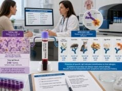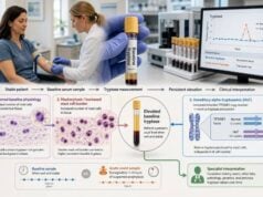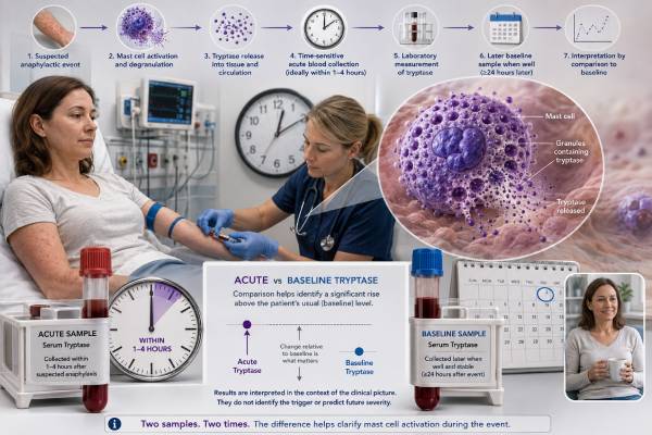
An acute tryptase test is a time-sensitive blood test used to look for evidence that mast cells released their contents during a suspected anaphylactic or severe systemic reaction. The number is most useful when it is compared with the same person’s baseline tryptase level after recovery. A clear rise can support mast cell activation, but a normal result cannot safely rule out anaphylaxis. Food reactions, reactions limited mainly to the skin, and samples collected too early or too late are among the situations in which tryptase may remain within the laboratory range. Because anaphylaxis is diagnosed and treated from symptoms, blood collection must never delay epinephrine or emergency care. The strongest interpretation comes from pairing the event-time sample with a later baseline sample, reviewing the exact timing, and considering the trigger, symptoms, treatment, kidney function, and any condition that can raise baseline tryptase.
- Best timing: Draw acute tryptase as soon as practical, ideally within 1–2 hours after symptoms begin, without delaying emergency treatment.
- Paired testing is essential: Compare the acute value with a baseline sample collected at least 24 hours after complete symptom resolution.
- A significant rise usually means: Acute tryptase is greater than the baseline multiplied by 1.2, plus 2 ng/mL.
- A normal acute level does not exclude anaphylaxis, especially after food-triggered reactions or when collection timing misses the release window.
- A persistently high baseline needs follow-up for hereditary alpha-tryptasemia, mastocytosis, kidney disease, or other causes.
- Emergency symptoms require epinephrine first; tryptase is a supporting test, not a reason to wait before treatment.
Table of Contents
- What an Acute Tryptase Test Measures
- Best Time to Collect an Acute Tryptase Sample
- Paired Samples and the 20% Plus 2 Formula
- What a Normal or Negative Result Means
- Causes of High Acute or Baseline Tryptase
- How Tryptase Fits Into Anaphylaxis Care
- Follow-Up After Acute Tryptase Testing
What an Acute Tryptase Test Measures
Tryptase is a protein enzyme stored mainly inside mast cells. Mast cells sit in the skin, airways, digestive tract, and around blood vessels, where they help coordinate immune responses. When mast cells activate strongly, they can release tryptase along with histamine, prostaglandins, leukotrienes, and other chemical mediators.
An acute tryptase test measures total tryptase in serum during or soon after a suspected reaction. It does not identify the allergen, prove that immunoglobulin E caused the event, or show how severe the next reaction will be. Its main purpose is to document a temporary increase above the person’s usual level.
That distinction is important because a single result can represent two different things:
- An acute rise caused by mast cell activation during anaphylaxis or another systemic event.
- A chronically elevated baseline caused by a higher mast cell burden, extra inherited copies of the alpha-tryptase gene, reduced clearance, or another underlying condition.
A person can have a baseline of 4 ng/mL, 10 ng/mL, or higher and still experience a meaningful event-related rise. Conversely, an acute value that falls inside the laboratory’s reference interval may still be significant if the person’s baseline is much lower. This is why interpretation should focus on change from baseline rather than whether the acute number is merely marked “high.”
Acute tryptase testing is most informative after reactions that include low blood pressure, fainting, cardiovascular collapse, severe breathing problems, or reactions to injected medicines and insect stings. In these settings, mast cell mediator release often produces a measurable rise. The test is less sensitive in some food-triggered reactions and in episodes dominated by hives or swelling without major cardiovascular or respiratory involvement.
Tryptase also cannot separate allergic anaphylaxis from every nonallergic pathway. Certain medicines, physical triggers, or direct mast cell activators can cause clinically similar mediator release. The history remains necessary to determine whether the episode was likely due to food, venom, medication, exercise, temperature, an infusion, or another cause.
For a broader view of the same biomarker outside an emergency episode, a tryptase blood test interpretation considers both acute reactions and persistent elevations. A dedicated baseline tryptase test focuses on the stable value measured after recovery.
Best Time to Collect an Acute Tryptase Sample
Timing strongly affects whether the test captures a rise. Tryptase begins increasing after mast cells degranulate, commonly reaches its highest concentration around 1–2 hours after symptoms begin, and then declines as it is cleared. The exact curve varies with the trigger, severity, treatment, baseline level, and individual biology.
Most current guidance favors collecting the acute sample as early as practical, ideally within 2 hours of symptom onset. A sample taken from roughly 30 minutes to 4 hours may still be useful. Later testing can occasionally detect an elevation after a severe event, but sensitivity falls as the level returns toward baseline.
| Time from symptom onset | Likely value of testing | Practical interpretation |
|---|---|---|
| First 15–30 minutes | May be too early | A normal level can precede the peak; repeat collection may be helpful if clinically feasible. |
| About 30 minutes to 2 hours | Highest-yield window | Often best for detecting a meaningful rise after systemic mast cell activation. |
| About 2–4 hours | Still useful | The level may be falling, but a significant rise can remain detectable. |
| More than 4–6 hours | Lower sensitivity | A normal result may reflect missed timing rather than absence of anaphylaxis. |
| At least 24 hours after complete recovery | Baseline sample | Provides the personal reference needed to judge the acute change. |
The recorded time should refer to the start of symptoms, not the time of arrival at the emergency department. If symptoms began at 2:00 p.m. and blood was drawn at 4:30 p.m., the relevant interval is 2.5 hours. Recording when epinephrine, intravenous fluids, antihistamines, corticosteroids, or other treatments were given also helps reconstruct the event, although treatment should never be withheld to preserve a laboratory signal.
Some protocols obtain more than one event-time specimen, such as one sample as soon as possible and another 1–2 hours later. This can help when the first sample was very early or the symptom onset is uncertain. The later baseline specimen remains necessary even when two acute samples were collected.
Routine fasting is not required. Eating does not meaningfully prepare or invalidate the assay. The practical challenge is access: many reactions occur outside normal laboratory hours, and emergency teams may need a predefined order set or specimen-handling process. A patient with a history of unexplained severe reactions can ask an allergist whether a written emergency plan should mention acute and baseline tryptase collection.
Paired Samples and the 20% Plus 2 Formula
The most widely accepted way to interpret acute tryptase is to compare it with a stable baseline. A significant event-related rise is generally defined as:
Acute tryptase greater than 1.2 × baseline tryptase + 2 ng/mL
This is often called the “20% plus 2” rule. It adjusts for the fact that people start from different baselines. A fixed cutoff such as 11.4 ng/mL would miss some meaningful increases in people with low baselines and could misclassify people whose baseline is naturally above that number.
Examples show how the calculation works:
| Baseline tryptase | Calculated threshold | Example acute result | Does it meet the criterion? |
|---|---|---|---|
| 3 ng/mL | 1.2 × 3 + 2 = 5.6 ng/mL | 7 ng/mL | Yes |
| 8 ng/mL | 1.2 × 8 + 2 = 11.6 ng/mL | 10.5 ng/mL | No |
| 15 ng/mL | 1.2 × 15 + 2 = 20 ng/mL | 24 ng/mL | Yes |
| 25 ng/mL | 1.2 × 25 + 2 = 32 ng/mL | 31 ng/mL | No |
Meeting the formula supports substantial systemic mast cell activation in the right clinical setting. It does not independently prove a specific trigger, and it does not replace the symptom-based diagnosis of anaphylaxis. A rise could also occur with other severe mast cell activation events.
Failing to meet the formula does not erase a convincing clinical episode. Sampling may have occurred outside the optimal window, the reaction may have produced little circulating tryptase, or the baseline may not truly represent the person’s stable level. Baseline samples taken during ongoing symptoms, soon after a second reaction, or during a period of changing health can complicate comparison.
Laboratories may report tryptase in ng/mL or mcg/L. These units are numerically equivalent: 1 ng/mL equals 1 mcg/L. The laboratory’s reference interval may differ by assay, so the report should be interpreted with its own method and range. Serial samples are ideally measured using the same assay platform, particularly when the difference is close to the calculated threshold.
A clinician may also consider the magnitude and pattern of the rise. A dramatic increase from 5 to 50 ng/mL is strongly supportive in a compatible event. A borderline change deserves closer review of timing, analytical variation, symptom severity, and whether the baseline sample was truly obtained after full recovery.
What a Normal or Negative Result Means
A normal acute tryptase does not rule out anaphylaxis. Anaphylaxis remains a clinical diagnosis based on the rapid development of characteristic symptoms, such as airway swelling, wheezing, low blood pressure, collapse, or involvement of more than one body system after a likely trigger.
Several factors can produce a normal or nondiagnostic result:
- Food-triggered anaphylaxis: Tryptase rises less consistently after food reactions than after injected drugs or insect venom.
- Limited mast cell burden or localized release: A reaction may involve enough mediator activity to cause symptoms without creating a large serum increase.
- Early collection: Blood drawn before the level rises may underestimate the peak.
- Late collection: A sample obtained after the level has declined may resemble baseline.
- Milder or skin-predominant events: Hives and swelling alone often produce smaller changes.
- Uncertain baseline: Without a stable comparison sample, a value labeled normal may still represent a meaningful personal increase.
For example, an acute result of 7 ng/mL may look comfortably within a laboratory range that ends near 11 ng/mL. If the later baseline is 2 ng/mL, however, the calculated event threshold is 4.4 ng/mL, so the rise is significant. The reverse can also occur: an acute result of 16 ng/mL may be flagged high, but if the person’s baseline is 15 ng/mL, it does not meet the event-related formula.
A “negative” result also should not be used to deny epinephrine, shorten observation solely on that basis, or dismiss a detailed reaction history. Tryptase is primarily a retrospective supporting test. Clinicians often receive the result after the immediate treatment decisions have already been made.
When recurrent episodes suggest mast cell mediator release but tryptase remains nondiagnostic, an allergist may review whether other testing is appropriate. A mast cell activation test panel may include urine or blood markers, but each has timing, handling, and specificity limits. Results should be matched to clearly documented episodes rather than used as broad screening in people with nonspecific symptoms alone.
Causes of High Acute or Baseline Tryptase
A high event-time tryptase can reflect acute mast cell degranulation, a high baseline, or both. The first step is to repeat the test after complete recovery and compare the values.
High acute value with a lower baseline
This pattern supports a temporary mast cell activation event. Common clinical settings include:
- Anaphylaxis caused by insect venom, medication, food, latex, or another allergen
- Severe perioperative reactions involving anesthetics, antibiotics, antiseptics, or other exposures
- Infusion reactions and direct mast cell activation by certain drugs
- Idiopathic anaphylaxis when no trigger is identified after evaluation
The test does not identify which exposure caused the event. Trigger evaluation may require a detailed timeline, skin testing, specific IgE testing, component testing, medication review, or supervised challenge when appropriate and safe.
High acute value with a high baseline
A person with an elevated baseline can still have an acute rise. The 20% plus 2 formula is designed for this situation. Persistently elevated baseline tryptase may be associated with:
- Hereditary alpha-tryptasemia: Extra copies of the alpha-tryptase–encoding gene can raise baseline levels. Some carriers have no symptoms, while others may have more severe mediator-related reactions.
- Systemic mastocytosis: A clonal mast cell disorder can increase mast cell burden. A baseline above 20 ng/mL is one minor diagnostic criterion, but the number alone neither confirms nor excludes the disease.
- Other myeloid blood disorders: Some hematologic conditions can increase tryptase.
- Chronic kidney disease: Reduced clearance or altered metabolism can contribute to higher concentrations.
- Normal biological variation and assay-related factors: Mild elevations require context and sometimes repeat confirmation.
A stable baseline above the laboratory range should not automatically be labeled mastocytosis. Clinical features such as recurrent anaphylaxis, unexplained fainting, characteristic skin lesions, severe venom reactions, osteoporosis, low-trauma fractures, enlarged organs, abnormal blood counts, or a detectable KIT mutation can change the level of concern. The baseline tryptase evaluation may include repeat testing, hereditary alpha-tryptasemia testing, peripheral blood KIT D816V testing, or referral to a specialist.
Mildly elevated value without symptoms
An isolated modest elevation found during unrelated testing is not evidence of a recent allergic reaction. Tryptase should not be treated like a general inflammation marker. It is more closely tied to mast cell biology, baseline genetics, and certain hematologic or renal conditions. Repeating the measurement when the person is well can confirm whether the finding is persistent.
How Tryptase Fits Into Anaphylaxis Care
Epinephrine is the first-line treatment for suspected anaphylaxis, and a tryptase draw must not delay it. A patient with rapidly worsening breathing difficulty, throat tightness, low blood pressure, collapse, or severe multisystem symptoms needs emergency assessment based on the clinical picture.
Tryptase helps later in several ways:
- It can support that a confusing or undocumented episode involved systemic mast cell activation.
- It may help distinguish anaphylaxis from fainting, panic, isolated asthma, flushing syndromes, or other mimics, although no laboratory result works alone.
- It can prompt evaluation for an elevated baseline or underlying mast cell disorder.
- It can strengthen the investigation of reactions during anesthesia or complex medication exposure.
- It can provide objective evidence when the trigger remains uncertain.
A high tryptase does not grade the reaction in a simple one-to-one way. More severe cardiovascular reactions are more likely to produce a rise, but the number cannot predict the exact clinical course or future risk by itself. A small rise may occur during a serious reaction, and a large rise can occur in someone who responds quickly to treatment.
Emergency care should include documentation of the symptom onset, suspected trigger, vital signs, organ systems involved, treatments and response, and blood collection times. This information may be more useful to the allergist than a laboratory value without context.
After stabilization, patients may need an epinephrine autoinjector, training on when and how to use it, trigger avoidance instructions, and referral for allergy evaluation. People should not wait for hives before using prescribed epinephrine; some severe reactions cause breathing or circulation symptoms without prominent skin findings.
An acute tryptase result also does not diagnose mast cell activation syndrome by itself. Consensus-based diagnosis generally requires recurrent compatible symptoms involving multiple organ systems, objective evidence of mediator release during episodes, and a meaningful response to therapies that target mast cell mediators, while excluding other explanations. Broad symptom lists without event-linked laboratory evidence are not enough. A histamine blood test has an even shorter collection window and additional handling limitations, so it should not be viewed as an easy substitute for properly timed paired tryptase samples.
Follow-Up After Acute Tryptase Testing
The most useful follow-up combines the laboratory result with a detailed reconstruction of the episode. Bring the acute report, baseline report, emergency records, photographs of skin findings, medication list, and a written timeline to the allergy appointment.
A practical review usually includes these steps:
- Confirm the sample times. Note the interval from symptom onset to each blood draw and when symptoms fully resolved.
- Calculate the personal threshold. Multiply baseline by 1.2 and add 2 ng/mL, then compare the acute result.
- Assess the clinical criteria. Review the speed of onset, systems involved, blood pressure, breathing symptoms, skin or gastrointestinal findings, and response to epinephrine.
- Investigate the likely trigger. Consider foods, stings, medicines, exercise, alcohol, infection, heat, or combinations of cofactors.
- Evaluate a persistent baseline elevation. Repeat testing or specialized workup may be appropriate when the stable value remains high.
- Create a prevention and emergency plan. This can include avoidance measures, epinephrine, medical identification, and testing tailored to the suspected exposure.
People with severe insect-sting reactions may need insect venom IgE testing and consideration of venom immunotherapy. Those with a suspected medication reaction may need a carefully selected drug allergy evaluation; commercially available IgE tests exist for only a limited number of drugs and cannot replace specialist assessment.
Seek urgent care for any new episode involving trouble breathing, throat or tongue swelling, fainting, confusion, blue or pale skin, rapidly falling blood pressure, or symptoms affecting multiple organ systems after a likely exposure. Use prescribed epinephrine promptly and call emergency services. Collecting tryptase can be requested after treatment begins, but the laboratory test is never the priority over airway, breathing, circulation, and epinephrine.
For future episodes, a written plan can reduce missed timing. It may specify that emergency clinicians collect an acute serum tryptase without delaying treatment, record the exact draw time, and arrange a baseline sample after recovery. This simple pairing often turns an isolated number into clinically useful evidence.
Keep copies of both reports because acute and baseline samples may be filed under different encounters or health systems. The comparison can be missed when an emergency department result is not visible to the outpatient allergist. If the baseline sample was never collected, it can still be obtained later when the person is well, although a long delay makes careful record matching more important. The result should include the assay value, units, collection date, and collection time rather than only a verbal statement that it was normal or elevated.
References
- Anaphylaxis: A 2023 practice parameter update 2024 (Guideline)
- EAACI guidelines: Anaphylaxis (2021 update) 2022 (Guideline)
- Updated Diagnostic Criteria and Classification of Mast Cell Disorders: A Consensus Proposal 2021 (Position Statement)
- Defining baseline variability of serum tryptase levels improves accuracy in identifying anaphylaxis 2022 (Research Article)
- Tryptase: The Silent Witness of Past and Ongoing Systemic Events 2024 (Review)
- Why the 20% + 2 Tryptase Formula Is a Diagnostic Gold Standard for Severe Systemic Mast Cell Activation and Mast Cell Activation Syndrome 2019 (Review)
Disclaimer
This article is for general education and cannot diagnose anaphylaxis or a mast cell disorder. Suspected anaphylaxis requires immediate epinephrine and emergency care based on symptoms; do not wait for a tryptase result. A qualified clinician should interpret acute and baseline values together with the event timeline, examination, and medical history.