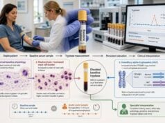
A tryptase blood test measures a protein released mainly by mast cells. The result can answer two different questions, depending on when the sample is drawn. During a sudden reaction, a temporary rise above the person’s usual level can support systemic mast cell activation, including anaphylaxis. When measured on an ordinary, symptom-free day, the baseline value can suggest increased mast cell burden, hereditary alpha-tryptasemia, or another condition that keeps tryptase persistently elevated. These uses should not be mixed. A value that falls within the laboratory range can still represent a meaningful acute rise, while a chronically high value does not prove that a recent episode was anaphylaxis. Timing, comparison with a baseline sample, symptoms, treatment, kidney function, and possible mast cell disease all affect interpretation. Epinephrine and emergency care must never be delayed while waiting for tryptase testing. The test is supporting evidence—not a substitute for recognizing and treating a dangerous reaction.
- For suspected anaphylaxis, draw acute tryptase as early as possible, ideally within 2 hours of symptom onset.
- Obtain a separate baseline sample after recovery, generally at least 24 hours after all symptoms have resolved.
- A significant rise is commonly defined as acute tryptase greater than baseline × 1.2 + 2 ng/mL.
- A normal acute value does not rule out anaphylaxis, especially after food reactions or delayed sampling.
- Persistent baseline elevation can occur with hereditary alpha-tryptasemia, mastocytosis, kidney disease, or myeloid disorders.
- Baseline tryptase above 20 ng/mL is one minor criterion for systemic mastocytosis, not a diagnosis by itself.
Table of Contents
- What the Tryptase Test Measures
- Acute and Baseline Sampling
- Interpreting an Acute Rise
- Causes of High Baseline Tryptase
- Tryptase and Mastocytosis
- Tryptase in Mast Cell Activation Syndrome
- Next Steps After Testing
What the Tryptase Test Measures
Tryptase is an enzyme stored in mast-cell granules. Mast cells live in tissues throughout the body, especially near the skin, airways, digestive tract, blood vessels, and nerves. They help defend against threats and take part in allergic inflammation, wound responses, and other immune functions.
Most routine laboratory assays report total serum tryptase in nanograms per milliliter, written as ng/mL. This total includes immature tryptase forms that mast cells release continuously and mature beta-tryptase released during degranulation. The test does not separately report each form in ordinary clinical practice.
That biology creates two distinct signals:
- Baseline serum tryptase mainly reflects steady release from the body’s mast-cell population and inherited tryptase production.
- Acute serum tryptase can rise when many mast cells activate over a short period.
A single number cannot reliably tell which signal is present. The collection time and a comparison sample are essential. For example, 10 ng/mL might be a normal baseline for one person, a substantial acute increase for someone whose baseline is 3 ng/mL, or a falling value if the peak was missed.
Tryptase is more stable in blood than histamine. Histamine rises and falls quickly, often before a patient reaches a clinic. Tryptase generally becomes detectable within about 30 to 60 minutes, peaks around 1 to 2 hours, and declines with a half-life near 2 hours. This wider window makes it the preferred blood marker for documenting systemic mast-cell involvement after many suspected anaphylactic events.
The test has important limits. Basophils contribute little to routine serum tryptase, and not every allergic pathway releases enough tryptase to produce a measurable rise. Food-triggered reactions, reactions dominated by breathing or skin symptoms, and less severe episodes may yield a normal result. A high result also does not identify the trigger or prove an IgE mechanism. Drugs, physical factors, and non-IgE pathways can activate mast cells.
The blood draw itself is simple. Fasting is usually unnecessary, and antihistamines do not remove tryptase from serum. Treatment should not be withheld for the sake of testing. The laboratory’s collection instructions still matter: serum should be separated from red blood cells promptly because prolonged contact can reduce analyte stability. Recording the exact symptom onset and draw time makes the result far more useful.
Acute and Baseline Sampling
The most informative anaphylaxis evaluation uses paired samples rather than one isolated result. One sample captures the event; the other establishes the person’s ordinary level.
Acute sample: Draw as early as practical after symptoms begin, ideally within 2 hours. A sample obtained between roughly 30 minutes and 2 hours has the best chance of capturing the rise. Testing may remain useful for several hours, but sensitivity falls as tryptase returns toward baseline.
Baseline sample: Draw when the person is well and the reaction has completely resolved, generally at least 24 hours later. Some clinicians obtain it days or weeks afterward during follow-up. The sample should represent the usual state rather than a prolonged or recurrent episode.
When feasible, save the following details with the laboratory record:
- time the first symptom started;
- time and nature of the likely exposure;
- blood-draw time;
- blood pressure and objective respiratory findings;
- skin, gastrointestinal, neurologic, or cardiovascular symptoms;
- epinephrine doses and timing;
- intravenous fluids, antihistamines, bronchodilators, or corticosteroids;
- whether symptoms recurred.
These details help distinguish a properly timed normal value from a sample collected too late. They also permit interpretation when the acute number is below the laboratory’s upper limit but still meaningfully above baseline.
| Sample | Suggested timing | Main purpose | Common problem |
|---|---|---|---|
| Acute | As soon as possible, ideally within 2 hours | Detect a temporary event-related rise | Drawn after the peak has passed |
| Later acute | Several hours after onset | May still support activation if elevated | A normal value becomes less informative |
| Baseline | At least 24 hours after full recovery | Establish the person’s usual level | Collected while symptoms are continuing |
| Repeat baseline | On another well day when needed | Confirm persistent elevation or assess variability | Different assay or changing kidney function |
A missed acute sample does not invalidate the clinical diagnosis. Anaphylaxis is diagnosed from the rapid pattern of symptoms and signs, not from a laboratory requirement. If a person develops throat tightness, breathing difficulty, faintness, low blood pressure, or rapidly progressive multisystem symptoms after a likely exposure, treatment takes priority. Drawing blood should never postpone intramuscular epinephrine, airway support, fluids, or emergency transport.
For planned procedures with elevated anaphylaxis risk—such as an operation in someone with a previous perioperative reaction—the team may document a baseline in advance. If another event occurs, this provides an immediate comparator. Hospitals can also build paired-tryptase instructions into anaphylaxis protocols so the second sample is not forgotten after discharge.
Interpreting an Acute Rise
The laboratory’s population cutoff is not the best way to judge an acute event. The preferred comparison asks whether the event value rose enough above that individual’s baseline.
The widely used consensus formula is:
Significant acute tryptase = greater than baseline × 1.2 + 2 ng/mL
Suppose baseline tryptase is 5 ng/mL. The threshold is 5 × 1.2 + 2, or 8 ng/mL. An acute result above 8 supports systemic mast-cell activation even if the laboratory flags only values above 11.5 ng/mL.
If baseline is 20 ng/mL, the threshold is 26 ng/mL. An acute result of 23 would be above many reference ranges but would not meet the event-related formula. It might reflect the person’s persistently high baseline rather than a demonstrable acute increase.
The formula supplies biochemical support; it does not diagnose anaphylaxis by itself. Interpretation still requires a compatible episode, such as sudden symptoms involving the airway, circulation, skin, or gastrointestinal tract. A rise can also occur during non-IgE mast-cell activation, allergen challenge, or some drug reactions. The test confirms activation more readily than it identifies the cause.
A negative result has several explanations:
- the sample was collected before tryptase rose or after it fell;
- the reaction released little tryptase;
- the trigger was food, which often produces smaller increases than insect-sting or perioperative reactions;
- the episode was localized rather than systemic;
- the symptoms came from a non-mast-cell condition;
- the true baseline was not measured correctly.
Therefore, “normal tryptase” should not be translated into “no anaphylaxis.” Clinical criteria and response to treatment remain central. Conversely, receiving epinephrine and improving does not prove anaphylaxis, because epinephrine can improve several forms of shock or airway symptoms.
The absolute height of the peak is not a reliable severity forecast for future reactions. Marked elevations are more common with hypotension and extensive systemic activation, but trigger, route, dose, medications, cardiovascular disease, asthma, and treatment delay also shape severity. A person with a small or absent rise can still have a dangerous reaction.
Serial measurements occasionally help in prolonged or perioperative events, but routine repeated draws every hour are not necessary for most patients. The useful pair is an appropriately timed event sample and a true baseline. Laboratories and clinicians should use the same units and, when practical, the same assay platform for comparison.
Causes of High Baseline Tryptase
A persistently elevated baseline result points to a different question from an acute rise. It may reflect inherited tryptase copy number, increased mast-cell burden, reduced clearance, or a hematologic condition.
Reference limits differ by laboratory. Many reports flag results at approximately 11.4 or 11.5 ng/mL, while mast-cell experts have proposed that a broader healthy baseline interval of about 1 to 15 ng/mL can include many people with hereditary alpha-tryptasemia. The printed reference interval should be used, but a value should not trigger or end an evaluation without context.
Common and clinically important causes include:
Hereditary alpha-tryptasemia. This autosomal dominant genetic trait results from extra alpha-tryptase–encoding copies of the TPSAB1 gene. It is present in roughly 5% to 7% of sampled European and North American populations. Many carriers are healthy. Others report flushing, itching, gastrointestinal symptoms, connective-tissue complaints, or severe reactions, although the strength and specificity of these associations vary. The trait can also modify anaphylaxis risk when it coexists with venom allergy or mastocytosis.
Systemic or cutaneous mastocytosis. Clonal mast cells accumulate in skin, bone marrow, or other organs. Baseline tryptase often rises with mast-cell burden, but indolent disease can occur with a value below 20 ng/mL. A high value supports evaluation; it neither confirms nor excludes the disease.
Myeloid neoplasms. Some leukemias, myelodysplastic or myeloproliferative disorders, and related conditions can raise tryptase. Abnormal blood counts, enlarged spleen, constitutional symptoms, or other findings guide this investigation.
Reduced kidney function. Chronic kidney disease can produce persistent elevation. Kidney function should be reviewed before assuming a mast-cell disorder.
Recent severe mast-cell activation. A supposedly “baseline” sample drawn too soon after an event may still be falling. Repeating the level when the person has been well can prevent misclassification.
Less common associations include helminth infection and marked eosinophilic disease. Laboratory variation also matters. Baseline tryptase has some natural fluctuation, especially at higher levels, so a small change should not be treated as proof of progression.
The pattern around the number determines the next step. An otherwise healthy person with stable mild elevation may need only confirmation and consideration of TPSAB1 copy-number testing. A patient with recurrent unexplained hypotension, severe insect-sting anaphylaxis, typical skin lesions, abnormal blood counts, bone pain, organ enlargement, or unexplained osteoporosis deserves a more directed mast-cell evaluation.
Tryptase and Mastocytosis
Systemic mastocytosis is a clonal disorder in which abnormal mast cells accumulate in one or more organs, most often bone marrow. Symptoms may come from mediator release—flushing, itching, abdominal cramping, diarrhea, low blood pressure, or anaphylaxis—or from organ infiltration in more advanced disease.
A baseline serum tryptase level above 20 ng/mL is one minor diagnostic criterion in current mastocytosis classifications. It is not the sole criterion, and it must be handled carefully when another myeloid neoplasm or hereditary alpha-tryptasemia is present. A value of 25 ng/mL cannot be used to diagnose systemic mastocytosis without the rest of the evaluation.
Diagnostic assessment may include:
- examination for typical brown-red skin lesions and a dermatology evaluation when present;
- complete blood count, liver tests, kidney function, and other targeted laboratory studies;
- sensitive testing for the KIT D816V mutation in peripheral blood;
- TPSAB1 copy-number testing when hereditary alpha-tryptasemia is plausible;
- bone density assessment when unexplained osteoporosis or fractures occur;
- abdominal examination or imaging when organ enlargement is suspected;
- bone marrow biopsy when the clinical probability is sufficient.
Most adults with systemic mastocytosis carry an activating KIT D816V mutation, but a negative peripheral-blood result does not absolutely exclude disease if the method lacks sensitivity or the mast-cell burden is low. Bone marrow examination evaluates cell clusters, morphology, immunophenotype, molecular findings, and other classification criteria.
Several clinical patterns raise suspicion even when baseline tryptase is not dramatically high. Severe insect-sting anaphylaxis with low blood pressure and little or no hives is one. Recurrent unexplained anaphylaxis is another. Typical skin lesions, unexplained osteoporosis in a younger adult, elevated alkaline phosphatase with bone symptoms, or persistent gastrointestinal and systemic findings may also prompt assessment.
Hereditary alpha-tryptasemia and systemic mastocytosis can coexist. In that situation, the inherited increase can inflate the measured baseline. Current diagnostic frameworks allow adjustment of tryptase for the extra gene copies when deciding whether the greater-than-20-ng/mL minor criterion is met. This calculation belongs in specialist care; dividing by an arbitrary number without confirmed genotyping can mislead.
Tryptase trends can contribute to monitoring diagnosed mastocytosis, especially when interpreted alongside symptoms, blood counts, organ findings, and treatment. A rising value is not automatically progression, and a stable value does not capture every clinical change. The disease subtype and full assessment determine prognosis and therapy.
Tryptase in Mast Cell Activation Syndrome
Mast cell activation syndrome, or MCAS, is not diagnosed by chronic symptoms alone or by one high baseline result. Consensus approaches require three elements: typical episodic symptoms involving at least two organ systems, objective evidence of mast-cell mediator release during an episode, and improvement with treatment that blocks mast-cell mediators or their effects.
Examples of compatible episodes include sudden flushing or hives together with wheezing, throat symptoms, cramping, diarrhea, faintness, or low blood pressure. Chronic fatigue, headache, pain, brain fog, or heart-rate changes may be real and disabling, but they are too nonspecific to establish MCAS without characteristic episodes and biochemical support.
For serum tryptase, the baseline × 1.2 + 2 ng/mL rule is the usual objective criterion. The sample must be timed correctly. A persistently high baseline may suggest hereditary alpha-tryptasemia or mastocytosis, but it does not document that mast cells caused a particular spell.
Other mediator tests—such as urinary N-methylhistamine, leukotriene E4, or a prostaglandin D2 metabolite—may be considered when tryptase is not captured or remains normal. These tests have their own collection windows, medication effects, and reference limitations. Mild isolated abnormalities should not be stacked together to create a diagnosis without a convincing clinical pattern.
MCAS can be classified according to the underlying setting. A clonal form is associated with mastocytosis or another clonal mast-cell condition. A secondary form occurs when a known allergy or other stimulus repeatedly activates otherwise normal mast cells. Idiopathic MCAS is considered only after relevant triggers and clonal disease have been evaluated.
Overdiagnosis can lead to extensive food restriction, unnecessary medications, fear of ordinary exposures, and missed alternative diagnoses. Underdiagnosis can leave recurrent anaphylaxis untreated. A balanced evaluation considers mimics such as vasovagal episodes, arrhythmia, asthma, panic attacks, endocrine tumors, hereditary angioedema, medication effects, autonomic disorders, and gastrointestinal disease.
A normal baseline does not exclude MCAS, because the criterion depends on an event-related rise. Likewise, a baseline above 8 or 11.5 ng/mL does not prove MCAS. The distinction between activation and mast-cell burden should remain explicit throughout the workup.
Next Steps After Testing
The appropriate response depends on whether the result came from an acute event or a symptom-free baseline.
After a suspected anaphylactic reaction:
- Document the symptoms, suspected trigger, timing, and treatment.
- Confirm whether the acute sample was drawn within a useful window.
- Arrange a baseline tryptase after full recovery if one is not already available.
- Calculate the event-related threshold using the paired values.
- Evaluate the likely trigger with history-directed skin testing, specific IgE, component testing, or supervised challenge when appropriate.
- Provide an emergency plan and epinephrine autoinjectors when the clinical risk warrants them.
After an elevated baseline result:
- Confirm that the sample was obtained when well and repeat it if timing or laboratory error is uncertain.
- Review kidney function, complete blood count, medications, skin findings, infection history, and prior reactions.
- Ask about severe venom reactions, recurrent unexplained fainting or hypotension, osteoporosis, fractures, flushing, and family patterns.
- Consider TPSAB1 copy-number testing, sensitive KIT D816V testing, or referral to allergy, immunology, hematology, or a mast-cell specialist based on the pattern.
- Avoid assuming that every symptom comes from the tryptase value.
Emergency symptoms require action regardless of the test. Use epinephrine promptly for suspected anaphylaxis according to the person’s action plan, and seek urgent medical care for breathing difficulty, throat swelling, collapse, severe low blood pressure, or rapidly progressing multisystem symptoms. Antihistamines can reduce itching and hives but do not replace epinephrine for airway or circulatory involvement.
Questions worth asking the clinician include:
- Was this an acute sample or a true baseline?
- What time was it drawn relative to symptom onset?
- Does the acute value exceed baseline × 1.2 + 2 ng/mL?
- Could kidney disease or hereditary alpha-tryptasemia explain the baseline?
- Does my history justify KIT testing or bone marrow evaluation?
- What trigger testing is needed?
- What should I do if another episode occurs, and where can tryptase be drawn quickly?
Do not use a home symptom flare, an online calculator, or a single borderline laboratory value to self-diagnose mastocytosis or MCAS. The value of tryptase comes from pairing the right sample with the right clinical question: temporary change for activation, stable baseline for mast-cell burden, and specialist evaluation when the two do not fit a simple explanation.
References
- Anaphylaxis: A 2023 practice parameter update 2024 (Guideline)
- Diagnostic Significance of Tryptase for Suspected Mast Cell Disorders 2023 (Review)
- Using the Right Criteria for MCAS 2024 (Review)
- Review and Updates on Systemic Mastocytosis and Related Entities 2023 (Review)
- Hereditary alpha tryptasemia and clinical implications 2025 (Review)
- TRYPT – Overview: Tryptase, Serum 2026 (Official Laboratory Guide)
Disclaimer
This article provides general information and cannot diagnose anaphylaxis, mast cell activation syndrome, hereditary alpha-tryptasemia, or mastocytosis. Suspected anaphylaxis requires immediate treatment based on symptoms; do not wait for a tryptase result before using epinephrine or seeking emergency care. Interpret acute and baseline values with a qualified clinician who can account for timing, laboratory method, and the full medical history.