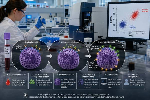
The basophil activation test, or BAT, is a specialized blood test that exposes a patient’s living basophils to a suspected allergen in the laboratory. Flow cytometry then measures whether the cells switch on activation markers such as CD63 or CD203c. This makes BAT a functional test: it asks whether allergen-bound IgE can activate the person’s basophils, rather than merely measuring whether specific IgE is present. BAT can help clarify selected food, drug, insect venom, and other immediate allergies when history, skin testing, and serum IgE do not agree. It may reduce the need for a risky challenge in some patients, but availability and standardization remain limited. A positive result supports clinically relevant sensitization only when it matches the reaction history. A negative result can be inconclusive if the sample was delayed, the allergen preparation was unsuitable, treatment suppressed cell responses, or the person’s basophils did not respond to laboratory controls.
- BAT measures living basophil responses to an allergen, usually by flow-cytometric detection of CD63, CD203c, or both.
- The blood sample must be processed quickly, commonly the same day and generally within 24 hours of collection.
- A positive test can support immediate IgE-mediated allergy, but laboratory cutoffs differ by allergen and testing center.
- CD63 mainly reflects degranulation, while CD203c rises with activation, and the markers are not interchangeable.
- A valid negative result requires working positive controls; nonresponding basophils can make the test uninterpretable.
- BAT may estimate reaction threshold or likelihood in selected allergies, but it cannot guarantee the severity of a future reaction.
Table of Contents
- How the Basophil Activation Test Works
- CD63, CD203c, and Other Test Readouts
- When BAT Is Used in Allergy Evaluation
- Interpreting Positive, Negative, and Invalid Results
- What BAT Can Say About Reaction Risk and Severity
- Sample Timing, Medications, and Limitations
- How BAT Fits With Other Allergy Tests
How the Basophil Activation Test Works
Basophils are rare white blood cells that carry high-affinity IgE receptors on their surface. In an IgE-mediated allergy, allergen molecules can link adjacent IgE antibodies and trigger the cell to release histamine and other mediators. BAT reproduces part of this process outside the body.
A laboratory usually follows these steps:
- Collect fresh venous blood into the required anticoagulant tube.
- Incubate small portions of the sample with several concentrations of the suspected allergen.
- Run a negative control without allergen to measure spontaneous activation.
- Run one or more positive controls that should activate responsive basophils.
- Stain the cells with fluorescent antibodies that identify basophils and detect activation markers.
- Analyze thousands of cells by flow cytometry and calculate the activated percentage or dose-response pattern.
Basophils may be identified with combinations such as CCR3, CD123 with exclusion of HLA-DR, CRTH2, or other laboratory-specific markers. The gating strategy matters because basophils make up less than 1% of circulating white blood cells and can be confused with other cell types if identification is poor.
The allergen is tested at several concentrations because too little may fail to activate sensitized cells, while too much can produce nonspecific effects or a declining response at the highest dose. A concentration curve is often more informative than one tube.
Sample preparation can use whole blood or isolated leukocytes. Whole-blood methods preserve the person’s serum factors and reduce handling, while washed-cell methods can help research specific mechanisms but may alter the response. Laboratories also choose whether to add interleukin-3, a priming cytokine that can strengthen activation. These technical choices affect the cutoff, so a result is valid only within the protocol that generated it.
The number of basophils collected is another quality issue. Because these cells are rare, a sample may contain too few events for a stable percentage, especially in a person with basopenia or after cell loss during processing. Reporting 50% activation from only a handful of cells is less reliable than the same percentage from several hundred well-identified basophils. Laboratories set minimum event counts and may reject or qualify a sparse sample.
The negative control shows the person’s baseline activation in the tube. If spontaneous activation is already high, a small allergen-related increase may be difficult to interpret. The positive control checks whether the cells are capable of responding. Common controls stimulate the IgE receptor pathway or activate basophils through a different receptor.
BAT differs from a specific IgE blood test. Specific IgE detects antibody binding to an allergen; BAT tests whether that recognition produces a cellular response under the assay conditions. This functional step can improve specificity in some clinical settings.
CD63, CD203c, and Other Test Readouts
CD63 and CD203c are the two most familiar BAT activation markers, but they describe related rather than identical cell events.
CD63 is found largely on membranes of intracellular granules. When a basophil degranulates and those granules fuse with the cell surface, CD63 becomes strongly detectable on the outer membrane. It is therefore commonly treated as a marker of anaphylactic degranulation and histamine release.
CD203c is present at a low level on resting basophils and rises rapidly after activation. It can increase even when full degranulation is limited. Some assays use CD203c both to help identify basophils and to measure activation, while others rely on a separate identification panel.
| Measurement | What it represents | Interpretive caution |
|---|---|---|
| Percentage CD63-positive basophils | Proportion showing substantial degranulation after stimulation | Cutoffs vary by allergen, concentration, and protocol. |
| CD203c increase | Upregulation of an activation marker relative to resting cells | May respond differently from CD63 and cannot be converted directly. |
| Stimulation index | Allergen response divided by or compared with the negative control | A large ratio can be misleading when the denominator is extremely low. |
| Basophil reactivity | Maximum proportion of activated cells | Describes response size under test conditions, not guaranteed clinical severity. |
| Basophil sensitivity | Allergen concentration needed to trigger a defined response | Requires a reliable dose-response curve and laboratory-specific calculation. |
Some laboratories report a single positive or negative interpretation. Others provide the percentage activated at each dose, an area under the dose-response curve, EC50, CD-sens, or a combined score. These outputs should be interpreted using validation data for the same allergen, marker, instrument settings, and patient population.
A CD63 cutoff validated for peanut cannot simply be applied to a drug or venom. Even for the same allergen, extracts, purified components, incubation time, and marker panels can change performance. This is one reason BAT remains concentrated in specialist and academic laboratories.
CD63 and CD203c can disagree. A weak stimulus may increase CD203c without causing much CD63 expression, while a strong degranulation response may make CD63 the clearer marker. Some allergens perform better with one endpoint than the other. Using both can add information, but it also requires separate validation and does not automatically make the test more accurate.
Results may be expressed after subtracting the negative-control percentage, as a stimulation index, or through a receiver-operating-characteristic cutoff selected in a validation cohort. These calculations are not interchangeable. A patient should therefore avoid comparing a “15% positive” report from one center with a “stimulation index 2.0” report from another as if they use the same scale.
When BAT Is Used in Allergy Evaluation
BAT is most helpful when it can resolve a specific diagnostic uncertainty and change management. It is not a general screening test for vague symptoms.
Food allergy
In selected food allergies, BAT can distinguish clinically reactive patients from people who have positive skin or IgE tests but tolerate the food. Evidence is strongest for several common foods, including peanut, cow’s milk, egg, and some tree nuts or plant foods. Current European food-allergy guidance allows BAT in specialist settings when standard testing leaves uncertainty and before proceeding to an oral food challenge in appropriate cases.
For example, a child may have peanut-specific IgE and a positive skin test but no clear ingestion history. A strongly positive BAT can increase confidence that a challenge would be high risk, while a valid negative BAT may support proceeding to a carefully supervised challenge. The exact decision still depends on age, components, asthma, prior reactions, and local test performance.
Drug allergy
BAT is used for selected immediate reactions to beta-lactam antibiotics, neuromuscular blocking agents, radiocontrast media, biologics, and other drugs. It can be valuable when skin testing is unavailable, unsafe, poorly validated, or negative despite a convincing history.
Drug BAT is technically difficult because the relevant antigen may be a metabolite rather than the parent drug, and some drugs directly activate or damage cells at high concentrations. A negative result cannot routinely clear a patient to take the medicine. Drug provocation remains the reference procedure in many situations, when the expected benefit outweighs the risk.
Insect venom allergy
BAT may help when a patient has a convincing sting reaction but skin and serum venom IgE results are negative, or when tests are positive to both honeybee and vespid venoms and the culprit is uncertain. Component testing and inhibition studies may also be used. An insect venom IgE test is more widely available, while BAT is generally reserved for complex cases.
Other uses
Specialist centers may use BAT for latex, occupational allergens, vaccines, excipients, chronic urticaria research, monitoring immunotherapy, or evaluating unusual reactions to biologic cancer treatments. Evidence and clinical cutoffs vary widely. A technically possible assay is not necessarily a clinically validated one.
Interpreting Positive, Negative, and Invalid Results
A positive BAT means the tested allergen activated the patient’s basophils above the laboratory threshold. In the setting of an immediate reaction to that allergen, it supports functional IgE-mediated hypersensitivity.
It does not prove that every future exposure will cause symptoms. Nonspecific activation, an unsuitable high allergen concentration, cross-reactivity, or a laboratory cutoff that performs poorly in the relevant population can produce false-positive results.
A negative BAT can mean several things:
- The patient is not clinically allergic to the tested allergen.
- IgE sensitization is present but does not activate basophils under the test conditions.
- The wrong allergen, component, drug concentration, or metabolite was tested.
- The blood was too old or poorly transported.
- Medication or biologic therapy suppressed the cellular response.
- The reaction occurred through a mechanism BAT does not measure.
- The patient is a basophil nonresponder.
A nonresponder has little or no activation with the positive control that uses the IgE receptor pathway. In that situation, a negative allergen response cannot be trusted. Some basophils may still respond to a non-IgE control, which confirms cell viability but not a functional IgE signaling pathway. Laboratories should report whether controls passed rather than issuing a simple negative label.
| Allergen response | Controls | Likely interpretation |
|---|---|---|
| Positive | Valid | Supports functional sensitization if the exposure history matches. |
| Negative | Valid | Lowers the probability of allergy, but sensitivity depends on the allergen and protocol. |
| Negative | IgE positive control fails | Inconclusive or nonresponder; allergy cannot be excluded. |
| High resting activation | Variable | May be uninterpretable because the negative-control background is excessive. |
A report should identify the allergen and concentrations, marker used, definition of positivity, background activation, positive-control response, and overall interpretation. A bare percentage without the laboratory’s cutoff and controls is difficult to use.
Borderline results deserve particular caution. If activation barely crosses the cutoff at one concentration but not at neighboring doses, the pattern may reflect noise rather than a convincing dose response. A consistent rise across several concentrations, followed by a plateau or expected high-dose decline, is usually more biologically persuasive. The laboratory may classify such findings as equivocal rather than forcing them into positive or negative categories.
Clinical probability changes the meaning of the same result. A borderline positive BAT after a documented immediate reaction to the tested drug is more supportive than the same result in a person who has never received that drug. Conversely, a valid negative test has greater reassurance when the original history was already weak than when the patient had objectively documented anaphylaxis.
What BAT Can Say About Reaction Risk and Severity
BAT can provide more information about functional reactivity than antibody concentration alone. In some food-allergy studies, stronger maximum activation has correlated with reaction severity, while greater basophil sensitivity has correlated with reacting to a smaller dose. These are useful group-level patterns and may improve risk stratification in selected clinics.
They do not create a reliable personal forecast. Reaction severity changes with dose, food preparation, exercise, alcohol, infection, asthma control, sleep, medications, and timing of epinephrine. A person with modest BAT activation can still have anaphylaxis, and a highly reactive laboratory pattern does not mean every exposure will be severe.
BAT should not be used to decide that someone can leave epinephrine at home, intentionally eat a suspected allergen, or undergo an unsupervised challenge. It also cannot specify the exact amount that will trigger symptoms outside the controlled assay.
The phrase “reaction risk” can refer to different questions:
- Probability of clinical allergy: BAT may improve discrimination between allergy and asymptomatic sensitization.
- Threshold dose: A dose-response curve may relate to how easily basophils activate, but clinical threshold prediction remains imperfect.
- Potential severity: Maximum activation may correlate with severity in some allergens and populations, but individual prediction is limited.
- Change during treatment: Reduced activation may occur during oral immunotherapy or biologic treatment, yet it does not always equal permanent tolerance.
A treatment can temporarily suppress basophil function while the underlying allergy remains. Challenge decisions after immunotherapy require a protocol designed to distinguish desensitization from sustained unresponsiveness; BAT alone cannot make that distinction.
Sample Timing, Medications, and Limitations
Freshness is a defining requirement. Basophils lose responsiveness after blood collection, so the sample generally needs controlled transport and processing within hours, usually no later than 24 hours. Weekends, long-distance shipping, temperature extremes, and delayed courier delivery can invalidate the test.
The patient does not usually need to fast. Standard H1 antihistamines act at the histamine receptor in the body and generally do not prevent laboratory basophil activation, so they are less problematic than for skin testing. However, the testing center should review all medicines.
Omalizumab reduces free IgE and IgE-receptor expression and can markedly suppress BAT responses. Allergen immunotherapy, oral immunotherapy, systemic corticosteroids, calcineurin inhibitors, and other immune treatments may also influence results depending on dose and timing. These therapies should not be stopped without medical direction merely to obtain a test.
Recent anaphylaxis may temporarily alter basophil responsiveness in some patients. The optimal waiting period is not fully standardized and may depend on the allergen and clinical urgency. The laboratory or allergist should advise on timing after a major reaction.
Other limitations include:
- No universally standardized protocol across laboratories
- Limited access and higher cost than routine specific IgE
- Need for experienced flow-cytometry staff
- Allergen extracts or drug preparations that differ between centers
- A subset of patients whose basophils do not respond through the IgE pathway
- Insufficient validation for many rare allergens and drugs
- Possible loss of sensitivity over time after a remote reaction
Because BAT uses the patient’s cells, it is an ex vivo assay and does not expose the patient to the allergen. The blood draw itself has ordinary minor risks such as bruising or lightheadedness, but the laboratory stimulation does not cause an allergic reaction in the patient.
Preanalytical details should be confirmed before collection. The testing center may require a particular tube type, room-temperature transport, a morning courier, or arrival before a stated cutoff. Refrigeration can impair some protocols, while excessive heat and vigorous shaking can activate or damage cells. Drawing the sample at a local laboratory that routinely ships only overnight may not be suitable even when the test appears on its order menu.
A recent complete blood count can be useful when basophils are extremely low, but routine basophil count does not predict whether the BAT will be positive. The assay evaluates cell function, not simply the number of basophils in circulation.
How BAT Fits With Other Allergy Tests
BAT is a complementary test positioned between sensitization tests and direct challenge. Its value is highest when those approaches point in different directions.
| Test | What it measures | Main strength | Main limitation |
|---|---|---|---|
| Serum specific IgE | Circulating allergen-binding IgE | Widely available and stable sample | Shows sensitization, not necessarily clinical reactivity |
| Skin-prick test | IgE-dependent skin response | Rapid and sensitive | Affected by skin condition, medicines, and extract quality |
| Basophil activation test | Functional activation of circulating basophils | Can improve specificity and avoid some challenges | Fresh sample, specialist laboratory, nonresponders |
| Supervised challenge | Clinical response to controlled exposure | Most direct answer to current reactivity | Time, cost, anxiety, and risk of an allergic reaction |
A sensible sequence starts with the history, then uses skin or blood IgE testing selected for the suspected trigger. BAT is added when it has validated performance and could change whether a challenge, avoidance plan, or immunotherapy is recommended. A broad BAT panel without a clinical hypothesis can create the same false-positive problems as broad IgE screening.
For food allergy, a focused food IgE evaluation and component testing are usually more accessible first steps. For suspected medication allergy, the relevant drug, reaction timing, and known test validation determine whether BAT is worth pursuing.
Patients should ask whether the laboratory is accredited for the assay, how quickly the sample must arrive, which controls are reported, and whether the chosen allergen has clinically validated cutoffs. The result should be reviewed by an allergist who can integrate it with exposure history and decide whether a supervised challenge remains necessary. Ask for the complete report rather than a verbal positive or negative summary. The most useful document includes raw activation percentages, control performance, tested concentrations, marker choice, and the laboratory’s interpretation. Keeping that report is important because BAT methods evolve, and a future specialist may need to understand exactly how the original conclusion was reached. It also helps prevent repeating an expensive test when the original controls and protocol were already adequate for clinical interpretation and follow-up.
References
- Basophil activation test; User’s manual 2025 (Review)
- Multiplex basophil activation tests for allergy diagnosis 2025 (Review)
- Basophil activation test: How many basophils are enough? 2024 (Review)
- The Role of the Basophil Activation Test in the Diagnosis of Drug Allergy 2024 (Review)
- EAACI guidelines on the diagnosis of IgE-mediated food allergy 2023 (Guideline)
- Basophil Activation Test for Allergy Diagnosis 2021 (Review)
Disclaimer
This article is for general education and does not diagnose or exclude an allergy. BAT results require a validated laboratory, successful controls, and specialist interpretation with the reaction history and other tests. Never test a suspected allergen by deliberate exposure at home after a serious reaction.