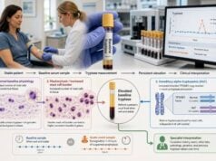
A mast cell activation test panel uses blood and urine markers to look for objective evidence that mast cells released inflammatory mediators during a symptomatic episode. The panel may include serum tryptase, plasma histamine, urinary N-methylhistamine, urinary prostaglandin D2 metabolites, and urinary leukotriene E4. These tests do not form one universal, standardized panel, and no single result diagnoses mast cell activation syndrome. The strongest evidence usually comes from a properly timed acute tryptase compared with the person’s stable baseline, supported when appropriate by urine mediator measurements collected near the same flare. Results must be matched to sudden, recurrent symptoms involving more than one body system and assessed against other explanations such as allergy, asthma, medication reactions, endocrine disorders, infection, and systemic mastocytosis. The testing process works best when planned before the next episode, because specimen timing, handling, medications, and incomplete urine collection can change the findings.
- A mast cell panel is a group of complementary tests, not one standardized laboratory package.
- Acute serum tryptase is most useful when drawn about 30 minutes to 4 hours after symptoms begin, then compared with a later baseline.
- The commonly used event threshold is baseline × 1.2 + 2 ng/mL, rather than one fixed “high” cutoff for everyone.
- Urinary N-methylhistamine, prostaglandin metabolites, and leukotriene E4 may support mediator release, especially when collected within several hours of a flare.
- Normal tests do not exclude every mast cell reaction, but chronic symptoms without event-related objective elevation do not establish MCAS.
- Anaphylaxis symptoms require immediate epinephrine and emergency care; testing must never delay treatment.
Table of Contents
- What a Mast Cell Activation Panel Is
- Symptoms and Diagnostic Context
- Tryptase, Histamine, Prostaglandin, and Leukotriene Markers
- How to Time Blood and Urine Collection
- How Clinicians Read Result Patterns
- MCAS, Mastocytosis, Allergy, and Related Diagnoses
- Common Testing Problems and Misinterpretations
- Building a Follow-Up Plan
What a Mast Cell Activation Panel Is
“Mast cell activation panel” is a practical name for several tests that examine different mediators or their metabolites. Laboratories may bundle some of them, but there is no single internationally standardized panel with identical specimens, cutoffs, and diagnostic performance. A clinician may order only tryptase, a combination of acute and baseline tryptase, urine markers, or a larger set based on the symptoms and local test availability.
Mast cells live in tissues near blood vessels, nerves, skin surfaces, airways, and the gastrointestinal tract. They store tryptase, histamine, and other substances in granules and can also produce prostaglandins and leukotrienes after activation. When released in a controlled way, these mediators help defend the body and coordinate healing. Sudden widespread release can cause hives, flushing, airway narrowing, abdominal cramping, low blood pressure, and anaphylaxis.
The tests do not count mast cells directly. They answer two different questions:
- Was there mediator release during this event? Acute tryptase and event-related urine markers address activation.
- Is there evidence of increased mast cell burden or a clonal disorder? Persistently elevated baseline tryptase, KIT mutation testing, bone marrow findings, and cell-surface markers help address this separate issue.
These questions overlap but are not interchangeable. A patient can have anaphylaxis with a normal baseline tryptase. A patient can also have a high baseline tryptase from hereditary alpha-tryptasemia or systemic mastocytosis without showing a large increase during every symptom episode.
A broad panel is not automatically more accurate. Each marker has a collection window, preanalytical weakness, and limited specificity. Ordering tests without a symptom timeline can produce isolated abnormal values that are difficult to use. The panel becomes clinically meaningful when a clinician defines the suspected episode, chooses the right specimens, and obtains a stable baseline for comparison. A written collection plan also reduces missed windows during stressful, fast-moving reactions.
Symptoms and Diagnostic Context
Mast cell activation syndrome is considered when severe, recurrent episodes resemble systemic allergic reactions and affect at least two organ systems. Symptoms may include skin flushing or hives, swelling, wheezing, throat symptoms, vomiting, diarrhea, abdominal cramping, rapid pulse, low blood pressure, dizziness, or fainting. The attacks should be distinct enough to identify a beginning, peak, and recovery rather than consisting only of constant nonspecific discomfort.
Common symptom combinations include:
- hives plus wheezing;
- flushing plus diarrhea and lightheadedness;
- abdominal cramping plus throat tightness;
- swelling plus low blood pressure;
- sudden nasal, skin, gastrointestinal, and cardiovascular symptoms after a trigger.
Many conditions can mimic parts of this pattern. Chronic urticaria can cause frequent hives without systemic mast cell activation. Panic attacks can cause rapid heart rate, shaking, and breathlessness. Postural tachycardia, medication effects, menopause, carcinoid syndrome, thyroid disease, pheochromocytoma, infection, food intolerance, and inflammatory bowel disease can create overlapping complaints. The laboratory panel does not replace this differential diagnosis.
Consensus-based evaluation generally looks for three elements: compatible recurrent systemic episodes, an objective rise in a validated mediator during symptoms, and improvement with treatments that block mast cell mediators or activation. Treatment response supports the picture but is not specific; antihistamines can improve ordinary urticaria, and leukotriene blockers can improve asthma without proving MCAS.
A single chronic symptom—fatigue, headache, brain fog, bloating, or itching—does not by itself define systemic mast cell activation. These symptoms may still deserve care, but labeling them as MCAS without episodic multisystem findings and objective evidence can delay the search for another cause.
The immediate priority during a severe episode is treatment. Epinephrine is first-line for anaphylaxis. Blood collection can follow when it is safe and feasible, but no diagnostic sample is worth delaying emergency action.
Tryptase, Histamine, Prostaglandin, and Leukotriene Markers
Each marker reflects a different part of mast cell biology. Their strengths and weaknesses explain why results can disagree.
| Marker | Typical specimen | What it reflects | Important limitation |
|---|---|---|---|
| Tryptase | Serum | Release from mast cell granules and baseline mast cell-related traits | May remain normal in some reactions; timing and baseline comparison are essential |
| Histamine | Plasma | Rapid histamine release | Very short collection window and demanding specimen handling |
| N-methylhistamine | Random or 24-hour urine | Histamine metabolism over a longer interval | Can be affected by diet, other cells, and laboratory-specific instructions |
| 2,3-dinor-11β-prostaglandin F2α | Random or 24-hour urine | A stable metabolite related to prostaglandin D2 production | Not specific to one disease and can be influenced by medicines |
| Leukotriene E4 | Random or 24-hour urine | Cysteinyl leukotriene production | Also rises in asthma, AERD, and eosinophilic inflammation |
Serum tryptase
Tryptase is the most established biomarker for severe systemic mast cell activation. An acute sample is compared with the person’s baseline. The widely used threshold is an acute level greater than baseline × 1.2 + 2 ng/mL. For a baseline of 5 ng/mL, the event threshold is above 8 ng/mL. For a baseline of 20 ng/mL, it is above 26 ng/mL. This individualized formula recognizes that a meaningful rise depends on where the person started.
A laboratory upper reference limit—often around 11 to 15 ng/mL depending on the assay—is not the same as the event formula. An acute value can remain inside the population reference range yet meet the person-specific threshold. A value can also be chronically above the reference range without proving that the current symptoms represent activation.
Histamine and N-methylhistamine
Histamine causes itching, hives, flushing, nasal symptoms, airway effects, abdominal symptoms, and blood-vessel dilation. Plasma histamine rises and falls quickly. The sample usually needs prompt collection, cooling, centrifugation, and freezing according to strict laboratory instructions. Tourniquet time, difficult phlebotomy, and handling can affect accuracy.
N-methylhistamine is a urinary metabolite and offers a longer sampling window. A random specimen obtained near symptoms may be compared with baseline, or a 24-hour collection may be used. A dedicated N-methylhistamine urine test is more practical than plasma histamine in many settings, but it still is not fully specific to mast cells.
Prostaglandin D2 pathway
Activated mast cells can generate prostaglandin D2, which contributes to flushing, blood-vessel changes, airway effects, and inflammation. Direct prostaglandin D2 measurement is technically difficult, so laboratories often measure a stable urinary metabolite such as 2,3-dinor-11β-prostaglandin F2α. Names differ across laboratory catalogs, which can lead to ordering the wrong analyte. A prostaglandin D2 pathway test should be confirmed by exact specimen and metabolite name.
Leukotriene E4
LTE4 is the stable urinary end product of cysteinyl leukotriene production. These mediators narrow airways, increase mucus, and promote swelling. Urinary LTE4 may rise in mast cell activation, but also in asthma and aspirin-exacerbated respiratory disease. A result near or above a common cutoff such as 104 pg/mg creatinine must be interpreted using the performing laboratory’s range.
How to Time Blood and Urine Collection
Timing often determines whether the panel answers the clinical question. A normal sample drawn when the patient is well cannot show what happened during a short episode. An acute sample without a baseline may also be difficult to interpret.
Acute tryptase plan
The usual plan is to draw serum tryptase as soon as practical after symptoms begin, ideally within about 30 minutes to 2 hours and generally no later than 4 hours when possible. Tryptase often peaks around one to two hours and then declines. A second sample may be obtained if the first timing is uncertain.
The baseline sample is collected after complete recovery, commonly at least 24 hours later and often on a separate symptom-free day. The same laboratory and assay are preferable. The tryptase blood test report should be interpreted as a pair, not as two unrelated numbers.
Urine mediator plan
Random urine samples for N-methylhistamine, prostaglandin metabolites, and LTE4 are often collected within three to six hours of symptom onset. Some laboratories use 24-hour collections started near a flare. The ordering instructions control which container, storage temperature, preservative, and reporting method apply.
A useful home plan, created with the clinical team, may include:
- Written criteria for when an episode is severe enough to collect.
- The location of the nearest laboratory and its hours.
- Standing orders if appropriate.
- Correct urine containers stored as instructed.
- A list of emergency actions that always come before sampling.
- A symptom and medication log.
- A scheduled baseline collection during a stable period.
For a 24-hour urine collection, the first morning void is usually discarded, then every void is collected for the next 24 hours, including the final sample at the ending time. Missing urine or collecting for the wrong duration can invalidate the study. Random specimens are easier, but an overly delayed sample may miss the event.
Do not stop antihistamines, mast cell stabilizers, steroids, aspirin, NSAIDs, leukotriene modifiers, or other medicines unless the clinician gives a safe, test-specific instruction. Continuing treatment may affect a result, but stopping treatment can provoke symptoms or create risk. Documentation is often safer and more informative than unsupervised withdrawal.
How Clinicians Read Result Patterns
The panel is interpreted as a pattern across symptoms, timing, baseline values, and technical quality. There is no score in which a certain number of positive markers automatically equals MCAS.
A clear acute tryptase rise with a compatible systemic episode provides strong evidence of mast cell activation. The trigger may still be IgE-mediated allergy, a drug reaction, venom, a clonal mast cell disorder, or an idiopathic event. The biomarker confirms activation, not its cause.
Normal tryptase with elevated event-related urine mediators may provide supportive evidence, especially when collection was well timed and the increase is substantial compared with baseline. The clinician asks whether asthma, AERD, eosinophilic inflammation, diet, medicines, or another process better explains the urinary result.
High baseline tryptase without an event-related rise points toward a baseline trait or increased mast cell burden rather than proof that the tested episode involved activation. Causes include hereditary alpha-tryptasemia, systemic mastocytosis, some myeloid disorders, and reduced kidney function. Baseline elevation may prompt repeat testing, TPSAB1 copy-number evaluation, KIT D816V testing, or hematology assessment depending on the value and clinical picture.
One mildly abnormal urine marker during a symptom-free period is weak evidence. It may justify repeating the same marker during a representative episode under controlled conditions, but it should not be treated as a definitive diagnosis.
All markers normal during a correctly timed severe event lowers the likelihood that systemic mast cell activation explains the episode. It does not make symptoms imaginary; it redirects the investigation toward other mechanisms. A normal result from a late or mishandled specimen is less informative.
Serial results should use the same laboratory whenever possible. A numerical change can reflect a new method, different unit, urine dilution, or specimen type rather than a biological shift. For urine tests, event-to-baseline ratios are being studied and may be helpful, but universally validated diagnostic cutoffs are not as established as the tryptase formula.
MCAS, Mastocytosis, Allergy, and Related Diagnoses
Mast cell activation is an event. MCAS is a syndrome defined by recurrent systemic activation meeting clinical, laboratory, and response criteria. Mastocytosis is a clonal disease involving abnormal accumulation of mast cells. Allergy is an immune mechanism that can activate otherwise normal mast cells. A patient may have one, more than one, or none of these conditions.
Secondary mast cell activation
Food, venom, medication, or environmental allergy can produce the same mediators measured in a mast cell panel. If a reaction consistently follows a specific exposure, targeted allergy testing and history may identify the cause. In this situation, mediator elevation confirms the reaction pathway but does not make the condition idiopathic MCAS.
Primary or clonal disease
Systemic mastocytosis is evaluated through criteria that include bone marrow or tissue findings, abnormal mast cell markers, KIT mutations, and baseline tryptase, among other features. A persistently high tryptase, severe venom anaphylaxis, unexplained fainting episodes, characteristic skin lesions, enlarged organs, abnormal blood counts, or osteoporosis may increase suspicion. A mediator panel alone cannot diagnose or exclude systemic mastocytosis.
Hereditary alpha-tryptasemia
Extra copies of the alpha-tryptase–encoding TPSAB1 gene can raise baseline tryptase. This inherited trait is relatively common and may modify the risk or severity of mediator symptoms in some people. It is not the same as systemic mastocytosis, and it does not mean every symptom is caused by mast cells.
Idiopathic MCAS
Idiopathic MCAS is considered when episodes meet accepted criteria but no allergic, clonal, or other trigger is found. “Idiopathic” should follow a structured evaluation; it should not be used as a shortcut for unexplained chronic symptoms.
The distinctions matter because management differs. Trigger avoidance and epinephrine planning are central for anaphylaxis. Clonal disease may require hematology-directed assessment and treatment. Asthma or chronic urticaria has its own evidence-based pathway. Accurate classification prevents both unnecessary restriction and missed risk.
Common Testing Problems and Misinterpretations
The most frequent failure is collecting only a baseline panel while the patient is well. That can identify persistent abnormalities but cannot document a transient rise. The opposite error is collecting only during symptoms and having no baseline for comparison.
Other common problems include:
- drawing tryptase too late;
- using a population upper limit instead of the individualized event formula;
- assuming normal baseline tryptase rules out anaphylaxis;
- assuming elevated baseline tryptase proves MCAS;
- ordering direct prostaglandin D2 when the laboratory’s validated test is a metabolite;
- using the wrong urine preservative or storage temperature;
- missing part of a 24-hour collection;
- comparing a random urine ratio with a 24-hour total as though they are the same test;
- stopping medicines without medical supervision;
- treating a low-level abnormality as disease despite no compatible episodes;
- using symptom improvement with antihistamines as the only diagnostic evidence.
Plasma histamine deserves particular caution. It has a short half-life and can be affected by collection and processing. A result may look precise while reflecting preanalytical error. The laboratory must receive and handle the sample under its validated protocol.
Food and supplement restriction before urine testing is another area of inconsistency. Some laboratories provide specific lists; others do not. Follow the performing laboratory’s current instructions. Broad self-imposed restriction can be burdensome and may not improve validity.
Commercial “mast cell panels” marketed directly to consumers may include unvalidated markers or interpretations that exceed the evidence. A result should be traceable to a clinically accredited laboratory, a named method, a specimen type, and a reference interval. Testing that cannot explain these basics is difficult to use safely.
Building a Follow-Up Plan
A useful follow-up plan begins with the exact episodes that need explanation. Write down the first symptom, progression, organ systems involved, suspected triggers, treatment, and recovery time. Photographs, emergency records, ambulance notes, anesthesia records, and medication timelines can be as important as the panel.
After testing, ask:
- Were samples collected inside the intended window?
- Was a stable baseline obtained?
- Did acute tryptase meet baseline × 1.2 + 2 ng/mL?
- Were urine results creatinine-adjusted and technically adequate?
- Could asthma, AERD, allergy, infection, kidney disease, or medicine effects explain the pattern?
- Does persistent baseline tryptase need a clonal disorder or hereditary alpha-tryptasemia evaluation?
- Which diagnosis best explains the full episode rather than one laboratory value?
- How will the result change emergency planning or long-term treatment?
Referral to an allergist or clinical immunologist is appropriate for recurrent anaphylaxis-like episodes, difficult result interpretation, or suspected MCAS. Hematology may be involved when clonal mast cell disease is suspected. Pulmonology, dermatology, gastroenterology, endocrinology, or cardiology may be needed when another organ-specific disorder fits better.
Treatment is individualized and may include trigger avoidance, H1 or H2 antihistamines, leukotriene modifiers, mast cell stabilizers, asthma therapy, or other targeted medicines. A response can support the diagnosis but should be judged against objective goals such as fewer attacks, less epinephrine use, improved breathing, or reduced emergency visits—not only a vague sense that “something helped.”
People at risk for anaphylaxis should have an emergency action plan and epinephrine if prescribed. The best panel is one that supports a safer, more accurate plan. It should narrow the diagnosis, not become a permanent search for abnormal numbers.
References
- Biomarkers in the diagnosis of mast cell activation 2024 (Review)
- Mast cell activation syndrome: Current understanding and research needs 2024 (Review)
- Using the Right Criteria for MCAS 2024 (Review)
- Diagnosis and management of mast cell activation syndrome (MCAS) in Canada: a practical approach 2025 (Review)
- Diagnostic Significance of Tryptase for Suspected Mast Cell Disorders 2023 (Review)
- Review and Updates on Systemic Mastocytosis and Related Entities 2023 (Review)
Disclaimer
This article provides general information about mast cell mediator testing and is not a diagnosis or personal testing plan. Collection timing, reference ranges, and medication instructions must come from the ordering clinician and performing laboratory. Suspected anaphylaxis requires immediate epinephrine when prescribed and emergency care; do not delay treatment to collect samples.