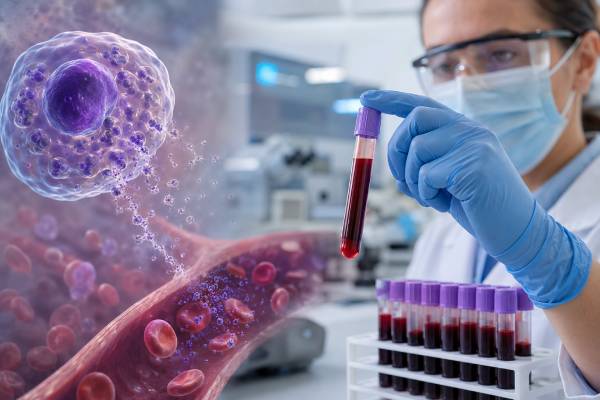
A histamine blood test measures histamine in plasma, whole blood, or another specified blood fraction. Histamine is a short-lived signaling molecule released by mast cells and basophils during allergic reactions, but it is also made in the stomach, nervous system, and other tissues. In a severe immediate reaction, plasma histamine can rise within minutes and then return toward baseline quickly. That narrow window, combined with demanding collection and handling requirements, makes the test difficult to use. A high value may support mediator release when the sample was collected correctly during compatible symptoms, but it does not identify the trigger and does not diagnose mast cell activation syndrome by itself. Basophils, diet, microbes, medicines, and sample processing can affect some measurements. A normal result is common if collection is delayed and does not rule out anaphylaxis or mast cell activation. Acute and baseline tryptase, and selected urinary mediator metabolites, are generally more established tools for evaluating systemic mast cell activation.
- Histamine disappears from plasma rapidly, with a half-life of only minutes, so timing is critical.
- Plasma, serum, and whole-blood histamine are not interchangeable and require different reference ranges.
- A high result is not specific for mast cells because basophils also store and release histamine.
- A normal result does not rule out anaphylaxis, allergy, or mast cell activation.
- No universally validated blood-histamine cutoff confirms mast cell activation syndrome.
- Tryptase and selected urinary mediator metabolites are usually more useful when collected through a planned protocol.
Table of Contents
- What Histamine Is and What the Test Measures
- Timing, Specimen Type, and Sample Handling
- Why Blood Histamine May Be High
- Histamine in Anaphylaxis and Immediate Allergy
- Histamine and Mast Cell Activation Syndrome
- Related Tests and Common Misinterpretations
- Planning Testing and Next Steps
What Histamine Is and What the Test Measures
Histamine is produced from the amino acid histidine by histidine decarboxylase. It acts through several receptor types and has roles in allergic inflammation, stomach acid secretion, wakefulness, nerve signaling, blood-vessel tone, and immune responses.
Mast cells store histamine in granules within tissues, especially near skin, blood vessels, airways, and the gastrointestinal tract. Basophils are related circulating white blood cells that also contain histamine. When IgE or another activating pathway triggers these cells, granules release histamine and many other mediators.
Histamine can cause itching, hives, flushing, swelling, nasal symptoms, bronchospasm, intestinal cramping, increased vascular permeability, and low blood pressure. These effects overlap with symptoms caused by leukotrienes, prostaglandins, platelet-activating factor, tryptase-associated pathways, and other mediators. Symptoms cannot be assigned to histamine alone from appearance.
The phrase “histamine blood test” can refer to different measurements:
- Plasma histamine: free histamine in the liquid portion of anticoagulated blood
- Whole-blood histamine: histamine in plasma plus blood cells, especially basophils
- Serum histamine: measured after clotting, which can permit cellular release and is method-dependent
These tests answer different questions. Plasma histamine is the most conceptually relevant to a brief systemic release event. Whole-blood histamine reflects the cellular histamine pool as well as circulating histamine and is not a direct snapshot of mast cell activation.
Results may be reported in nanograms per milliliter, nanomoles per liter, or other units. The specimen type and laboratory interval must be read together. A number copied without those details cannot be interpreted safely.
The test is not an allergen-specific IgE test. It cannot determine whether food, venom, a medicine, latex, or another exposure caused a reaction. Trigger evaluation requires a separate history and targeted allergy testing.
Histamine is also not the same as tryptase. Tryptase is a mast cell protease that remains elevated longer after many systemic reactions. Histamine rises earlier and falls more quickly, which makes it both biologically interesting and clinically difficult to capture.
Timing, Specimen Type, and Sample Handling
Plasma histamine can peak within minutes after the start of anaphylaxis and often returns toward baseline within approximately 15 to 30 minutes. Its circulating half-life is commonly described as about one to two minutes because enzymes rapidly metabolize it.
This means a sample drawn after transport, registration, stabilization, or routine laboratory processing may miss the rise. Emergency treatment must never be delayed to collect it. Epinephrine and supportive care take priority.
Collection protocols are laboratory-specific but may require:
- A pre-chilled tube containing the correct anticoagulant
- Immediate placement on ice
- Prompt centrifugation at a controlled temperature
- Separation of plasma from cells without delay
- Freezing and transport under specified conditions
- Avoidance of hemolysis or cell disruption
If cells remain in the specimen, basophils can release histamine after the draw. Difficult venipuncture, temperature changes, delayed separation, microbial contamination, and incorrect storage can create unreliable values. A technically high result may therefore reflect the tube rather than the patient’s circulation.
Serum values are especially affected by clotting because cells can release histamine during that process. A serum reference interval cannot be used for plasma, and neither can be compared directly with whole blood.
The timing of a “baseline” sample also matters. A sample obtained while the patient still has symptoms or soon after a reaction may not represent baseline. If comparison is planned, the clinician should define when the recovered sample will be collected.
Histamine is not routinely available around the clock in many hospitals. The sample may need to be shipped to a specialty laboratory, increasing preanalytical risk. Before ordering, the care team should confirm that the collection site can meet the laboratory’s instructions.
A normal result from a delayed or mishandled specimen is not reassuring. The report must be interpreted with exact symptom onset, draw time, tube type, and processing record. Without that information, the test may have little diagnostic value.
The practical clock starts when symptoms begin, not when the patient reaches the laboratory. A draw labeled “during the reaction” may still be too late if flushing and hypotension started 40 minutes earlier. Conversely, an early sample taken before symptoms fully develop may not capture the peak. Exact times should be written in the medical record rather than estimated later.
Repeatability is another problem. Two samples collected during similar-looking episodes may differ because the first was chilled immediately and the second remained with cells. A change cannot be called biological unless the collection pathway was comparable. For this reason, institutions that use plasma histamine need a rehearsed protocol involving emergency staff, phlebotomy, centrifugation, storage, and the referral laboratory.
| Situation | Likely effect | Interpretive problem |
|---|---|---|
| Plasma collected within minutes of symptoms | Best chance of capturing a transient rise | Still nonspecific and technically demanding |
| Sample collected 30–60 minutes later | Histamine may already be near baseline | False-negative result is likely |
| Whole blood or serum used instead of plasma | Cellular histamine contributes | Cannot use plasma cutoffs |
| Delayed separation or warm transport | Ex-vivo release or degradation may occur | High or low artifact is possible |
Why Blood Histamine May Be High
A properly captured plasma rise can occur after mast cell or basophil activation. Possible clinical settings include anaphylaxis, immediate IgE-mediated allergy, some non-IgE infusion reactions, physical urticarias, and other mediator-release events.
The result is not specific to a disease. Basophils can contribute to circulating histamine during IgE-mediated reactions. Some medications and laboratory procedures can activate cells. Rare disorders involving abnormal basophils or mast cells may alter histamine measurements.
Histamine can also rise in situations that resemble allergy but use different pathways. Radiocontrast, infusion medicines, physical stimuli, complement activation, and direct mast cell activation may produce mediator symptoms without classic allergen-specific IgE. The blood result cannot distinguish those mechanisms. The exposure timeline, prior tolerance, and targeted testing determine whether the event was allergic, non-IgE-mediated, or unrelated.
A high whole-blood value may reflect an increased basophil number or a larger cellular histamine content rather than current release. A complete blood count and specimen description can help distinguish this from an acute plasma measurement.
Food contains variable amounts of histamine, especially fermented, aged, spoiled, or improperly stored products. Dietary histamine and microbial production can influence symptoms and some urine measurements, but a single high blood result does not diagnose “histamine intolerance.” Absorbed histamine is normally metabolized rapidly, and the relationship between diet, diamine oxidase activity, symptoms, and blood levels is complex.
Microbes can produce histamine in a specimen or food. Contamination and storage therefore matter. A result should not be attributed to mast cell disease until technical factors have been excluded.
Possible interpretations of a high result include:
- Correctly timed systemic mediator release
- Basophil activation rather than mast cell activation alone
- Increased cellular histamine in whole blood
- Incorrect specimen type or reference range
- Delayed processing or cell disruption
- Dietary or microbial influence, depending on assay and collection
- Analytical interference or laboratory variation
The number does not measure how many mast cells a person has. Mastocytosis is evaluated with clinical features, baseline tryptase, KIT mutation testing, bone marrow or tissue assessment when indicated, and established diagnostic criteria.
A high result also does not predict the severity of the next reaction. Future risk depends on trigger, dose, route, asthma, cardiovascular disease, mast cell burden, prior anaphylaxis, cofactors, and access to epinephrine.
Histamine in Anaphylaxis and Immediate Allergy
Anaphylaxis is a clinical diagnosis. It can involve skin or mucosal symptoms together with breathing, circulation, or severe gastrointestinal features, or it can present with sudden low blood pressure or airway compromise after a likely trigger. Laboratory confirmation is not required before treatment.
Histamine rises early enough to contribute to flushing, itching, hives, swelling, bronchospasm, and hypotension. Because it falls so quickly, routine emergency blood collection often misses it. This limitation is one reason serum tryptase is used more commonly.
An acute tryptase test should ideally be collected as early as practical, commonly within two hours of symptom onset, and compared with a later baseline. Tryptase can also be normal in genuine anaphylaxis, particularly some food reactions, but it has a longer useful window than plasma histamine.
A high acute histamine result can support mediator release if:
- The symptoms meet or strongly suggest anaphylaxis
- The draw occurred within the brief expected window
- Plasma was collected and processed correctly
- The value exceeds the laboratory’s appropriate range
- Alternative technical explanations are unlikely
It cannot identify the culprit. After recovery, allergen-specific IgE, skin tests, component tests, medication evaluation, or venom testing may be selected from the event history.
A normal histamine result should never be used to deny that anaphylaxis occurred. It may simply mean the sample was late. Epinephrine response is also not a diagnostic laboratory test; improvement after epinephrine is clinically important but not specific enough to prove the mechanism.
The absence of hives does not exclude anaphylaxis either. Some severe reactions present mainly with airway or circulatory collapse, and skin findings can be absent, unnoticed, or masked. In those events, clinical documentation of blood pressure, oxygenation, wheeze, swelling, gastrointestinal symptoms, and treatment timing is especially valuable. Laboratory tests may support the retrospective diagnosis but cannot replace those observations.
Likewise, a high histamine result in a patient with isolated flushing does not automatically establish anaphylaxis. Flushing has many causes, and the diagnostic criteria require the full clinical pattern. A mediator result should increase or decrease confidence only in proportion to the quality of the event history and sample.
Antihistamines block some histamine receptors and can reduce hives or itching. They do not reverse airway obstruction or shock reliably and must not delay epinephrine in anaphylaxis. A low or high laboratory histamine concentration does not change that emergency principle.
Histamine and Mast Cell Activation Syndrome
Mast cell activation syndrome, or MCAS, is not diagnosed from nonspecific chronic symptoms plus one abnormal mediator. Consensus approaches require recurrent episodes compatible with systemic mast cell mediator release, objective biochemical evidence during an event, and improvement with appropriate mediator-targeted treatment, while other explanations are assessed.
The most validated biochemical criterion is a transient rise in serum tryptase above the individual baseline using the event-related formula of baseline multiplied by 1.2, then plus 2 ng/mL. This is often called the 20% plus 2 rule.
Blood histamine has important weaknesses for MCAS:
- It is unstable and clears in minutes
- Basophils also release it
- Collection and transport are demanding
- Diet and microbes may affect some measurements
- Laboratories use different methods and ranges
- A validated event-to-baseline cutoff for blood histamine is lacking
Selected urinary metabolites can be more practical because they accumulate over a longer period. N-methylhistamine, a histamine metabolite, may be measured in a timed or spot urine sample. Urinary leukotriene E4 and a prostaglandin D2 metabolite are also used in some centers. These tests remain less specific and less standardized than tryptase and require careful timing, medication review, and laboratory instructions.
An elevated urinary N-methylhistamine does not prove that mast cells are the only source, and normal urine testing does not exclude an event. Comparing a sample collected during symptoms with a true baseline may be more informative than comparing one value with a population range, but proposed ratios are not universally validated.
Urine testing has its own preanalytical requirements. The laboratory may request a spot sample within several hours of symptoms or a timed collection, often normalized to creatinine. Certain medicines can affect mediator pathways or analytical results, and diet can influence histamine-related measurements. Patients should follow the performing laboratory’s written instructions rather than improvising a collection during a flare.
Many conditions mimic mediator symptoms, including chronic urticaria, panic episodes, autonomic disorders, medication effects, endocrine disease, menopause, infections, carcinoid syndrome, pheochromocytoma, hereditary angioedema, and gastrointestinal disorders. Testing should be directed by the pattern rather than assuming every episode is MCAS.
A persistently elevated baseline tryptase can suggest hereditary alpha-tryptasemia, mastocytosis, kidney disease, or other causes. It represents a different question from a brief histamine rise and may guide clonal mast cell evaluation.
Related Tests and Common Misinterpretations
Plasma histamine versus histamine release testing
A plasma histamine test measures histamine present in the patient’s plasma at collection. A histamine release test stimulates cells in the laboratory and measures how much histamine they release. The second is a functional assay and does not require the patient to be sampled during a spontaneous reaction.
Histamine versus diamine oxidase
Diamine oxidase, or DAO, helps metabolize histamine in the intestine. Commercial blood DAO testing is sometimes marketed for histamine intolerance, but values are not a validated stand-alone diagnosis. Symptoms after histamine-rich food require assessment of dose, storage, alcohol, medicines, gastrointestinal disease, and alternative causes.
Histamine versus food-specific IgE
Histamine can be released during food allergy, but a histamine value cannot identify the food. Food-specific IgE and clinical history evaluate sensitization. An oral food challenge may be needed when diagnosis remains uncertain.
Histamine versus chronic urticaria testing
Chronic spontaneous urticaria involves mast cell activation in the skin, but routine plasma histamine is not a standard diagnostic test. Diagnosis is based mainly on recurring hives or angioedema for more than six weeks and a focused evaluation. A normal histamine result does not challenge that diagnosis.
Histamine versus tryptase
Tryptase is more stable, more mast-cell-focused, and supported by a validated event-related formula. It is still imperfect. Histamine may rise in events where tryptase does not, but its timing and specificity problems make it difficult to use as a replacement.
High histamine without symptoms
An isolated high result in a person without compatible symptoms should trigger review of specimen type and handling before a disease label. Repeating the same poorly controlled test can reproduce the artifact. The laboratory may need to discuss the collection directly with the clinician.
Planning Testing and Next Steps
Testing is most effective when planned before the next event. A clinician can provide written orders, identify the laboratory, specify the acute and baseline windows, and explain which medicines should or should not be changed. Patients should not stop essential treatment to improve a test result.
For suspected anaphylaxis, the plan usually prioritizes acute tryptase and a later baseline rather than relying on plasma histamine. Emergency staff should treat first and draw blood only when it does not interfere with care.
If histamine testing is requested, ask:
- Is the specimen plasma, serum, or whole blood?
- How soon after symptom onset must it be collected?
- Which tube and temperature are required?
- How quickly must cells be separated?
- Is a paired baseline needed?
- Could medicines, diet, or contamination affect the assay?
- How will either result change diagnosis or management?
Record the exact start and end time of symptoms, suspected exposures, foods, medications, exercise, infection, menstruation, alcohol, vital signs, treatment, and response. Photographs and emergency records often provide more diagnostic value than a late blood sample.
A useful plan also identifies where the baseline sample will be drawn and who will interpret paired results. Emergency departments may not know a specialty laboratory’s protocol, while outpatient laboratories may be closed during an episode. Written instructions can prevent a patient from carrying an unprocessed tube between facilities or delaying treatment in an attempt to obtain the test. The plan should make clear that missing the sample is safer than postponing epinephrine or emergency assessment under any circumstances at all.
Seek emergency care for throat or tongue swelling, trouble breathing, faintness, low blood pressure, or rapidly progressing symptoms. Use epinephrine promptly when prescribed. Do not wait for laboratory testing or try to obtain a “high histamine” result before treatment.
For chronic flushing, itching, digestive symptoms, or fatigue without clear episodes, a broad mediator panel may not be the best first step. A clinician should review common and serious alternatives, medication effects, and objective findings before selecting tests.
The safest conclusion is that blood histamine is a narrow-window supportive marker with substantial technical limitations. It can contribute to a well-documented acute evaluation, but it cannot independently label a person with allergy, histamine intolerance, mastocytosis, or MCAS.
References
- Biomarkers in the diagnosis of mast cell activation 2024 (Review)
- Mast cell activation syndrome: Current understanding and research needs 2024 (Review)
- Anaphylaxis: A 2023 practice parameter update 2024 (Practice Parameter)
- Detecting Changes in Mast Cell Numbers Versus Activation in Mast Cell Disease 2024 (Review)
- Diagnosis and Investigation of Mast Cell Activation Disorders and Syndrome 2025 (Position Paper)
- Diagnosis and management of mast cell activation syndrome in Canada 2025 (Review)
Disclaimer
This article is educational and does not diagnose allergy, anaphylaxis, histamine intolerance, mastocytosis, or mast cell activation syndrome. Blood histamine requires exact timing and laboratory handling, and results must be interpreted with symptoms and other tests by a qualified clinician. Use epinephrine as prescribed and seek emergency care for suspected anaphylaxis.