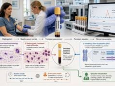
A histamine release test is a functional laboratory assay that measures how much histamine basophils release after they are exposed to an allergen, anti-IgE stimulus, drug, patient serum, or another activating substance. Unlike a specific IgE blood test, which detects antibody binding, a release assay asks whether living cells actually respond under controlled laboratory conditions. It can provide additional evidence in selected food, drug, venom, and environmental allergies when routine history, skin testing, and specific IgE leave uncertainty. A related version used in chronic spontaneous urticaria exposes healthy donor basophils to the patient’s serum to look for factors that activate the IgE receptor pathway. These are specialized tests with major limitations. Basophils must remain functional, allergen preparation and concentration matter, some people’s cells are nonresponders, and laboratories use different protocols and cutoffs. A positive result supports functional activation but does not prove clinical allergy or predict reaction severity. A negative result does not safely exclude allergy.
- The test measures cell function, not the amount of histamine already circulating in blood.
- Direct assays usually stimulate the patient’s fresh basophils with a suspected allergen.
- Urticaria assays may test whether patient serum activates healthy donor basophils.
- Positive and negative controls are essential because some basophils do not respond normally to IgE stimulation.
- Results depend on fresh samples, allergen concentration, cell quality, and laboratory method.
- The assay can support difficult diagnoses but does not replace a clinical history or supervised challenge.
Table of Contents
- What a Histamine Release Test Measures
- How the Laboratory Assay Works
- Direct, Passive, and Urticaria Test Formats
- How Results Are Calculated and Interpreted
- Clinical Uses in Allergy and Urticaria
- Limitations, False Results, and Nonresponders
- Histamine Release Versus Other Allergy Tests
- Preparing for Testing and Next Steps
What a Histamine Release Test Measures
Basophils are rare circulating white blood cells that carry the high-affinity IgE receptor, FcεRI. Allergen-specific IgE can bind to these receptors. When a matching allergen links adjacent IgE molecules, intracellular signals trigger degranulation and histamine release.
The histamine release test recreates part of this process outside the body. A blood sample supplies living basophils, and the laboratory exposes them to a series of allergen concentrations. Histamine in the fluid around the cells is then measured and compared with the total histamine available in those cells.
The result is often expressed as a percentage:
Percent histamine release = stimulated histamine minus spontaneous histamine, divided by total histamine minus spontaneous histamine, multiplied by 100.
The exact formula and corrections vary by laboratory. Spontaneous release is histamine that escapes without the specific stimulus. Total release is measured after disrupting cells or using a strong releasing condition. These controls help distinguish true allergen-induced release from fragile cells or low histamine content.
A release result contains more functional information than antibody binding alone. Specific IgE may be present but unable to trigger meaningful cell activation at tested concentrations. Conversely, a functional response may occasionally be detected when a commercial specific IgE assay is negative because the relevant allergen structure is represented differently.
The assay does not duplicate an allergic reaction in the whole person. It lacks mast cells in tissues, airway and cardiovascular responses, digestion, metabolism, cofactors, and the actual exposure route. It therefore cannot determine the severity of a future reaction.
The test should not be confused with a plasma histamine blood test. Plasma histamine attempts to capture a short-lived mediator rise during symptoms. A release test intentionally stimulates cells after collection and can be performed when the patient is well.
How the Laboratory Assay Works
Most assays require fresh anticoagulated blood because basophil function declines with time and improper temperature. The sample is transported under validated conditions and processed within the laboratory’s accepted window.
A typical direct procedure includes:
- Identify or enrich basophils in whole blood or a cell preparation.
- Incubate aliquots with several concentrations of the suspected allergen.
- Include a buffer control to measure spontaneous release.
- Include positive controls, often anti-IgE or another receptor-dependent stimulus.
- Include a non-IgE stimulus when the method uses one, to test broader cell viability.
- Stop the reaction after a defined incubation.
- Separate cells from supernatant.
- Measure released and total histamine by an immunoassay, fluorometric method, or another validated technique.
A concentration series is important because too little allergen may not activate cells, while an excessive concentration can produce nonspecific effects or a reduced response after maximal stimulation. The shape of the dose-response curve can be more informative than one concentration.
Reliable laboratories also define acceptance criteria before interpreting the allergen wells. Replicate measurements should agree within the method’s allowable variation, spontaneous release must remain below a specified limit, and total histamine must be sufficient for a stable denominator. A failed positive control, damaged sample, or erratic concentration curve should lead to an invalid or indeterminate report rather than a reassuring negative result. These quality checks matter because a numerical percentage can look precise even when the underlying cells or controls were unsuitable.
Positive controls answer whether the patient’s basophils can respond through the IgE pathway. If anti-IgE produces little or no activation, a negative allergen result is difficult to trust. Some protocols also use formyl-methionyl-leucyl-phenylalanine or another non-IgE activator to show that the cells remain capable of releasing histamine through a separate pathway.
The allergen reagent must preserve clinically relevant structures and remain noncytotoxic. Food extracts, purified components, venoms, and drug preparations behave differently. A drug may need metabolism or protein binding to become allergenic, which is hard to reproduce in vitro.
Timing, temperature, anticoagulant, dilution, incubation, and calculation rules are not fully standardized across laboratories. A result from one method cannot always be transferred to another laboratory’s cutoff.
Direct, Passive, and Urticaria Test Formats
Direct histamine release
The direct test uses the patient’s own basophils and their naturally bound IgE. It is the closest counterpart to a direct basophil activation test. It can be used to examine responses to foods, venoms, inhalants, drugs, or other suspected triggers in specialist settings.
The result depends on both sensitization and the intrinsic responsiveness of the patient’s basophils. Treatment that lowers free IgE or FcεRI expression, recent strong activation, and individual signaling differences can change the response.
Passive sensitization or passive release
In passive testing, donor basophils are prepared so that patient serum antibodies can bind to them. The sensitized donor cells are then exposed to allergen. This approach can separate the antibody component from the patient’s own basophil responsiveness.
Passive methods are technically demanding. Donor selection, removal of existing IgE, receptor expression, complement, serum factors, and the sensitization procedure can alter results. They are used mainly in specialized or research laboratories.
Basophil histamine release assay for chronic urticaria
A different assay asks whether serum from a patient with chronic spontaneous urticaria can make healthy donor basophils release histamine. Functional serum factors may include IgG autoantibodies directed against FcεRI or IgE, and other activating influences can contribute.
This assay may be called BHRA, histamine-releasing urticaria test, or urticaria-inducing activity test. It does not test whether a food or environmental allergen causes the hives. Chronic spontaneous urticaria is usually not driven by a hidden external allergen.
A positive BHRA can support type IIb autoimmune chronic spontaneous urticaria when interpreted with other evidence, such as an autologous serum skin test and autoantibody testing. The tests do not always agree, and no single result defines the disease in every guideline.
Positive basophil tests have been associated in some studies with higher disease activity and different treatment response patterns. They are not universally available and are not required for routine diagnosis or standard first-line treatment.
| Format | Cells and stimulus | Main question |
|---|---|---|
| Direct allergen release | Patient basophils plus suspected allergen | Do the patient’s cells release histamine to this trigger? |
| Passive sensitization | Donor basophils plus patient serum, then allergen | Can serum antibodies transfer allergen responsiveness? |
| Urticaria BHRA | Healthy donor basophils plus patient serum | Does patient serum contain functional basophil-activating factors? |
How Results Are Calculated and Interpreted
A laboratory may report maximum percent release, release at each allergen concentration, a threshold concentration, or a measure of basophil sensitivity. The report should state its own positivity criterion.
Maximum release describes the greatest response observed across the concentration series. It reflects cellular reactivity under test conditions but can be low in nonresponders.
Sensitivity describes how little allergen is needed to produce a defined fraction of the maximum response. Some laboratories use an EC50-like value or a calculated metric. A more sensitive response may require a lower allergen concentration, but it does not directly translate into the patient’s real-world threshold dose.
A positive result is most persuasive when:
- The patient had a compatible immediate reaction to that trigger
- The allergen caused a concentration-dependent response
- Spontaneous release was acceptable
- Positive controls confirmed functional cells
- The reagent and cutoff are validated for that clinical use
- Other test results and exposure history support the same conclusion
A negative result can mean that the patient is not functionally sensitized, but it can also reflect nonresponsive basophils, degraded cells, incorrect allergen concentration, an inadequate reagent, treatment effects, or a non-IgE reaction mechanism.
Spontaneous release that is too high can make the assay uninterpretable. Fragile cells may release histamine during transport or processing, leaving little reserve for specific stimulation. Low total histamine can also limit calculation accuracy.
The report should not be reduced to “allergic” or “not allergic.” It should preserve the dose-response data, controls, method, and interpretive comment. Results from research protocols may not have validated clinical cutoffs.
A positive value does not predict anaphylaxis severity. Basophil response is only one part of a reaction. Mast-cell distribution, exposure route, allergen dose, asthma, cardiovascular health, medicines, and cofactors influence clinical outcomes.
Likewise, a low activating concentration in the tube is not the same as a low clinical reaction threshold. The laboratory concentration is delivered directly to isolated blood cells, whereas real exposure may involve digestion, absorption, metabolism, skin penetration, or tissue distribution. Exercise, alcohol, infection, sleep loss, and anti-inflammatory medicines can further change a person’s threshold on a particular day. Functional results can help rank evidence, but challenge testing and real-world history remain the clinical reference points when they can be used safely.
Clinical Uses in Allergy and Urticaria
Food allergy
Functional basophil tests can improve specificity for selected foods, including peanut, tree nuts, milk, egg, and wheat, and may reduce some oral food challenges in expert centers. Histamine release assays have been used less widely than flow-cytometric basophil activation tests.
A positive functional response can be helpful when specific IgE is positive but clinical allergy is uncertain. A negative result may lower probability but does not authorize home ingestion after a serious reaction.
Drug allergy
Drug-specific IgE assays exist for few medications and often have limited sensitivity. Functional basophil testing can add evidence for selected beta-lactams, neuromuscular blocking agents, radiocontrast media, biologics, and chemotherapy drugs. Performance varies substantially by drug and protocol.
The assay may fail when a metabolite or drug-protein conjugate is the true allergen. Cytotoxic or irritating concentrations can produce nonspecific release. Testing requires drug-specific expertise.
Insect venom allergy
Basophil functional assays may help when venom skin tests and specific IgE are negative or conflicting despite a convincing systemic sting reaction, or when tests are positive to more than one venom. They are adjuncts to, not replacements for, standard venom evaluation.
Respiratory allergy
Histamine release can be measured after pollen, mite, or animal allergen stimulation. Routine diagnosis usually relies on history, skin testing, and specific IgE because they are more accessible. Functional testing may be used in research, immunotherapy monitoring, or complex discordant cases.
Chronic spontaneous urticaria
BHRA evaluates functional serum activity rather than external allergy. A positive result can contribute to identifying a type IIb autoimmune pattern and may have prognostic associations. Routine chronic urticaria care still begins with symptom control and a limited, history-directed workup.
The test should not be ordered as a broad explanation for flushing, fatigue, food intolerance, or nonspecific symptoms. Its value depends on a narrowly defined clinical question.
Limitations, False Results, and Nonresponders
A significant proportion of people have basophils that respond poorly to IgE-dependent positive controls. These “nonresponders” may have intact basophils but altered signaling molecules downstream of FcεRI. An allergen-specific negative result in this setting cannot exclude allergy.
Responsiveness can also vary over time. Recent allergen exposure, seasonal disease, infection, severe reaction, and biologic treatment may alter receptor density or signaling. Omalizumab lowers free IgE and FcεRI expression and can reduce IgE-mediated basophil responses. Other immune-targeted treatments may have different effects.
Preanalytical limitations include:
- Delay between collection and testing
- Temperature extremes during transport
- Wrong anticoagulant or tube
- Low basophil count
- High spontaneous release
- Insufficient total cellular histamine
- Cell damage during preparation
Analytical limitations include unstandardized allergen extracts, batch differences, cytotoxic drug concentrations, different positive controls, variable incubation, and laboratory-specific thresholds.
Biological false positives can occur when a stimulus activates basophils nonspecifically or when a cross-reactive allergen produces laboratory release without clinically relevant exposure. False negatives can occur if the tested extract lacks the relevant molecular structure.
The assay evaluates basophils, not tissue mast cells. Although the cells share IgE receptors and mediators, their signaling and environment are not identical. A person may have a clinically important mast-cell reaction that is not reproduced by circulating basophils.
Very low circulating basophil numbers create another practical problem. A direct release assay may still contain too little total histamine for dependable measurement, while a flow-cytometric assay may collect too few events for confident gating. Laboratories should report whether minimum cell or event requirements were met. Repeating the sample can sometimes resolve a technical failure, but repeated failure may reflect the patient’s biology rather than collection error. In that situation, clinicians should rely more heavily on history, validated specific IgE or skin testing, and supervised challenge when appropriate.
A negative test does not rule out non-IgE mechanisms, including complement activation, direct mast cell activation, delayed T-cell reactions, food protein-induced enterocolitis syndrome, and many infusion reactions.
For chronic urticaria BHRA, donor basophil selection is a major source of variation. Some donor cells respond strongly to activating serum, while others do not. Complement handling and serum storage can also affect release. Results across laboratories may not be directly comparable.
Histamine Release Versus Other Allergy Tests
Specific IgE blood testing detects antibody binding to an extract or component. It is widely available and stable but does not prove functional activation.
Skin prick testing demonstrates mast-cell-associated sensitization in the skin. It provides quick results but is affected by antihistamines, skin disease, technique, and extract quality.
Basophil activation testing uses flow cytometry to measure surface markers such as CD63 or CD203c after stimulation. CD63 is closely associated with degranulation and histamine release. BAT can identify cells and activation in a small blood volume without directly measuring histamine in supernatant.
A basophil activation test and histamine release assay are related but not identical. They can disagree because marker expression, mediator secretion, gating, and calculation capture different parts of the response. Neither is universally superior for every allergen.
Oral or drug challenge tests clinical tolerance by controlled exposure. When safe and appropriate, it provides a more direct diagnostic answer than an in-vitro assay. It carries reaction risk and requires medical supervision.
Acute tryptase can support systemic mast cell activation during a real reaction but does not identify the trigger. A histamine release test examines a suspected trigger in vitro but does not confirm that a systemic event occurred.
The best test is selected from the question. A routine positive specific IgE plus a classic immediate reaction may not need a release assay. A complex drug reaction with no validated IgE test may benefit more, if a specialized laboratory has experience with that drug.
Preparing for Testing and Next Steps
Before testing, confirm which assay is being ordered. “Histamine release” may refer to direct allergen stimulation, passive sensitization, or an urticaria serum assay. The specimen, interpretation, and clinical meaning differ.
Ask the laboratory about sample timing, transport temperature, accepted collection days, and maximum transit time. A Friday afternoon draw may be unsuitable if living cells cannot be processed promptly. The testing center may need to schedule collection with the referral laboratory.
Provide the exact suspected allergen or drug, reaction timing, dose, route, and conventional test results. A generic order such as “allergy histamine release” does not tell the laboratory what to stimulate.
Medication instructions are assay-specific. Antihistamines generally block histamine receptors rather than preventing release and may not affect an in-vitro release assay the way they affect skin testing. Omalizumab and other biologics can alter basophil biology. Do not stop prescribed medication unless the ordering clinician and laboratory provide a safe plan.
When results return, review the positive controls first. A negative allergen response with failed anti-IgE control is often inconclusive rather than truly negative. Review spontaneous release, total histamine, concentration-response pattern, and the laboratory’s validation statement.
The next step may be continued avoidance, component testing, repeat functional testing, a supervised challenge, or no change at all. A result that does not match regular real-life tolerance should not automatically create an allergy label.
For chronic urticaria, a positive BHRA may help phenotype disease but does not mean that ordinary foods or environmental allergens are releasing histamine. Treatment decisions should follow urticaria severity, guideline steps, comorbidities, and specialist judgment.
Seek emergency care for breathing difficulty, throat swelling, faintness, or a rapidly worsening reaction. A specialized cell test is never used to decide whether acute symptoms need epinephrine.
References
- Basophil Activation and Histamine Release Tests in Relation to Omalizumab Treatment of Chronic Spontaneous Urticaria 2025 (Clinical Study)
- IgE Nonresponders in Basophil Activation Tests 2025 (Review)
- Applications of basophil activation test in paediatric allergic diseases 2024 (Review)
- Basophil activation test: User’s manual 2025 (Review)
- Performance Characteristics of Basophil Activation Tests for Diagnosing Penicillin Allergy 2024 (Clinical Study)
- Positive Basophil Tests Are Linked to High Disease Activity and Poor Treatment Response in Chronic Spontaneous Urticaria 2023 (Clinical Study)
Disclaimer
This article is educational and does not diagnose allergy or autoimmune chronic urticaria. Histamine release assays require specialist laboratories and must be interpreted with controls, sample quality, reaction history, and conventional testing. Do not attempt exposure at home or delay epinephrine and emergency care because of a laboratory result.