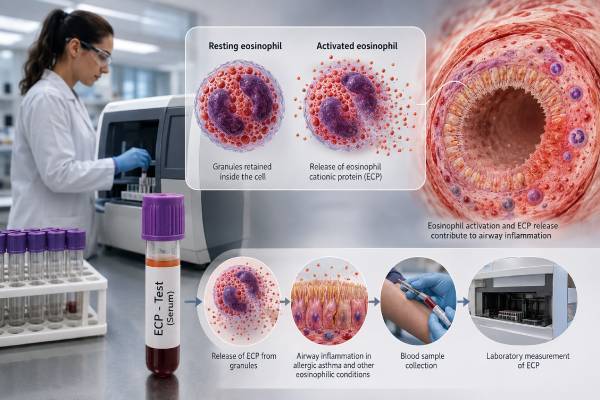
An eosinophil cationic protein test measures ECP, a granule protein released mainly by activated eosinophils. Eosinophils are white blood cells involved in type 2 inflammation, a common pathway in allergic asthma, allergic rhinitis, eczema, and several eosinophilic disorders. ECP can rise when eosinophils are active even if the blood eosinophil count is only modestly elevated, so the test is sometimes used as an additional marker of inflammatory activity. Its interpretation is more difficult than a routine blood count. Serum and plasma values are different, sample clotting and processing can strongly affect results, and reference ranges vary by laboratory. A high result does not diagnose asthma, identify a specific allergen, or prove that symptoms are caused by eosinophils. A low result does not rule out airway inflammation. In most asthma guidelines, blood eosinophils, exhaled nitric oxide, spirometry, symptoms, and exacerbation history have more established roles. ECP is therefore best treated as a supporting test used for a specific clinical question.
- ECP reflects eosinophil activation and degranulation, not simply the number of eosinophils in blood.
- Serum ECP is highly sensitive to collection and clotting conditions, so comparison requires standardized handling.
- A high ECP result is nonspecific and can occur in several allergic, respiratory, skin, gastrointestinal, infectious, and inflammatory conditions.
- ECP does not confirm asthma or reveal the responsible allergen.
- Blood eosinophils, FeNO, sputum eosinophils, and clinical outcomes usually guide asthma treatment more directly.
- Trends may be more informative than one value when the same laboratory method and timing are used.
Table of Contents
- What Eosinophil Cationic Protein Is
- How Eosinophils Create Inflammation
- How the ECP Test Is Collected
- What High and Low ECP Results Mean
- ECP in Asthma, Allergy, and Other Conditions
- ECP Versus Other Type 2 Biomarkers
- When Testing May Help and Next Steps
What Eosinophil Cationic Protein Is
Eosinophil cationic protein is a positively charged protein stored in eosinophil granules. It is also known as ribonuclease 3. When eosinophils become activated, they can release ECP along with major basic protein, eosinophil peroxidase, and eosinophil-derived neurotoxin. These substances participate in host defense but can also injure tissue and amplify inflammation.
The ECP blood test is different from an eosinophil count. A count answers how many eosinophils are circulating at the time of the blood draw. ECP is intended to reflect how much granule protein has been released or becomes available during sample preparation. The two often rise together, but they can diverge.
For example, a person may have many eosinophils that are relatively inactive, producing a high count with a less impressive ECP result. Another person may have a moderate count but strongly activated cells that release more ECP. Treatment can also change cell number and activation differently.
ECP can be measured in serum, plasma, sputum, nasal secretions, saliva, bronchoalveolar lavage fluid, and other research samples. Routine clinical reports most often involve serum. Results are generally given in micrograms per liter, written as µg/L, but the specimen and assay must be checked before comparing values.
Serum ECP is not simply the concentration circulating inside the patient. During clotting, eosinophils can continue to release ECP into the tube. The final number therefore reflects both in-vivo activation and a controlled ex-vivo release process. This is why the collection protocol is unusually important.
The test does not measure IgE and is not allergen-specific. ECP may be elevated in allergic and nonallergic eosinophilic inflammation. A person with a high result still needs separate testing if the clinical question is whether pollen, dust mites, a food, or another source triggers symptoms.
How Eosinophils Create Inflammation
Eosinophils develop in the bone marrow under the influence of signals including interleukin-5. They enter the bloodstream and can migrate into the lungs, nose, skin, or gastrointestinal tract when chemokines and adhesion signals attract them.
In type 2 inflammation, epithelial alarm signals and immune cells generate interleukins such as IL-4, IL-5, and IL-13. These pathways can promote IgE production, mucus, airway hyperresponsiveness, and eosinophil survival. Allergic disease is one common cause, but type 2 inflammation can occur without detectable allergen-specific IgE.
Activated eosinophils release granule contents by several processes. They may discharge selected proteins, release larger granule packages, or undergo cell death that leaves extracellular traps and intact granules in tissue. ECP can damage epithelial cell membranes, influence nerves, alter mucus, and interact with microbes and other immune cells.
In asthma, this activity may contribute to airway swelling, cough sensitivity, mucus production, and variable airflow obstruction. In the nose, it can accompany allergic rhinitis or chronic rhinosinusitis with nasal polyps. In skin, eosinophil products may be present in atopic dermatitis and some drug reactions. In the gastrointestinal tract, tissue eosinophilia can occur in eosinophilic esophagitis and other eosinophilic gastrointestinal diseases.
A blood result cannot show where the protein was released. ECP from airway inflammation, skin disease, systemic eosinophilia, or release in the collection tube all contributes to the measured value. The test therefore lacks organ specificity.
Eosinophils are also not the only determinant of symptoms. Asthma can involve airway smooth muscle, infection, mucus plugging, obesity, smoking, pollution, neutrophilic inflammation, and altered breathing patterns. A high ECP may support one pathway without explaining the whole disease.
Because ECP itself is biologically active, it is sometimes described as a marker of tissue-damaging potential. That description should not be converted into a personal prediction. The blood concentration does not quantify permanent organ injury and is not a direct measure of future lung-function loss.
How the ECP Test Is Collected
ECP testing usually requires venous blood. Fasting is not generally necessary, but the laboratory’s instructions should be followed. The most important variables are specimen type, collection tube, clotting time, temperature, centrifugation, and delay before separation.
For standardized serum testing, blood may need to clot for a defined period before centrifugation. If it remains in contact with blood cells too long, eosinophils can continue releasing ECP and create a falsely high or poorly comparable value. If clotting is too brief or conditions differ, release may be lower. Exact requirements vary by assay and laboratory.
Plasma is collected with an anticoagulant, so clotting-related release is reduced. Plasma concentrations are usually lower than serum concentrations and cannot be interpreted with a serum reference interval. A result must identify which material was used.
Preanalytical factors include:
- Time from collection to centrifugation
- Clotting duration and temperature
- Tube material and separator gel
- Vigorous handling or transport delay
- Storage temperature and freeze-thaw cycles
- Time of day and recent allergen exposure
- Current infection, asthma flare, or systemic corticosteroid treatment
A result from one laboratory should not be compared casually with a result from another. Different assays, calibrators, reference groups, and handling protocols can produce different ranges. For monitoring, the same laboratory and specimen protocol are preferable.
The report should include the numeric result, units, reference interval, specimen type, and collection date. “ECP positive” is not an adequate medical-record entry because the test is quantitative and method-dependent.
Reference intervals deserve special caution. A laboratory may establish or verify its own range using a particular kit, tube, clotting time, and healthy population. Values published elsewhere may be much higher or lower because the method differs. Children and adults may also have different distributions. The correct comparison is therefore the range printed by the performing laboratory, not a cutoff copied from a website. Even then, being just above the upper limit does not prove active disease. Clinicians look for a coherent pattern and may repeat an unexpected result after confirming collection conditions.
Medication can change the biology being measured. Inhaled corticosteroids may reduce eosinophilic airway inflammation over time. Oral or injected corticosteroids can lower eosinophils and related biomarkers more strongly. Biologic medicines that target IL-5, the IL-5 receptor, IL-4 receptor alpha, IgE, or upstream epithelial signals can alter eosinophil activity in different ways.
Antihistamines do not directly invalidate an ECP assay, but they may reduce symptoms without fully suppressing eosinophilic inflammation. Recent treatment, adherence, and timing should therefore be documented rather than assuming a value represents untreated disease.
What High and Low ECP Results Mean
A high ECP result means the measured concentration exceeds that laboratory’s reference interval. It supports increased eosinophil activation or release under the test conditions, but it is not a diagnosis.
Possible reasons include:
- Allergic or eosinophilic asthma
- Allergic rhinitis during active exposure
- Atopic dermatitis, especially when active
- Chronic rhinosinusitis with nasal polyps
- Eosinophilic gastrointestinal disease
- Drug hypersensitivity with eosinophilic inflammation
- Parasitic infection or other causes of eosinophilia
- Certain immune, hematologic, or inflammatory disorders
- Improper or delayed sample processing
The value should be interpreted beside the absolute eosinophil count. A high ECP with a high count can support active eosinophilic inflammation. A high ECP with a normal count may reflect activated cells, tissue migration, a recent change in treatment, or preanalytical release. Neither pattern identifies the cause by itself.
A normal or low result means the measured ECP was within the laboratory range under those collection conditions. It does not exclude asthma, allergy, or tissue eosinophilia. Inflammation can be intermittent or localized, treatment can suppress the marker, and not all asthma is eosinophilic.
There is no universal cutoff that separates controlled from uncontrolled asthma. Published thresholds differ by age, assay, specimen, population, and study purpose. A value used in one pediatric study should not be transferred to an adult laboratory report or a different platform.
Changes over time can be useful only when sampling is comparable. A fall after consistent anti-inflammatory treatment may support reduced eosinophil activity, especially if symptoms, exacerbations, and lung function also improve. A small change may reflect normal analytical variation or different clotting conditions.
| Pattern | Possible interpretation | Needed context |
|---|---|---|
| High ECP and high blood eosinophils | Active systemic eosinophilic pattern is possible | Symptoms, medicines, infection, allergy, parasite and organ evaluation |
| High ECP with normal eosinophil count | Cell activation, tissue disease, treatment effect, or sample artifact | Specimen handling and repeat standardized testing |
| Normal ECP with asthma symptoms | Eosinophilic activity may be suppressed, intermittent, or absent | Spirometry, FeNO, count, adherence, and alternative diagnoses |
| Falling ECP during treatment | May support reduced activity | Clinical improvement and comparable collection method |
ECP does not predict the exact severity of the next asthma attack or allergic reaction. Emergency risk assessment depends on prior exacerbations, lung function, asthma control, medication use, comorbidities, and access to treatment.
ECP in Asthma, Allergy, and Other Conditions
Asthma
Research has linked higher ECP with type 2 inflammation, poorer asthma control, airflow limitation, and exacerbations in some groups, particularly children with atopic asthma. ECP may add information when counts and symptoms do not fully describe eosinophil activity.
It cannot establish the asthma diagnosis. Asthma requires a history of variable respiratory symptoms and evidence of variable expiratory airflow limitation when possible. Spirometry with bronchodilator testing, peak-flow variability, or other objective tests serve that purpose.
Current asthma strategies emphasize blood eosinophils, fractional exhaled nitric oxide, sputum eosinophils where available, allergen-driven history, exacerbations, and treatment response. ECP is not commonly a required biomarker for selecting biologic therapy. Payer and regulatory criteria generally use better-standardized measures.
This distinction matters in severe asthma. A high ECP result may strengthen the impression that eosinophils are active, but it cannot replace documentation of exacerbations, maintenance treatment, adherence, blood eosinophil thresholds, FeNO, or local eligibility rules. Likewise, a normal ECP should not disqualify a patient from effective treatment when established criteria are met. The biomarker should support, not override, a complete severe-asthma assessment.
In milder asthma, repeated ECP testing is not a substitute for asking whether symptoms wake the patient, limit activity, require reliever medication, or follow missed controller doses. Clinical control and prevention of attacks remain the goals. A laboratory number can improve understanding only when linked to those outcomes.
Allergic rhinitis
ECP can rise during pollen exposure or active perennial rhinitis, but it does not identify the allergen. An environmental specific IgE panel or skin testing is used to establish sensitization. Nasal symptoms, season, and exposure determine clinical relevance.
Nasal ECP has been studied as a local marker, but routine collection and reference standards are limited. A serum value may reflect inflammation outside the nose.
Atopic dermatitis
Some patients with active eczema have elevated ECP, especially with extensive disease and eosinophilia. The result is not a standard measure of eczema severity and does not reveal food triggers. Clinical examination and validated severity tools are more useful.
Eosinophilic gastrointestinal disease
ECP can be elevated in eosinophilic gastrointestinal conditions, but blood testing cannot replace endoscopy and tissue biopsy when those are required. Studies have shown substantial overlap between affected patients and controls. Symptoms and organ-specific criteria remain central.
Infection, parasites, and systemic disease
Parasitic infections can activate eosinophils, although the pattern depends on the organism and tissue migration. Drug reactions, vasculitis, hypereosinophilic syndromes, and some blood cancers can also raise eosinophil-related markers. Persistent marked eosinophilia or organ symptoms require a broader evaluation rather than labeling the result “allergy.”
ECP Versus Other Type 2 Biomarkers
No single biomarker captures all type 2 inflammation. Each test samples a different part of the pathway.
Blood eosinophil count is inexpensive, widely standardized, and used in severe-asthma evaluation and biologic eligibility. It measures cell number rather than activation and varies with time of day, infection, corticosteroids, and other diseases.
FeNO measures nitric oxide in exhaled breath. It reflects IL-4 and IL-13-related airway signaling and can support type 2 airway inflammation or inhaled corticosteroid response. Smoking, technique, nitrate intake, infection, and treatment affect it. FeNO and eosinophils can disagree because they represent different pathways.
Sputum eosinophils more directly assess airway inflammatory cells and can guide treatment in specialized centers. Induced sputum is time-consuming and not widely available.
Allergen-specific IgE identifies sensitization to a source. It answers “what might trigger allergy,” not “how active are eosinophils today.” Total IgE is used for limited purposes, including dosing criteria for some therapies, but is a poor general activity marker.
ECP adds a degranulation or activation perspective. Its disadvantages are strong preanalytical dependence, variable reference intervals, lower availability, and fewer guideline-based treatment thresholds.
A combined pattern may be more informative than any isolated number. High blood eosinophils, high FeNO, compatible symptoms, and recurrent exacerbations provide stronger evidence of active type 2 asthma than ECP alone. When markers disagree, clinicians should review treatment adherence, corticosteroid timing, infection, smoking, comorbid nasal disease, and sample quality.
Different biomarkers also change at different speeds. A short course of oral corticosteroid can quickly lower circulating eosinophils, while symptoms or airway inflammation may not normalize at the same moment. FeNO can respond to inhaled corticosteroid use and adherence, whereas serum ECP may be influenced by both biological change and the blood-tube process. Sampling all markers on different days without recording treatment can create an apparent contradiction that is entirely explainable.
For this reason, a monitoring plan should specify which outcome is primary. If the goal is fewer exacerbations, attack frequency is more important than normalizing ECP. If the goal is checking inhaled corticosteroid response, symptoms, technique, FeNO, and lung function may be more actionable. Biomarkers are tools for decisions, not treatment targets in isolation.
Biomarkers should not replace fundamentals. Incorrect inhaler technique, missed medication, ongoing smoke exposure, vocal cord or laryngeal problems, obesity, reflux, sleep apnea, and cardiac disease can all cause poor control despite a reassuring or abnormal biomarker profile.
When Testing May Help and Next Steps
ECP may be considered when a clinician wants an additional measure of eosinophil activity and the laboratory can provide standardized collection. It may be used in selected pediatric allergy or respiratory clinics, research-supported monitoring programs, or cases in which the eosinophil count does not match the clinical picture.
Before ordering, define how the result will change care. Will it help decide whether to repeat airway testing, review anti-inflammatory treatment, investigate a systemic eosinophilic disorder, or monitor a previously high value under the same conditions? If no action follows either a high or low result, the test may add little.
A high result should prompt questions rather than an automatic increase in treatment:
- Was the serum processed according to the laboratory protocol?
- Is asthma objectively confirmed and currently controlled?
- What are the blood eosinophil count, FeNO, spirometry, and exacerbation history?
- Are inhaled medicines taken correctly and consistently?
- Is active rhinitis, eczema, sinus disease, infection, or another organ condition present?
- Are oral corticosteroids or biologics suppressing some markers?
- Is persistent eosinophilia severe enough to require parasite, drug, immune, or hematology evaluation?
Do not use an ECP value to self-adjust inhaled or oral corticosteroids. Excess treatment can cause harm, while undertreatment can increase asthma risk. Changes should be based on symptoms, objective assessment, and an agreed action plan.
Seek urgent care for severe breathlessness, difficulty speaking, blue or gray lips, faintness, rapid deterioration, or poor response to rescue medication. ECP testing has no role in deciding whether an acute asthma attack needs emergency treatment.
For follow-up, keep the original report and note whether the sample was serum or plasma. If a trend is clinically important, repeat through the same laboratory at a similar time and under comparable treatment conditions. The result should be one line in a larger picture, not the final diagnosis.
Patients can make the visit more useful by bringing inhalers, a medication list, recent steroid dates, spirometry reports, exacerbation records, and earlier eosinophil or FeNO results. Note whether the blood was drawn during a flare, respiratory infection, pollen season, or a period of stable health. These details often explain more than the isolated ECP number.
They also help avoid repeating an abnormal test under different conditions and mistaking the difference for treatment failure. When uncertainty remains, clinicians may prioritize a standardized repeat or a more established organ-specific test instead of expanding the laboratory panel further unnecessarily.
References
- Global Strategy for Asthma Management and Prevention 2026 (Guideline)
- Serum Eosinophil Cationic Protein as a Biomarker for Distinguishing Pediatric Allergic Airway Diseases 2026 (Clinical Study)
- Eosinophilic Cationic Protein and Immunoglobulin E in Children with Chronic Cough 2024 (Review)
- Increased serum eosinophilic cationic protein in children with chronic cough 2023 (Clinical Study)
- Serum Eosinophilic Cationic Protein Is a Reliable Biomarker for Childhood Asthma 2022 (Clinical Study)
- Performance of Eosinophil Cationic Protein as a Biomarker in Asthmatic Children 2021 (Clinical Study)
Disclaimer
This article is educational and does not diagnose asthma, allergy, or an eosinophilic disorder. ECP results require interpretation with specimen handling, symptoms, blood counts, lung testing, medications, and other biomarkers by a qualified clinician. Seek emergency care for severe or rapidly worsening breathing symptoms.