
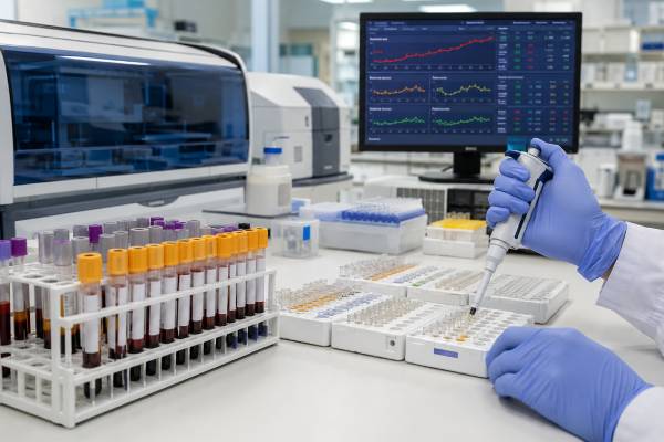
A macrophage activation syndrome blood test panel is not one single laboratory test. It is a coordinated set of urgent studies used when clinicians suspect macrophage activation syndrome (MAS), a life-threatening hyperinflammatory state most often associated with Still’s disease, systemic juvenile idiopathic arthritis, lupus, and other rheumatic illnesses. Typical testing includes ferritin, complete blood count, liver enzymes, triglycerides, fibrinogen, coagulation studies, D-dimer, lactate dehydrogenase, C-reactive protein, and sometimes soluble IL-2 receptor, IL-18, CXCL9, or broader cytokine testing. No result confirms or excludes MAS by itself. The strongest clue is a rapidly changing pattern: rising ferritin and liver injury, falling platelets or other blood-cell counts, worsening clotting abnormalities, persistent fever, and new organ dysfunction. MAS overlaps biologically and clinically with secondary hemophagocytic lymphohistiocytosis (HLH), severe infection, sepsis, liver failure, and cytokine release syndromes. Because deterioration can be fast, testing must occur alongside immediate clinical assessment and treatment planning rather than as a delayed outpatient screen.
- Ferritin is a central screening and trend marker, but even a very high value is not specific for MAS.
- Falling platelets, fibrinogen, or erythrocyte sedimentation rate can be more informative than whether a result is already below normal.
- IL-18 and CXCL9 can help characterize inflammasome and interferon-gamma activity, but they are specialized tests.
- Bone marrow hemophagocytosis is neither required nor sufficiently specific to diagnose MAS.
- Suspected MAS with organ dysfunction is a medical emergency.
Table of Contents
- What Macrophage Activation Syndrome Is
- Core Blood Tests in a MAS Panel
- How Ferritin Is Interpreted
- IL-18, CXCL9, and Cytokine Testing
- Diagnostic Criteria and Probability Scores
- MAS, HLH, Sepsis, and Other Overlap Syndromes
- Why Serial Testing Matters
- Urgent Next Steps and Follow-Up
What Macrophage Activation Syndrome Is
MAS is an acquired form of uncontrolled immune activation in which cytotoxic lymphocytes, macrophages, and inflammatory signaling loops fail to switch off appropriately. It is generally considered part of the broader HLH spectrum, but the term MAS is used especially when the syndrome occurs with rheumatic or autoinflammatory disease. Systemic juvenile idiopathic arthritis and adult-onset Still’s disease are classic settings, although MAS also occurs with systemic lupus erythematosus, dermatomyositis, Kawasaki disease, vasculitis, and other inflammatory disorders.
The biology involves excessive activation of T cells and macrophages, impaired cytotoxic control, and high production of interferon-gamma and other inflammatory mediators. Activated macrophages consume and damage blood cells, release ferritin and cytokines, alter lipid metabolism, and contribute to liver injury and coagulation abnormalities. Endothelial activation and tissue inflammation can affect the brain, lungs, kidneys, heart, and bone marrow.
Common clinical features include persistent high fever, enlarged liver or spleen, rapidly worsening fatigue, bleeding or bruising, confusion, seizures, breathing difficulty, low blood pressure, and failure of one or more organs. A patient whose underlying inflammatory disease previously caused high white-cell and platelet counts may show an unexpected fall toward “normal.” That downward shift can be an early warning even before counts cross below the laboratory range.
MAS is not diagnosed by the presence of macrophages in the blood, and routine testing does not directly count activated macrophages. The name describes the syndrome’s immune biology. A bone marrow, liver, spleen, or lymph-node sample may show hemophagocytosis, in which histiocytes contain blood cells, but this finding can be absent early and can occur in infection, transfusion, critical illness, and other conditions.
The trigger may be a flare of the underlying inflammatory disease, a viral or bacterial infection, a medication change, surgery, or another immune stress. More than one trigger can coexist. Infection is especially important because it can initiate MAS and also mimic it; finding an infection does not automatically end the MAS evaluation.
Core Blood Tests in a MAS Panel
The first-line panel is designed to detect the characteristic combination of inflammation, blood-cell consumption, liver injury, altered lipid metabolism, and impaired coagulation. A complete blood count with differential tracks platelets, hemoglobin, neutrophils, and total white cells. MAS often produces cytopenias, but the earliest clue may be a rapid decline from a previously elevated baseline. Platelets commonly fall first.
Ferritin is usually measured immediately and repeated frequently. Aspartate aminotransferase and alanine aminotransferase assess hepatocellular injury; bilirubin, albumin, alkaline phosphatase, and gamma-glutamyl transferase add context. Lactate dehydrogenase may rise with tissue injury, cell turnover, and liver involvement. Kidney function, electrolytes, glucose, and lactate help assess organ stress and critical illness.
Fasting triglycerides may rise because inflammatory signaling suppresses lipoprotein lipase and changes hepatic lipid handling. Fibrinogen may fall because of consumption and increased fibrinolysis, although it can remain normal or high early because it is also an acute-phase protein. Prothrombin time, activated partial thromboplastin time, D-dimer, and sometimes thrombin time help evaluate disseminated coagulation activation and bleeding risk.
C-reactive protein commonly rises, but the erythrocyte sedimentation rate can behave differently. ESR depends partly on fibrinogen; as fibrinogen falls, ESR may drop despite worsening inflammation. In a patient with active Still’s disease or another condition that usually produces a high ESR, an unexpected decline alongside rising ferritin can be a warning pattern.
Additional tests are selected by context. These can include soluble IL-2 receptor alpha, also called soluble CD25; natural killer-cell number or function; soluble CD163; CXCL9; IL-18; broad cytokine panels; blood cultures; viral polymerase chain reaction tests; Epstein–Barr virus and cytomegalovirus testing; hepatitis studies; and imaging. A cytokine panel may support mechanistic assessment but is not a substitute for the core emergency laboratory trend.
A peripheral smear can identify abnormal cells, schistocytes, blasts, parasites, or other clues. Bone marrow examination is considered when malignancy, marrow failure, infection, or another diagnosis remains possible. It should not delay treatment when the clinical probability of MAS or HLH is high and the patient is deteriorating.
How Ferritin Is Interpreted
Ferritin stores iron inside cells, but circulating ferritin also behaves as an acute-phase reactant. In MAS, levels can rise dramatically because of macrophage and hepatocyte activation, cellular injury, altered iron handling, and inflammatory signaling. The magnitude can be striking, yet there is no universal ferritin threshold that proves MAS across every age, disease, and clinical setting.
A ferritin result should be interpreted in three ways: absolute value, rate of change, and relationship to other findings. A rapidly rising value over hours or days may carry more urgency than a stable chronically elevated result. Very high ferritin increases concern for severe hyperinflammation, but major elevations also occur in adult-onset Still’s disease without MAS, severe infection, acute hepatitis, liver failure, hematologic cancer, iron overload, repeated transfusion, kidney disease, and catastrophic inflammatory states.
In children with systemic juvenile idiopathic arthritis, classification criteria developed for research use include ferritin plus changes in platelets, triglycerides, fibrinogen, and aspartate aminotransferase. These thresholds can help organize evidence, but they were not designed to replace clinical judgment in every disease. Adults, patients with lupus, critically ill patients, and people already receiving immunomodulators may show different patterns.
Ferritin also has limits as a severity marker. A patient can be critically ill before ferritin reaches an extreme level, and the highest number does not always identify who will have the worst outcome. Organ dysfunction, neurologic involvement, shock, respiratory failure, coagulopathy, and the need for intensive care are more direct measures of immediate danger.
Treatment changes ferritin kinetics. Corticosteroids, IL-1 blockade, IL-6 pathway inhibition, JAK inhibition, interferon-gamma blockade, chemotherapy, or treatment of an infectious trigger can alter the curve. Ferritin may fall before every other biomarker normalizes, or remain elevated during recovery. Clinicians therefore follow the whole pattern instead of waiting for ferritin to become normal.
Ratios such as ferritin divided by ESR or ferritin divided by platelets have been studied, but they are not universal stand-alone tests. They may be helpful within a validated population and laboratory method. A ratio should never override a clinically worsening patient whose individual trends are concerning.
IL-18, CXCL9, and Cytokine Testing
Interleukin-18 is an inflammasome-related cytokine that can promote interferon-gamma production, especially in cooperation with IL-12. Markedly increased IL-18 is associated with Still’s disease and MAS biology. In selected centers, IL-18 can help identify a highly inflammatory phenotype, estimate MAS risk, or distinguish Still’s disease from some competing diagnoses. However, assays are not standardized across all laboratories, turnaround may be slow, and proposed cutoffs depend on the method and population.
Most clinical assays report total IL-18 rather than the biologically unbound fraction. IL-18 binding protein naturally neutralizes part of circulating IL-18, so total concentration does not equal active signaling. An IL-18 test therefore adds context but cannot diagnose MAS independently or reliably separate every case of MAS from genetic HLH.
CXCL9 is a chemokine induced strongly by interferon-gamma. High CXCL9 can serve as a downstream indicator of interferon-gamma pathway activity and may help identify an IFN-gamma-dominant hyperinflammatory pattern. It is not specific to MAS and may rise in infection, immune activation, and other inflammatory diseases. The combination of IL-18, CXCL9, soluble CD25, and clinical features may be more informative than any one value.
Soluble CD25 reflects shedding of the alpha chain of the IL-2 receptor from activated lymphocytes. It is part of the HLH-2004 criteria and can support a diagnosis of immune activation. Results often take longer than routine blood tests, and high values also occur in lymphoma, leukemia, infection, autoimmune disease, and other conditions. It is useful as supporting evidence, not as an emergency gatekeeper.
Broader cytokine panels may include IL-1 beta, IL-6, IL-10, TNF-alpha, interferon-gamma, GM-CSF, and chemokines. These profiles are mainly used in specialized care or research because concentrations vary by timing, specimen type, assay platform, treatment, and sample handling. Cytokines can be produced locally in tissue and disappear quickly from blood. A normal serum concentration does not exclude active signaling.
The practical priority remains fast, widely available tests. Ferritin, blood counts, liver enzymes, fibrinogen, triglycerides, coagulation studies, and organ-function measures usually guide immediate decisions while specialized biomarkers are pending. Cytokine results are most valuable when they answer a defined question and are interpreted by a team familiar with the assay.
Diagnostic Criteria and Probability Scores
Several criteria sets and scores are used because MAS and HLH lack a single definitive test. Each tool was developed for a particular population. Applying it outside that setting can create false reassurance or overdiagnosis.
The 2016 classification criteria for MAS complicating systemic juvenile idiopathic arthritis use fever and elevated ferritin together with selected abnormalities in platelets, AST, triglycerides, and fibrinogen. They are useful for recognizing a characteristic laboratory pattern in febrile patients with known or suspected systemic juvenile idiopathic arthritis. They do not cover every early, treated, atypical, adult, or non-Still’s presentation.
HLH-2004 criteria include fever, splenomegaly, cytopenias, hypertriglyceridemia or low fibrinogen, hemophagocytosis, low or absent NK-cell activity, ferritin elevation, and increased soluble CD25. Meeting five of eight supports HLH in the appropriate setting, while a molecular diagnosis associated with familial HLH can establish the condition. These criteria originated in pediatric HLH protocols and may be incomplete early in adults with secondary disease.
The HScore estimates the probability of reactive HLH using immunosuppression, fever, organ enlargement, cytopenias, ferritin, triglycerides, fibrinogen, AST, and bone marrow findings. It can organize adult inpatient evidence, but it is not a replacement for repeated assessment. Missing data, treatment before sampling, and different disease populations affect performance.
Recent EULAR/ACR guidance emphasizes recognizing a pattern of persistent fever, rising or high ferritin, falling blood counts, liver inflammation, coagulation activation, splenomegaly, and organ dysfunction. No score should postpone action in a rapidly worsening patient. Conversely, a numerical score cannot identify the underlying trigger; infection, malignancy, rheumatic disease, and genetic susceptibility still require investigation.
Criteria are especially challenging after cytokine-targeted therapy. IL-6 blockade may suppress CRP and fever, corticosteroids can change blood counts, transfusions alter cytopenias, and fibrinogen replacement changes coagulation values. The clinician must reconstruct the trend and consider what each intervention has already modified.
MAS, HLH, Sepsis, and Other Overlap Syndromes
MAS and secondary HLH share the same broad hyperinflammatory spectrum. The choice of term often reflects the clinical setting rather than a completely separate mechanism. MAS is commonly used for rheumatic-disease-associated disease; HLH is used more broadly for genetic, infection-associated, malignancy-associated, and other secondary forms. Patients can also carry variants that increase susceptibility without having classic infantile familial HLH.
Sepsis is one of the hardest distinctions because fever, shock, cytopenias, liver injury, coagulopathy, high ferritin, and organ failure occur in both. Severe infection can trigger secondary HLH or MAS, so the conditions are not mutually exclusive. Cultures, molecular pathogen testing, imaging, source evaluation, and antimicrobial treatment often proceed at the same time as hyperinflammation assessment.
Malignancy-associated HLH is particularly important in adults. Lymphoma, leukemia, and other cancers may produce cytokine activation and marrow abnormalities. Clues can include persistent lymph-node enlargement, abnormal cells, disproportionate lactate dehydrogenase elevation, unexplained weight loss, clonal findings, or poor response to initial therapy. Tissue diagnosis may be necessary when clinically safe.
Cytokine release syndrome after CAR T-cell or other immune-engaging therapy can resemble MAS. Some patients develop an immune-effector-cell-associated HLH-like syndrome with very high ferritin, liver injury, cytopenias, hypofibrinogenemia, and coagulopathy after the initial cytokine release syndrome. Timing relative to treatment and therapy-specific criteria guide classification.
Acute liver failure, thrombotic microangiopathy, catastrophic antiphospholipid syndrome, severe lupus flare, disseminated infection, and drug hypersensitivity can produce overlapping laboratory patterns. A broad differential is safer than assuming that hyperferritinemia automatically equals MAS.
The distinction matters because treatment must address both the inflammatory loop and its cause. Immunosuppression without adequate infection therapy can be dangerous, while waiting for every infection test to return can allow uncontrolled hyperinflammation to progress. Management usually requires rheumatology, hematology, infectious diseases, critical care, and the relevant organ specialists.
Why Serial Testing Matters
MAS is dynamic. A single panel is a snapshot, and early results may not meet formal thresholds. Serial measurements reveal direction and speed. In a hospitalized patient with significant concern, complete blood count, ferritin, liver enzymes, fibrinogen, triglycerides, coagulation markers, and organ-function tests may be repeated daily or more often according to severity.
The most concerning pattern is convergence: ferritin climbing while platelets and fibrinogen fall, AST rises, D-dimer increases, and the patient develops neurologic, respiratory, hepatic, or circulatory dysfunction. Each individual result may initially be only mildly abnormal, yet the combined trajectory can be diagnostic.
Response monitoring also requires trends. Improvement may include defervescence, stabilization of blood pressure, recovering platelets, lower AST, improving fibrinogen, and falling ferritin. Specialized markers normalize at different speeds. CXCL9 and soluble CD25 may remain elevated after routine measures begin to improve, while IL-18 can decline slowly in Still’s-associated disease.
Apparent improvement must be interpreted cautiously after transfusion, plasma products, fibrinogen replacement, dialysis, or liver-support measures. These interventions can temporarily change laboratory values without ending immune activation. Similarly, a falling CRP after IL-6 blockade does not prove that MAS has resolved.
Baseline values are helpful. People with active Still’s disease may begin with leukocytosis, thrombocytosis, high CRP, and high fibrinogen. A fall into the laboratory’s normal range may represent a major pathologic shift for that person. Reviewing earlier records can expose this relative cytopenia or fibrinogen decline.
Outpatient retesting is inappropriate when red-flag symptoms are present. Persistent fever with confusion, breathing trouble, bleeding, severe weakness, jaundice, low blood pressure, rapidly falling counts, or escalating liver injury requires urgent hospital-level evaluation. Serial testing should occur where treatment and organ support are available, with immediate escalation when the trajectory worsens or new clinically apparent organ dysfunction appears.
Urgent Next Steps and Follow-Up
When MAS is suspected, clinicians stabilize the patient, collect urgent laboratory studies, search for triggers, and involve specialists early. Blood cultures and antimicrobial therapy may be needed immediately. Imaging, lumbar puncture, marrow examination, or tissue biopsy are chosen according to stability and the differential diagnosis; none should delay lifesaving treatment in a rapidly deteriorating patient.
Therapy depends on the cause and severity. High-dose corticosteroids are frequently used in rheumatic-disease-associated MAS. IL-1 blockade, calcineurin inhibition, JAK inhibition, interferon-gamma blockade, etoposide-based therapy, or other targeted approaches may be selected by the treating team. Treatment of infection, malignancy, or the underlying inflammatory disease is essential. There is no safe home treatment based only on a ferritin number.
Before interpreting a panel, confirm when the sample was collected relative to fever, transfusion, steroids, biologic medicines, anticoagulation, and organ-support therapies. Ask whether ferritin is rising, whether platelets are falling from baseline, and whether fibrinogen is dropping despite inflammation. Review the full coagulation and liver pattern instead of focusing on a single flagged result.
After recovery, follow-up may include repeated blood counts, ferritin, liver tests, coagulation studies, inflammatory markers, and disease-specific assessments. Persistent or recurrent episodes can prompt genetic evaluation for familial HLH pathways or other inborn errors of immunity, especially in children, young adults, consanguineous families, or patients with unusual infections and recurrent hyperinflammation.
Patients and families should receive a clear action plan. They need to know which symptoms require emergency care, which laboratory trends are being followed, and which medicines should not be stopped abruptly. A prior MAS episode increases concern during future inflammatory flares or infections, but it does not mean every fever is a recurrence.
A useful MAS panel answers a sequence of questions: Is systemic hyperinflammation present? Are blood cells, liver, coagulation, and organs becoming involved? Is the pattern worsening quickly? What trigger or overlapping disorder is present? Do the results and clinical status justify immediate immune-directed treatment? The safest interpretation combines rapid routine testing, selected specialized biomarkers, repeated trends, and bedside assessment.
References
- The 2022 EULAR/ACR points to consider at the early stages of diagnosis and management of suspected haemophagocytic lymphohistiocytosis/macrophage activation syndrome (HLH/MAS) 2023 (Consensus Guidance)
- EULAR/PReS recommendations for the diagnosis and management of Still’s disease, comprising systemic juvenile idiopathic arthritis and adult-onset Still’s disease 2024 (Clinical Recommendations)
- Heterogeneity of macrophage activation syndrome and treatment progression 2024 (Review)
- Immunopathology of and potential therapeutics for macrophage activation syndrome 2024 (Review)
- Serum levels of interleukin-18, CXCL9 and IFN-γ in Still’s disease complicated by macrophage activation syndrome 2024 (Research Article)
- Hyperferritinemia and Macrophage Activation Syndrome in Septic Shock: A Narrative Review 2025 (Review)
Disclaimer
This article is for general education and cannot diagnose or rule out macrophage activation syndrome, HLH, sepsis, or cancer. Suspected MAS requires urgent interpretation by clinicians using symptoms, serial laboratory trends, treatment history, and the search for an underlying trigger. Persistent fever with confusion, breathing difficulty, bleeding, low blood pressure, jaundice, or rapidly worsening laboratory results warrants emergency care.





