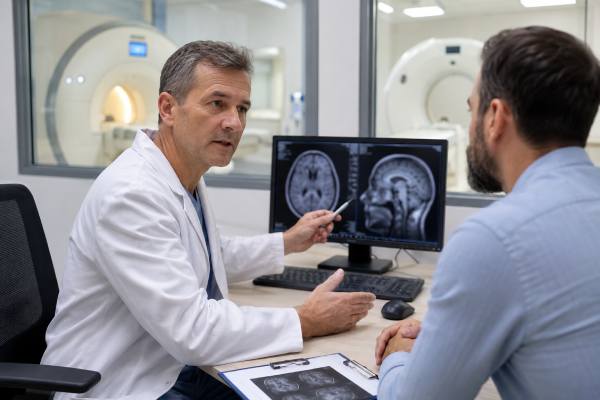
When someone has headaches, confusion, memory changes, dizziness, weakness, seizures, or other brain-related symptoms, imaging may be part of the medical workup. The common question is whether an MRI or CT scan is “better.” The practical answer is that neither test is always better. CT is often better for urgent situations where speed matters, especially possible bleeding, major head injury, or emergency stroke evaluation. MRI is often better when doctors need a more detailed look at brain tissue, small strokes, inflammation, tumors, demyelinating disease, or unexplained symptoms that have developed over time.
The right test depends on the symptom pattern, timing, exam findings, medical history, safety considerations, and what the doctor is trying to rule in or rule out. A normal scan can be reassuring, but it does not automatically explain every symptom. An abnormal scan also needs clinical interpretation, because some findings are incidental or age-related rather than the cause of the problem.
Table of Contents
- Which Scan Is Usually Better?
- How CT and MRI See the Brain
- When a CT Scan Is Usually Preferred
- When Brain MRI Is Usually Preferred
- Symptoms That Change the Imaging Choice
- Safety, Contrast, and Practical Differences
- What Happens After Imaging Results?
- Questions to Ask Before the Test
Which Scan Is Usually Better?
The better test is the one that answers the clinical question fastest and most safely. CT is usually preferred for many emergencies, while MRI is usually preferred for more detailed evaluation when the person is stable and the question requires finer brain-tissue detail.
A CT scan is often the first test in an emergency department because it is fast, widely available, and very good at detecting many urgent problems, especially fresh bleeding in or around the brain. A non-contrast head CT can often be completed quickly, which matters when doctors are deciding whether symptoms could be from bleeding, a skull fracture, swelling, or another condition that needs immediate action.
MRI takes longer but shows more detail in many types of brain tissue. It can detect smaller or subtler abnormalities that CT may miss, including small ischemic strokes, demyelinating lesions, inflammation, some infections, many tumors, and certain patterns of injury. MRI can also use different sequences, such as diffusion-weighted imaging, fluid-sensitive images, and contrast-enhanced images, to answer different diagnostic questions.
A useful way to think about the difference is this: CT is often the “rapid safety check” for urgent structural problems, while MRI is often the “high-detail tissue exam.” That is not a perfect rule, because CT can be sophisticated and MRI can be urgent in some settings. But it captures why doctors may choose CT first and MRI later, or choose MRI directly when the situation is not time-critical.
| Question | CT is often stronger when… | MRI is often stronger when… |
|---|---|---|
| Speed | Urgent decisions are needed within minutes | The person is stable enough for a longer exam |
| Bleeding | Fresh bleeding or head trauma is suspected | Older, subtle, or complex blood products need further characterization |
| Stroke | Emergency stroke triage needs rapid exclusion of hemorrhage | Small or early ischemic stroke, stroke mimics, or posterior circulation stroke is suspected |
| Tumor or inflammation | A quick first look is needed or MRI is not possible | Detailed tissue contrast and contrast enhancement are important |
| Radiation | Radiation exposure is acceptable for the clinical need | A radiation-free test is preferred and MRI is safe for the person |
In real care, the question is rarely “MRI or CT in isolation.” Doctors also consider the physical exam, blood pressure, fever, medications such as blood thinners, cancer history, immune status, pregnancy, kidney function, implanted devices, and whether symptoms started suddenly or gradually.
How CT and MRI See the Brain
CT and MRI create images in very different ways, which is why they answer different questions. CT uses X-rays to show density differences, while MRI uses magnetic fields and radiofrequency signals to show detailed tissue characteristics.
A CT scan is excellent at quickly showing dense structures and major acute changes. Bone, fresh blood, calcification, swelling, large masses, and major shifts in brain structures often stand out well. This is why CT is so useful after trauma and in emergency evaluations. It can also be paired with CT angiography, which looks at blood vessels after iodine-based contrast is injected, or CT perfusion, which helps estimate blood flow in selected stroke evaluations.
MRI is more flexible in the kinds of information it can show. Different MRI sequences highlight different tissue properties. Some sequences are sensitive to water movement, which helps identify many acute ischemic strokes. Others highlight fluid, inflammation, scarring, old injury, tumors, or abnormal enhancement after contrast. MRI can also be paired with MR angiography or MR venography to evaluate arteries or veins.
For people comparing tests, the most important distinction is not simply “MRI is clearer.” MRI is clearer for many tissue questions, but CT may be clearer, faster, or more practical for other questions. A dedicated brain CT scan explanation can help clarify what CT is designed to detect, while a focused brain MRI overview is useful for understanding why MRI is often ordered for detailed neurological evaluation.
The test result also depends on the protocol. A “brain MRI” is not always the same exam for every person. A doctor may request MRI without contrast, MRI with and without contrast, MR angiography, thin cuts through the inner ear canals, seizure protocol imaging, pituitary imaging, or another targeted protocol. CT also has protocols, including non-contrast CT, CT angiography, CT venography, and CT with contrast.
That is why the reason for the scan matters. A vague order such as “headache” may not be enough if the real concern is thunderclap headache, papilledema, cancer history, fever, new neurological deficit, or a positional headache. The more precise the clinical question, the more likely the imaging protocol will match the problem.
When a CT Scan Is Usually Preferred
CT is usually preferred when the situation is urgent and doctors need a rapid answer. It is especially important when bleeding, significant head injury, skull fracture, sudden severe symptoms, or emergency stroke treatment decisions are possible.
In many emergency settings, a non-contrast head CT is the first imaging test because it can be done quickly and can identify problems that may change immediate treatment. For example, a person with sudden weakness on one side of the body may need rapid imaging to help distinguish bleeding from ischemic stroke before certain treatments are considered. A person who fell and hit their head while taking blood thinners may need urgent CT to check for bleeding, even if symptoms seem mild at first.
CT is commonly used first for:
- Sudden neurological deficits, such as facial droop, one-sided weakness, speech trouble, or sudden vision loss
- Significant head injury, especially with loss of consciousness, worsening headache, vomiting, confusion, seizure, or blood thinner use
- Sudden “worst headache” or thunderclap headache, particularly when bleeding around the brain is a concern
- Acute confusion or reduced alertness when doctors are concerned about bleeding, mass effect, or stroke
- Suspected skull fracture or major trauma involving the head or face
- Situations where MRI is unsafe, unavailable, or would delay urgent care
CT is not always enough. A CT can be normal early in some ischemic strokes, especially small strokes or strokes in the brainstem or cerebellum. It may also miss subtle inflammation, small tumors, demyelinating lesions, or certain seizure-related abnormalities. In those cases, MRI may follow if symptoms persist or the first CT does not explain the clinical picture.
The biggest strength of CT is speed. The biggest limitation is that it uses ionizing radiation and has less soft-tissue detail than MRI for many non-emergency brain conditions. That tradeoff is often reasonable when the alternative is delaying urgent care.
Some symptoms should be treated as emergencies rather than reasons to wait for outpatient imaging. New weakness, trouble speaking, sudden severe headache, seizure with prolonged confusion, head injury with worsening symptoms, fainting with neurological signs, or a major change in consciousness should be assessed urgently. For more detail on when symptoms need emergency evaluation, see ER-level neurological warning signs.
When Brain MRI Is Usually Preferred
MRI is usually preferred when doctors need a detailed look at brain tissue and the situation is not so urgent that CT must come first. It is often the stronger test for unexplained, progressive, recurrent, or subtle neurological symptoms.
MRI is especially useful when the concern involves small or complex changes in the brain. This includes small strokes, multiple sclerosis or other demyelinating conditions, inflammation, infection, many tumors, pituitary or posterior fossa problems, seizure-related structural causes, and some patterns of traumatic brain injury that are not visible on CT. MRI is also often used when symptoms continue despite a normal CT.
Doctors may favor MRI for:
- New seizures or recurrent unexplained seizure-like episodes
- Gradual or progressive weakness, numbness, imbalance, or coordination problems
- Persistent headaches with concerning features, especially if the neurological exam is abnormal
- Memory loss or cognitive decline when structural causes need evaluation
- Suspected brain tumor, metastasis, inflammation, or infection
- Symptoms suggesting multiple sclerosis or another demyelinating disorder
- Unexplained symptoms after a CT scan does not answer the question
- Certain vision, hearing, pituitary, brainstem, or cranial nerve symptoms
MRI is not a general-purpose answer to every brain symptom. Many people with brain fog, dizziness, fatigue, anxiety, poor concentration, or mild forgetfulness have normal brain imaging because the cause is metabolic, sleep-related, medication-related, psychiatric, hormonal, cardiovascular, or functional rather than a visible structural lesion. In those situations, imaging may still be appropriate when red flags are present, but the workup often includes history, exam, labs, sleep evaluation, cognitive testing, medication review, or mental health assessment.
For memory symptoms, imaging is only one part of the evaluation. Cognitive screening, functional history, medication review, mood assessment, sleep assessment, and lab testing often matter just as much. A separate discussion of brain imaging for memory loss can help explain when MRI, PET, or other testing enters the workup.
MRI also has practical limits. Some people cannot have MRI because of certain implanted devices, metal fragments, or severe claustrophobia. Others may need sedation, an open MRI, or a shorter protocol. MRI is more sensitive, but that sensitivity can reveal incidental findings that are unrelated to symptoms, which may lead to follow-up scans or specialist visits.
Symptoms That Change the Imaging Choice
The same symptom can lead to different imaging choices depending on timing, severity, and associated signs. A mild recurring headache with a normal exam is very different from a sudden severe headache with neck stiffness, weakness, fever, cancer history, or a new seizure.
Headache is a good example. Many headaches do not require brain imaging, especially when they fit a stable migraine or tension-type pattern and the neurological exam is normal. Imaging becomes more important when the headache is sudden, new after age 50, progressively worsening, linked with fever or cancer history, associated with neurological deficits, triggered by exertion, occurring after trauma, or accompanied by signs of raised intracranial pressure such as papilledema.
Stroke-like symptoms are different. Sudden weakness, numbness, speech trouble, facial droop, loss of vision, severe dizziness with inability to walk, or sudden confusion needs urgent assessment. CT is often used first because treatment decisions are time-sensitive, and the first priority is often to identify or exclude bleeding. MRI may be used when the diagnosis remains uncertain or when it provides information that changes management.
Seizures also depend on context. A first seizure, seizure after head trauma, seizure with fever or immune suppression, or seizure followed by persistent neurological deficits may need urgent imaging. MRI is often preferred for a more detailed structural evaluation after a first unprovoked seizure, but CT may come first in the emergency setting. An EEG test may also be part of seizure evaluation because imaging and electrical brain testing answer different questions.
Dizziness and vertigo can be tricky. Many cases come from the inner ear and do not require brain imaging. However, sudden severe dizziness with trouble walking, double vision, slurred speech, new weakness, numbness, severe headache, or stroke risk factors may need urgent imaging. MRI is often better for small posterior circulation strokes, but CT may still be used first in emergency triage.
After concussion or mild traumatic brain injury, CT is used selectively to look for bleeding or fracture when risk factors are present. MRI is not usually needed for every concussion, but it may be considered when symptoms are persistent, worsening, or unexplained. For symptom-based evaluation beyond imaging, see concussion testing after mild brain injury.
Safety, Contrast, and Practical Differences
The safest test is the one with a clear reason, the right protocol, and risks that are justified by the expected benefit. CT, MRI, and contrast agents are generally safe when used appropriately, but each has different considerations.
CT uses ionizing radiation. A single medically necessary head CT has a low individual risk, but unnecessary repeated imaging should be avoided when it is unlikely to change care. Radiation matters more in children, pregnancy, and people who have had many prior scans, although urgent imaging should not be delayed when serious conditions are possible.
MRI does not use ionizing radiation. Its safety concerns are different. Because MRI uses a strong magnet, people must be screened for implanted devices, aneurysm clips, cochlear implants, pacemakers, stimulators, metal fragments, shrapnel, or certain older surgical materials. Many modern implants are MRI-conditional, meaning MRI may be possible under specific conditions, but the imaging team needs exact device information.
Contrast also differs. CT contrast usually contains iodine and is often used for blood vessels, tumors, infection, or specific vascular questions. MRI contrast usually contains gadolinium and is used when doctors need to evaluate enhancement patterns, inflammation, tumors, infection, or breakdown of the blood-brain barrier. Not every scan needs contrast. Many urgent head CT scans are done without contrast, and many brain MRIs can answer certain questions without contrast.
Kidney function may matter before contrast, especially in people with known kidney disease, diabetes with kidney complications, dehydration, older age with medical complexity, or recent kidney-related lab abnormalities. Prior allergic-type reactions to contrast should also be reported. An iodine allergy label, shellfish allergy, or vague “contrast allergy” history needs careful clarification because not all reactions mean the same thing.
Practical comfort matters too. CT is short and usually easier for people who are in pain, confused, medically unstable, or claustrophobic. MRI is longer, louder, and more sensitive to movement. Some MRI exams require lying still for 20 to 60 minutes, depending on the protocol. People with anxiety or claustrophobia may need coaching, music, mirrors, open MRI options, medication, or in select cases sedation.
Cost and access also influence real-world decisions. CT is often more available in emergency settings. MRI may have longer scheduling delays, although urgent MRI is available in many hospitals when needed. The “best” test on paper is not always the best first test if it delays care or cannot be done safely.
What Happens After Imaging Results?
A scan result is only one part of the diagnosis. Doctors interpret MRI or CT findings alongside symptoms, exam results, timing, labs, medications, and the person’s medical history.
If imaging is normal, that can be very useful. It may help rule out major bleeding, a large mass, hydrocephalus, significant swelling, or other structural problems, depending on the test and protocol. But normal imaging does not rule out every cause of brain symptoms. Migraine, concussion, early or small stroke, seizure disorders, sleep apnea, medication effects, thyroid disease, vitamin deficiencies, mood disorders, anxiety, functional neurological disorder, and many metabolic problems may not appear clearly on routine brain imaging.
If imaging is abnormal, the next step depends on the finding. Some results are urgent, such as bleeding, mass effect, acute stroke, infection, or dangerous swelling. Others require outpatient follow-up, such as a small meningioma, old stroke, white matter changes, sinus findings, cysts, or nonspecific spots. Some abnormalities are incidental, meaning they were found on the scan but may not explain the symptoms.
Common next steps may include:
- Reviewing the scan with the ordering clinician to connect the finding to the symptoms.
- Comparing with prior imaging, if available.
- Ordering a more targeted MRI, CT angiogram, MR angiogram, or contrast-enhanced scan.
- Referring to neurology, neurosurgery, neuroradiology, oncology, ENT, ophthalmology, or another specialist.
- Adding non-imaging tests such as blood work, EEG, lumbar puncture, cognitive testing, vestibular testing, or sleep studies.
- Repeating imaging after a defined interval when a finding needs surveillance.
It is reasonable to ask whether the finding explains the symptoms, whether it is urgent, whether it was new or old, and what follow-up is needed. A report may use terms such as “nonspecific,” “chronic,” “mild,” or “incidental,” which can sound alarming without context. A dedicated discussion of abnormal brain scan follow-up can help make those next steps easier to understand.
Imaging is also not the same as a mental health diagnosis. Brain scans can sometimes detect medical or neurological conditions that affect mood, cognition, behavior, or perception, but routine MRI or CT does not diagnose depression, anxiety, ADHD, autism, bipolar disorder, or most psychiatric conditions by itself. Those diagnoses are based mainly on clinical evaluation, symptom history, functional impact, and standardized assessment when appropriate.
Questions to Ask Before the Test
Good questions help make sure the scan matches the symptom and avoids unnecessary imaging. The goal is not to challenge the test, but to understand what it is expected to answer.
Before an MRI or CT scan, consider asking:
- What specific condition are we trying to rule out or confirm?
- Is this urgent, or can it be scheduled safely as an outpatient test?
- Why is CT or MRI preferred in my situation?
- Do I need contrast? What would contrast add?
- Are there reasons I should avoid contrast or have kidney function checked first?
- Could my implanted device, metal exposure, pregnancy, or claustrophobia affect the test choice?
- What symptoms would mean I should seek urgent care before the scheduled scan?
- What happens if the scan is normal but symptoms continue?
- Who will explain the report and decide on follow-up?
- Should prior scans be sent to the radiologist for comparison?
It is also helpful to bring a clear symptom timeline. Note when symptoms started, whether they were sudden or gradual, whether they are worsening, and whether they include weakness, numbness, speech changes, vision changes, seizures, fever, head injury, cancer history, immune suppression, or blood thinner use. Those details can change the imaging choice.
For many brain symptoms, the decision is not simply “MRI versus CT.” It is “Which test answers the safest and most important question right now?” In emergencies, CT may be the right first step because minutes matter. In more detailed neurological workups, MRI may provide the information CT cannot. In some cases, both tests are used at different stages. In others, neither scan is the first priority because the likely cause is better evaluated with labs, sleep testing, cognitive assessment, medication review, or a clinical examination.
References
- ACR Appropriateness Criteria® Head Trauma: 2021 Update 2021 (Guideline)
- Head injury: assessment and early management 2023 (Guideline)
- 2026 Guideline for the Early Management of Patients With Acute Ischemic Stroke: A Guideline From the American Heart Association/American Stroke Association 2026 (Guideline)
- Altered Mental Status, Coma, Delirium, and Psychosis 2024 (Guideline)
- Use of Computed Tomography of the Head in Patients With Acute Atraumatic Altered Mental Status: A Systematic Review and Meta-analysis 2022 (Systematic Review and Meta-analysis)
- ACR Appropriateness Criteria® Brain Tumors 2025 (Guideline)
Disclaimer
This article is for general educational purposes only and is not a substitute for professional medical advice, diagnosis, or treatment. Brain symptoms such as sudden weakness, trouble speaking, severe sudden headache, seizure, head injury with worsening symptoms, or major confusion need urgent medical evaluation.
Share this article on Facebook, X, or your preferred platform to help others understand when MRI or CT may be used for brain symptoms.