
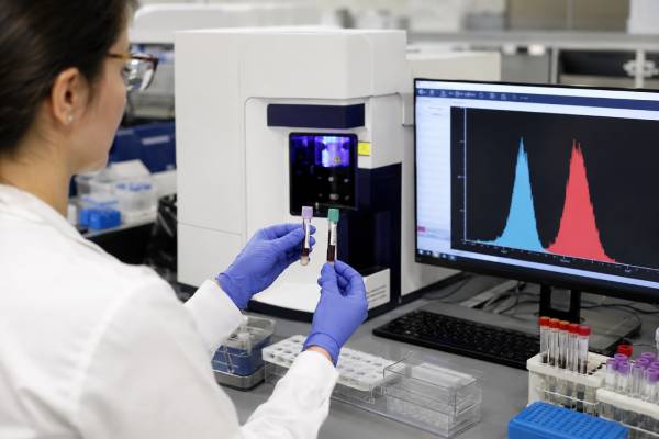
A neutrophil oxidative burst test measures whether neutrophils can generate reactive oxygen species after stimulation. The most widely used method is the dihydrorhodamine 123, or DHR, flow-cytometry assay. It is the standard screening test for chronic granulomatous disease (CGD), an inherited disorder of the phagocyte NADPH oxidase system. The test can show absent, reduced, mixed, or near-normal oxidative activity and may help identify female carriers of X-linked CGD. It does not measure every neutrophil function: migration, ingestion, granule release, and nonoxidative killing require different assessments. An abnormal DHR pattern must be interpreted with symptoms, infection history, medications, specimen quality, controls, and confirmatory genetic testing. Complete myeloperoxidase deficiency can produce a misleading abnormal DHR response even when NADPH oxidase activity is intact. Recent transfusion or granulocyte infusion can create a mixed pattern, and delayed transport can reduce cell function. Because CGD can cause severe bacterial and fungal infections and inflammatory complications, a strongly abnormal result or a highly suspicious clinical history requires prompt specialist evaluation.
- DHR flow cytometry measures the fluorescence shift produced when stimulated neutrophils oxidize a dye.
- CGD usually causes absent or reduced oxidative burst because the NADPH oxidase complex is defective.
- A two-population pattern can indicate an X-linked carrier, mixed donor cells, or recent granulocyte transfusion.
- Myeloperoxidase deficiency and poor specimen handling can mimic reduced oxidative activity.
- Functional abnormalities should be confirmed with molecular testing and interpreted with the infection phenotype.
Table of Contents
- What the Oxidative Burst Does
- How the DHR Test Works
- Why the Test Is Ordered
- DHR Patterns in Chronic Granulomatous Disease
- Carrier Testing and Mixed Cell Populations
- False-Abnormal and Indeterminate Results
- Specimen Handling, Timing, and Treatment Effects
- Confirmation, Follow-Up, and Practical Meaning
What the Oxidative Burst Does
Neutrophils are short-lived phagocytes that move rapidly to sites of infection. They attach to microbes, engulf them into phagosomes, release antimicrobial granule contents, and activate the NADPH oxidase complex. This enzyme transfers electrons to oxygen and generates superoxide, which is converted into hydrogen peroxide and other reactive oxygen species. Myeloperoxidase then uses hydrogen peroxide and chloride to help create additional antimicrobial oxidants.
This surge in oxygen-derived chemistry is called the respiratory or oxidative burst. It is especially important for killing certain catalase-positive bacteria and fungi. When NADPH oxidase is severely defective, phagocytes may ingest microbes but cannot generate a normal oxidative response. Organisms survive inside inflammatory cells, and the immune system forms granulomas in attempts to contain infection.
The NADPH oxidase complex has membrane and cytosolic components. Disease-causing variants in CYBB, which encodes gp91-phox, produce the common X-linked form of CGD. Autosomal recessive forms involve genes such as CYBA, NCF1, NCF2, NCF4, and CYBC1. Different variants leave different amounts of residual oxidase function.
Residual activity matters. People with some preserved superoxide production often have later onset, fewer severe infections, and better survival than those with almost no activity, although substantial complications can still occur. A quantitative DHR result therefore provides more information than a simple positive-or-negative screen.
CGD affects more than infection defense. Dysregulated inflammation can cause granulomatous obstruction, inflammatory bowel disease-like colitis, liver complications, lung inflammation, urinary or gastrointestinal obstruction, and poor wound healing. The test evaluates the enzyme defect underlying the disorder but cannot predict every inflammatory complication.
Oxidative burst is only one part of neutrophil biology. A normal DHR response does not rule out leukocyte adhesion deficiency, chemotaxis disorders, specific granule deficiency, severe congenital neutropenia, complement defects, antibody deficiency, or other causes of recurrent infection. It also cannot identify the organism causing an active infection or determine whether tissue damage has already developed in the affected organ. The clinical pattern determines whether additional immune testing is needed.
How the DHR Test Works
Fresh anticoagulated blood is incubated with dihydrorhodamine 123, a minimally fluorescent dye that enters neutrophils. The laboratory stimulates the cells, most commonly with phorbol 12-myristate 13-acetate, or PMA. PMA activates protein kinase C and drives assembly of the NADPH oxidase complex, producing reactive oxygen species.
As the oxidative burst proceeds, DHR is converted to fluorescent rhodamine 123. A flow cytometer measures fluorescence in thousands of individual neutrophils. Unstimulated cells establish the baseline, and stimulated cells should shift strongly toward higher fluorescence. The laboratory also runs a healthy control because cell transport and assay conditions affect performance.
Reports may include the percentage of DHR-positive neutrophils, mean fluorescence intensity, a stimulation index, or a neutrophil oxidative index. Some laboratories test PMA alone; others add formyl-methionyl-leucyl-phenylalanine, or fMLP, which activates a receptor-mediated pathway and may reveal selected partial defects. Numerical results are method-specific and should be interpreted against that laboratory’s controls and reference interval.
Flow plots provide information beyond one number. A normal sample shows one major population with a strong fluorescence shift after stimulation. Classic severe CGD shows little or no shift. Partial defects show a smaller shift, and carriers may show two distinct populations—one oxidizing normally and one poorly.
The DHR method largely replaced the nitroblue tetrazolium test. NBT relies on visual scoring of cells that reduce dye to blue formazan. It can distinguish broadly normal from abnormal burst but is less quantitative and less sensitive to mosaic carrier patterns. DHR is faster, objective, and better suited to measuring residual activity.
DHR fluorescence depends on several biochemical steps, including NADPH oxidase-generated hydrogen peroxide and myeloperoxidase-related oxidation. This dependence explains why complete myeloperoxidase deficiency can reduce the DHR signal even though superoxide generation is not absent. The pattern and confirmatory studies help separate these conditions.
A result may also include monocyte or lymphocyte gates as internal checks. Lymphocytes should not show a neutrophil-like oxidative shift, while monocytes can provide a lower-level positive response. Accurate gating prevents debris, eosinophils, dead cells, or abnormal populations from distorting interpretation.
Why the Test Is Ordered
The test is ordered when the infection pattern raises concern for CGD. Classic organisms include Staphylococcus aureus, Serratia marcescens, Burkholderia cepacia complex, Nocardia species, and Aspergillus species. Recurrent pneumonia, lymph-node infection, liver abscess, osteomyelitis, deep skin or soft-tissue infection, or invasive mold disease can prompt testing.
The pattern is often more important than the number of infections. A child with an unusual Aspergillus infection, Serratia liver abscess, or recurrent catalase-positive bacterial disease may warrant testing after a small number of events. Conversely, frequent uncomplicated viral upper-respiratory infections are not a typical CGD presentation.
CGD may present in infancy, childhood, adolescence, or adulthood. Residual oxidase activity, prophylaxis, environmental exposure, and the causative variant affect timing. Female carriers of X-linked CGD can develop inflammatory or autoimmune manifestations and, when X-chromosome inactivation is highly skewed, may also have clinically important infection risk.
Noninfectious findings can lead to testing. Granulomatous obstruction of the gastrointestinal or urinary tract, inflammatory colitis, unexplained granulomatous lesions, recurrent sterile inflammation, or a family history of CGD may raise suspicion. The test can also evaluate an asymptomatic relative before genetic results are available.
After hematopoietic cell transplantation, DHR can estimate restoration of NADPH oxidase function and demonstrate mixed donor and recipient neutrophil populations. It may complement donor-chimerism studies because genetic chimerism and functional oxidase recovery answer related but different questions.
The assay may be performed before gene therapy, after gene therapy, or during evaluation of residual function. In these settings, the laboratory and treating center often use protocol-specific analysis. A routine diagnostic reference interval may not capture all treatment-monitoring goals.
Testing should be coordinated with an immunologist or infectious-disease specialist when possible. Starting antimicrobial treatment for a serious infection should not be delayed while arranging DHR testing, and a normal result should not end the evaluation when the organism or phenotype strongly suggests another immune defect.
DHR Patterns in Chronic Granulomatous Disease
Severe X-linked CGD often produces a nearly flat stimulated histogram with little fluorescence increase. This indicates that most neutrophils cannot generate a measurable oxidative burst under strong PMA stimulation. Some autosomal recessive forms can look similar when the defect removes nearly all NADPH oxidase activity.
Hypomorphic variants produce reduced but not absent fluorescence. The percentage of responding cells may be high while the intensity per cell is low, or both percentage and intensity may be reduced. This residual activity can correlate with phenotype, but the DHR pattern alone does not identify every genotype with certainty.
Certain patterns can suggest the affected component. For example, some p47-phox deficiencies produce a characteristic partial shift, while gp91-phox defects may show absent or markedly reduced activity. However, overlapping patterns, laboratory differences, and uncommon variants prevent definitive genetic assignment from flow cytometry alone.
The strength of the stimulus matters. PMA bypasses many cell-surface signaling steps and directly activates intracellular pathways. A person with a receptor or signaling defect upstream of oxidase assembly may respond to PMA even if physiologic responses are impaired. Conversely, some residual CGD variants may appear closer to normal with strong PMA but show abnormal responses to fMLP or other receptor-dependent stimuli.
The laboratory should evaluate both how many neutrophils respond and how strongly each responds. A high responding percentage with reduced intensity means something different from a small normal-intensity population. Graphical plots and comments are often more informative than a single cutoff.
Clinical severity cannot be read from one fluorescence value alone. Residual oxidase production is an important prognostic factor, but infection exposure, prophylaxis, adherence, inflammatory disease, liver damage, and access to care also influence outcome. Even patients with measurable residual activity require specialist management.
An abnormal result confirms impaired oxidative burst under the assay conditions, not CGD by itself. Myeloperoxidase deficiency, specimen deterioration, treatment effects, and rare acquired conditions must be considered. Molecular confirmation establishes the affected gene and supports family testing, prognosis, and transplant or gene-therapy decisions.
Carrier Testing and Mixed Cell Populations
Females with one pathogenic CYBB variant usually have two neutrophil populations because random X-chromosome inactivation leaves some cells expressing the healthy copy and others expressing the altered copy. On DHR flow cytometry, this can appear as one normally shifting population and one nonshifting or weakly shifting population.
The proportion is not always close to 50:50. X-inactivation can be skewed, and the balance may change with age or hematopoietic selection. A carrier with a very small normal oxidizing population can have increased risk of serious infection. Inflammatory and autoimmune symptoms may occur even when the normally functioning fraction is larger.
Carrier detection is one of DHR’s major advantages over NBT. Flow cytometry shows the distribution at the single-cell level rather than averaging all neutrophils together. Still, subtle carrier patterns can be missed if the populations overlap, residual activity is substantial, or the sample quality is poor.
A mixed pattern is not automatically carrier status. Recent granulocyte transfusion can add donor neutrophils with normal function to a patient with CGD. Hematopoietic cell transplantation or gene therapy can also create mixed functional populations. The report must be interpreted with transfusion and treatment dates.
Somatic mosaicism and mixed donor chimerism are additional possibilities. In post-transplant care, the functional fraction can help assess whether enough oxidase-positive neutrophils are present, but it should be considered together with lineage-specific chimerism and clinical recovery.
Genetic testing is recommended for carrier confirmation. It identifies the familial variant, supports counseling, enables prenatal or preimplantation options when desired, and allows accurate testing of relatives. A normal DHR pattern does not exclude every carrier state, particularly for variants with preserved function or unusual lyonization.
Carrier counseling should avoid assuming that women are unaffected. Infection history, inflammatory symptoms, autoimmune features, oral ulcers, photosensitive or lupus-like manifestations, and changes in the oxidizing fraction may warrant specialist follow-up.
False-Abnormal and Indeterminate Results
Complete myeloperoxidase deficiency is the best-known biological mimic. DHR oxidation is reduced because the assay signal depends partly on myeloperoxidase chemistry. The flow pattern may be abnormal despite intact NADPH oxidase production. Other assays, such as superoxide measurement, NBT reduction, direct myeloperoxidase staining, or genetic testing, can resolve the distinction.
The DHR pattern in myeloperoxidase deficiency is often different from classic CGD, with more residual fluorescence, but this is not reliable enough to skip confirmation. Myeloperoxidase deficiency is commonly asymptomatic, although selected patients with diabetes or other risk factors may have increased susceptibility to Candida infection.
Poor cell viability can cause weak stimulation. Delayed delivery, refrigeration or heat exposure outside laboratory instructions, clotting, incorrect anticoagulant, and prolonged storage reduce neutrophil responsiveness. A simultaneously shipped healthy control helps reveal a transport problem affecting both samples.
Severe neutropenia can limit the number of events available for analysis. If too few viable neutrophils are collected, the result may be indeterminate or have wide uncertainty. Testing should not be forced into a normal-or-abnormal category when the cell count is inadequate.
Acute critical illness can alter neutrophil activation state. Cells may be primed, exhausted, immature, or exposed to inflammatory mediators that change baseline and stimulated fluorescence. This usually does not mimic the profound stable defect of classic CGD, but it can complicate borderline results.
Drugs that affect neutrophils or cellular metabolism can influence function. Corticosteroids, chemotherapy, immunosuppressants, and investigational agents should be documented. The antibiotic or antifungal treatment used for an infection generally should not be stopped simply to obtain a DHR test.
Some NCF1 variants can be technically challenging for routine genetic sequencing because of highly similar pseudogenes. A clearly abnormal functional result with negative initial sequencing may require deletion analysis, copy-number testing, specialized molecular methods, or broader genomic evaluation.
A normal PMA-stimulated DHR result does not rule out every phagocyte disorder. It also may not detect rare stimulus-specific signaling defects. When the clinical phenotype remains highly suggestive, specialists may request alternative stimulants, chemotaxis assays, phagocytosis testing, adhesion markers, granule-protein studies, or broader immune evaluation.
Specimen Handling, Timing, and Treatment Effects
DHR is a living-cell assay and usually requires fresh whole blood. The exact tube varies by laboratory, commonly sodium heparin, acid-citrate-dextrose, citrate, or another approved anticoagulant. Collection staff should follow the performing laboratory’s instructions rather than substituting a tube based on another center’s protocol.
Many laboratories require arrival within 24 hours, and some require same-day testing. Samples are often kept at room temperature and must not be refrigerated. Collection may be restricted to certain weekdays so the specimen does not sit through a weekend. Advance coordination is particularly important for patients who live far from the reference laboratory.
The date and time of collection should be recorded. A healthy control may need to be collected and shipped under similar conditions. If both the patient and control show weak oxidative responses, transport or assay failure is more likely than a true patient-specific defect.
Granulocyte transfusion can obscure the patient’s native pattern for as long as donor neutrophils remain in circulation. The laboratory and immunologist should know the timing and may recommend delaying nonurgent testing or interpreting the mixed population explicitly. Red-cell or platelet transfusion usually has a different effect, but any major treatment history should be documented.
After hematopoietic cell transplantation, DHR is expected to change as donor myeloid cells engraft. A mixed histogram can be clinically meaningful rather than artifactual. Serial tests may show increasing functional donor neutrophils, but the desired threshold and testing schedule depend on the transplant protocol.
Gene therapy can create a population of corrected oxidase-positive cells alongside uncorrected cells. Specialized centers may quantify expression and function in ways that differ from routine carrier analysis. Results should be trended in the same laboratory when possible.
Testing during active infection is acceptable and often necessary. The need for rapid diagnosis outweighs ideal baseline conditions. However, borderline abnormalities may need confirmation after recovery. Severe infection should be treated immediately regardless of whether the functional study has been completed.
Confirmation, Follow-Up, and Practical Meaning
A strongly abnormal DHR result should lead to prompt evaluation by clinical immunology. Confirmatory molecular testing identifies the causative gene and variant. The genetic result clarifies inheritance, allows testing of relatives, and may influence prognosis, transplant planning, conditioning choices, or eligibility for gene-based therapies.
The clinical workup includes a detailed infection history, culture and pathology records, imaging, inflammatory complications, growth, family history, and examination for granulomatous disease. Baseline blood counts, liver and kidney function, inflammatory markers, immunoglobulins, and other immune studies help identify complications or an alternative diagnosis.
If the pattern suggests myeloperoxidase deficiency, targeted MPO protein or genetic testing and an oxidase assay less dependent on MPO can distinguish it from CGD. If a female carrier pattern is suspected, CYBB sequencing and deletion or duplication analysis are appropriate. A mixed pattern after transplantation or transfusion should be correlated with chimerism and treatment records.
Patients diagnosed with CGD typically require antimicrobial prophylaxis, rapid evaluation of fever or focal symptoms, avoidance of selected high-risk exposures, and specialist monitoring for inflammatory disease. Interferon-gamma may be used in some settings. Hematopoietic cell transplantation can be curative, and gene therapy is an evolving option at specialized centers.
The DHR result should be retained as a graph and not only as a final label. The fluorescence distribution, responding fraction, residual intensity, stimulant, control performance, and collection timing can be important for future comparison. Repeating the test in the same laboratory improves trend interpretation.
A practical report answers four questions. Did most neutrophils respond? How strong was the response per cell? Was there one population or a mixture? Were controls and specimen quality acceptable? Those answers separate absent oxidase function, partial activity, carrier mosaicism, treatment-related mixtures, and unreliable testing.
Urgent care is needed for fever, breathing difficulty, severe pain, neurologic symptoms, rapidly spreading skin infection, or suspected invasive fungal disease in a person with known or possible CGD. A normal DHR result can be reassuring about NADPH oxidase function, but it does not replace microbiologic diagnosis or broader immune evaluation when severe infection is present.
References
- Diagnosis of Chronic Granulomatous Disease: Strengths and Challenges in the Genomic Era 2024 (Review)
- Functional Assays for the Diagnosis of Chronic Granulomatous Disease 2025 (Review)
- How I Treat: Chronic granulomatous disease 2026 (Clinical Review)
- Dihydrorhodamine Flow Cytometric Test, Blood 2026 (Laboratory Test Guide)
- Neutrophil Oxidative Burst Assay (DHR) 2026 (Laboratory Test Guide)
- Chronic granulomatous disease: Diagnosis and treatment 2025 (Patient Clinical Guidance)
Disclaimer
This article is for general education and does not diagnose chronic granulomatous disease, myeloperoxidase deficiency, or another immune disorder. DHR results require specialist interpretation with controls, specimen timing, transfusion and treatment history, infection pattern, and confirmatory genetic or biochemical testing. Serious bacterial or fungal infection in a person with known or suspected CGD requires urgent medical care.





