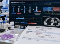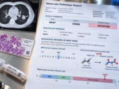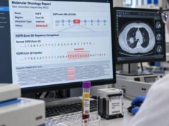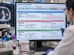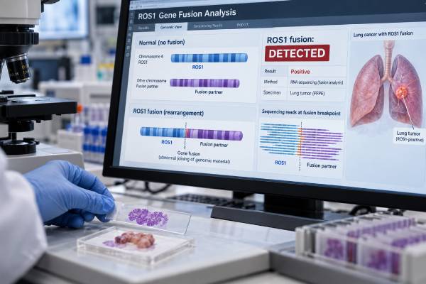
A ROS1 fusion test looks for a rearrangement that joins the ROS1 gene to another gene and creates an abnormal growth-driving kinase. In non-small cell lung cancer, a confirmed ROS1 fusion is an actionable molecular finding: it identifies a distinct tumor subtype that can respond to ROS1-targeted therapy. ROS1 fusions occur in roughly 1%–2% of NSCLC, most often in adenocarcinoma, and are more common in people with little or no smoking history, although clinical characteristics should never replace testing. The fusion can be detected with RNA- or DNA-based next-generation sequencing, fluorescence in situ hybridization, and in some settings immunohistochemistry as a screening method. RNA-based sequencing is particularly useful because it demonstrates the expressed fusion transcript and identifies the fusion partner. A positive result should name or otherwise confirm a functional ROS1 rearrangement, while a negative result must be interpreted in light of sample quality and the method used.
- What a positive ROS1 result means: The tumor contains a ROS1 gene fusion that is likely acting as an oncogenic driver and may make the cancer eligible for ROS1-targeted treatment.
- How common it is: ROS1 fusions are found in about 1%–2% of non-small cell lung cancers, especially adenocarcinomas.
- Best testing approach: Broad NGS, especially RNA-based fusion testing, can detect ROS1 while also evaluating other actionable lung cancer alterations.
- What a negative result means: No ROS1 fusion was detected by that assay; a negative result can be unreliable if the sample has too little tumor, degraded RNA, or incomplete fusion coverage.
- What is not the same as a fusion: ROS1 protein expression, a ROS1 point mutation, or an uncertain rearrangement is not automatically equivalent to a confirmed actionable ROS1 fusion.
Table of Contents
- What a ROS1 Fusion Is
- Who Should Be Tested for ROS1
- How ROS1 Fusions Are Detected
- Understanding a Positive ROS1 Result
- Understanding a Negative or Uncertain Result
- ROS1 and Other Lung Cancer Biomarkers
- Practical Next Steps After Testing
What a ROS1 Fusion Is
ROS1 is a gene that encodes a receptor tyrosine kinase, a signaling protein that can tell cells to grow and survive. In normal cells, its activity is tightly controlled. In some cancers, a chromosome rearrangement breaks ROS1 and joins its kinase-containing portion to part of another gene. The resulting fusion gene can produce a continuously active ROS1 fusion protein.
That abnormal protein sends growth signals without the normal regulatory controls. For this reason, a ROS1 fusion can function as an oncogenic driver: a central molecular event that helps sustain the cancer.
Several ROS1 fusion partners have been described in lung cancer. Common examples include CD74, EZR, SDC4, SLC34A2, TPM3, and CCDC6. The exact partner can affect laboratory detection and may have biologic implications, but the most important routine clinical question is usually whether a functional ROS1 kinase fusion is present.
ROS1 fusions are uncommon. Reviews consistently place them in about 1%–2% of NSCLC, with somewhat higher rates in selected adenocarcinoma populations. They are enriched in younger patients and never-smokers or light smokers, but they can occur outside those groups. Age, sex, and smoking history therefore should not be used to decide that testing is unnecessary when molecular profiling is otherwise indicated.
A ROS1 fusion is different from a point mutation. It is also different from simple overexpression of the ROS1 protein. Modern molecular reports may list many alterations, so the wording matters. A result that says “ROS1 fusion,” “ROS1 rearrangement,” or names a specific partner such as CD74–ROS1 is much more clinically meaningful than a nonspecific ROS1 variant with uncertain significance.
Who Should Be Tested for ROS1
ROS1 testing is part of modern molecular evaluation for appropriate patients with non-small cell lung cancer, particularly advanced non-squamous NSCLC and lung adenocarcinoma. Broad molecular profiling is favored because it can assess ROS1 alongside other actionable genes without consuming tissue through a long sequence of single-gene tests.
A lung cancer NGS panel can evaluate multiple driver mutations and gene fusions in one workflow. This is especially important in small biopsies, where tissue may also be needed for histologic stains and PD-L1 testing.
Testing should be driven by the cancer diagnosis and treatment setting rather than by a stereotyped patient profile. ROS1 fusions are more frequent in adenocarcinoma and in people who never smoked, but smokers can still have ROS1-positive disease. Restricting testing to young never-smokers would miss some patients who could benefit from targeted treatment.
ROS1 assessment may also become relevant again at disease progression. A patient with a known ROS1-positive cancer can develop resistance mechanisms after targeted therapy. Repeat tissue or liquid-biopsy molecular testing may help identify a ROS1 resistance mutation, bypass pathway, or another molecular change that can influence later treatment strategy.
In early-stage disease, the exact role and timing of broad molecular testing can depend on local guidelines, planned therapy, and specimen availability. In metastatic disease, identifying a targetable driver before finalizing first-line systemic treatment is especially important because driver status can change the preferred treatment sequence.
The NSCLC biomarker profile therefore includes more than ROS1. A complete workup may evaluate EGFR, ALK, BRAF, KRAS G12C, MET exon 14 skipping, RET, NTRK, HER2/ERBB2 and other alterations, as well as PD-L1 expression.
How ROS1 Fusions Are Detected
There are several ways to detect ROS1 rearrangements, and each method answers the question differently. The most useful result is one that reliably demonstrates a functional fusion and fits the tumor’s overall molecular profile.
RNA-based next-generation sequencing
RNA-based NGS is particularly well suited to gene-fusion detection. It tests the RNA transcript produced by the fusion, which helps confirm that the rearrangement is expressed and can identify the partner gene and fusion junction. Modern expert reviews increasingly favor RNA-based sequencing for difficult kinase fusions when adequate material is available.
The main limitations are RNA quality and quantity. Formalin-fixed tissue can contain degraded RNA, and tiny samples may not yield enough material. A failed RNA assay is not the same as a negative assay.
DNA-based next-generation sequencing
DNA NGS can test many genes at once and may detect ROS1 rearrangements, but fusion sensitivity depends on panel design. ROS1 breakpoints can occur in large intronic regions that are technically difficult to cover completely. A DNA-negative result can therefore warrant RNA testing when clinical or pathologic suspicion remains or when the panel reports limited fusion coverage.
Fluorescence in situ hybridization
FISH uses labeled probes that bind on either side of the ROS1 locus. A break-apart pattern indicates rearrangement. FISH has a long history in ROS1 testing and does not require knowing the partner gene in advance.
However, FISH usually does not identify the partner or prove that the rearrangement creates an expressed, in-frame fusion. Borderline patterns can be difficult to interpret, and unusual genomic events may require confirmation with sequencing.
Immunohistochemistry
ROS1 IHC detects increased ROS1 protein expression. It can be a useful screening method because it is fast and tissue-efficient, but ROS1 protein positivity is not completely specific for a true gene fusion. Positive or equivocal IHC generally needs molecular confirmation before the tumor is treated as ROS1 fusion-positive.
Liquid biopsy using circulating tumor DNA can also detect some ROS1 alterations when tissue is unavailable or inadequate. A positive plasma finding can be useful, but a negative blood test does not reliably exclude a fusion because some tumors shed little DNA into circulation and some assays have limited fusion sensitivity.
Understanding a Positive ROS1 Result
A confirmed positive result means that the cancer contains a ROS1 rearrangement expected to produce an active fusion kinase. This has both biologic and therapeutic meaning.
Biologically, the result identifies the tumor as part of the ROS1-driven NSCLC subgroup. ROS1 fusions are often mutually exclusive with other dominant kinase drivers, although co-alterations can occur. The fusion activates signaling pathways involved in proliferation and survival, making the ROS1 kinase a rational drug target.
Therapeutically, ROS1 positivity can make a patient eligible for approved ROS1 tyrosine kinase inhibitors. The treatment landscape has changed quickly, with newer agents designed to improve central nervous system activity and overcome some resistance mutations. The exact drug choice depends on disease setting, prior treatments, brain involvement, resistance profile, regulatory approvals, comorbidities, and current oncology guidelines.
A molecular report may provide the fusion partner and breakpoints. For example, a report might state CD74::ROS1 fusion detected. That is generally clearer than a report that says only “ROS1 rearrangement suspected.” If the finding is unusual, the molecular laboratory may classify it according to whether the fusion preserves the ROS1 kinase domain and is expected to be functional.
| Report wording | Typical meaning | What may be needed |
|---|---|---|
| ROS1 fusion detected with named partner | Strong evidence of a molecularly defined ROS1-positive tumor | Oncology review for targeted-treatment implications |
| ROS1 rearrangement detected by FISH | Supports ROS1 rearrangement | Sequencing may clarify an unusual or borderline result |
| ROS1 IHC positive | Protein expression raises suspicion for a fusion | Molecular confirmation is usually appropriate |
| ROS1 variant of uncertain significance | Not automatically an actionable fusion | Interpret variant type, evidence, and laboratory classification |
A positive result does not provide the lung cancer stage. Stage is determined by the anatomic extent of disease, including the primary tumor, lymph nodes, and distant metastases. ROS1 describes tumor biology, not how far the cancer has spread.
Understanding a Negative or Uncertain Result
A negative result means that the assay did not detect a reportable ROS1 fusion. It does not always mean the tumor has been definitively proven ROS1-negative.
The confidence of a negative result depends on three things: the specimen, the assay, and quality-control metrics. A sample with high tumor content and successful RNA-based NGS provides a more convincing negative result than a tiny biopsy with degraded nucleic acid or a DNA panel that incompletely covers ROS1 introns.
Reasons a fusion can be missed include:
- too few tumor cells in the tested section;
- low or degraded RNA quality;
- an uncommon partner or breakpoint outside the assay’s target regions;
- low circulating tumor DNA in a plasma sample;
- technical failure or insufficient sequencing depth;
- a complex rearrangement that is difficult for one platform to characterize.
If a report says “quantity not sufficient,” “RNA quality inadequate,” or “test failed,” it should not be summarized as negative. A second method or another specimen may be appropriate when the answer could change treatment.
An equivocal FISH or IHC result also needs care. Discordance between IHC, FISH, DNA sequencing, and RNA sequencing is possible. When results conflict, RNA-based testing can often clarify whether an expressed ROS1 fusion is present.
A negative ROS1 result also redirects attention to other biomarkers. It does not mean that no targetable driver exists. Broad testing may identify an EGFR mutation, ALK fusion, RET fusion, MET exon 14 skipping alteration, BRAF V600E, KRAS G12C, HER2 alteration, NTRK fusion, or another clinically relevant change.
ROS1 and Other Lung Cancer Biomarkers
ROS1 belongs to a group of kinase gene fusions that includes ALK, RET, and NTRK. These alterations can look similar at a high level—each creates an abnormal signaling protein—but they are different molecular targets and require distinct test interpretation.
An ALK fusion is not interchangeable with a ROS1 fusion even though some drugs have activity against both kinases. Likewise, a RET rearrangement or NTRK fusion defines a different molecular subtype with its own targeted treatment evidence.
PD-L1 is another important lung cancer biomarker, but it measures immune-related protein expression rather than a driver gene. A ROS1-positive tumor can have a PD-L1 result, yet the presence of a targetable oncogenic driver can strongly influence treatment sequencing. This is one reason clinicians try to obtain the full molecular profile before selecting systemic therapy in advanced NSCLC.
Tumor mutational burden is also conceptually different. TMB estimates the number of somatic mutations per megabase of analyzed DNA; it does not identify a specific kinase driver. A tumor can have a ROS1 fusion and a measurable TMB, but the ROS1 result is the more direct molecular explanation for ROS1-driven growth.
The practical lesson is that a biomarker report is not a list in which every positive line has equal weight. A confirmed ROS1 fusion is a high-value driver alteration because it defines a druggable molecular subtype. Other findings may describe immune phenotype, co-mutations, resistance biology, or variants whose significance is uncertain.
Practical Next Steps After Testing
When a ROS1 test is ordered, no special fasting or preparation is needed. The key practical issues are obtaining a good tumor specimen, preserving enough tissue, and choosing a method that can reliably detect fusions.
After the result, the following questions are useful:
- Was a true fusion or rearrangement detected? Read the exact wording rather than relying only on “ROS1 positive.”
- Which method was used? RNA NGS, DNA NGS, FISH, IHC, and plasma testing have different strengths and limitations.
- Was the sample adequate? Check for comments about tumor percentage, sequencing quality, RNA failure, or limited coverage.
- Was a fusion partner identified? A named in-frame partner can strengthen confidence in the molecular finding.
- Has the full biomarker panel been completed? Treatment planning usually depends on the entire molecular and PD-L1 profile.
- Is repeat testing needed at progression? Molecular reassessment may help identify resistance mechanisms after ROS1-targeted treatment.
Patients should not interpret a ROS1 result as a hereditary cancer test. Most ROS1 fusions in lung cancer are somatic, meaning they arose in tumor cells rather than being inherited through the family. Germline genetic counseling is based on personal and family history and on specific inherited variants, not on a routine tumor ROS1 fusion alone.
A ROS1-positive result is also not a prognosis by itself. Outcomes depend on stage, overall health, sites of disease, response to therapy, brain involvement, resistance mechanisms, and access to effective treatment. What the result does provide is something highly actionable: a molecular explanation for tumor growth that can be directly targeted.
For a negative result, the most important question is whether the test was technically capable of excluding the fusion. If it was, the workup moves to other drivers. If it was not, repeating fusion testing with a stronger method can prevent a clinically important alteration from being missed. In modern lung cancer care, the quality of the molecular test matters almost as much as the name of the gene being tested.
References
- Gene fusions in non-small cell lung carcinoma (NSCLC): Expert perspectives and practical considerations for routine testing 2026 (Review)
- ROS1-positive non-small cell lung cancer: from genomics to treatment decisions 2026 (Review)
- Advances and future directions in ROS1 fusion-positive lung cancer 2024 (Review)
- ROS1-rearranged non-small cell lung cancer: Understanding biology and optimizing management in the era of new approvals 2024 (Review)
- Current status of molecular diagnostics for lung cancer 2024 (Review)
- Gene Fusion Detection in NSCLC Routine Clinical Practice: Targeted-NGS or FISH? 2023
Disclaimer
ROS1 testing should be interpreted by the oncology and molecular pathology team in the context of the complete diagnosis and assay quality. A positive ROS1 fusion can have major treatment implications, while a technically limited negative result may require repeat or alternative testing. Do not start, stop, or change cancer treatment based on a molecular report without specialist review.