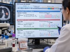
A lung cancer NGS panel is a molecular test that examines many cancer-related genes at the same time. In non-small cell lung cancer (NSCLC), especially advanced non-squamous disease, it is used to find driver mutations, gene fusions, copy-number changes, and other genomic alterations that can influence treatment. NGS stands for next-generation sequencing, a technology that can analyze far more DNA or RNA than older single-gene tests from a relatively small tissue or blood sample. The main benefit is efficiency: one broad test can look for multiple actionable biomarkers instead of using limited tumor material one marker at a time. Results must still be interpreted carefully. A panel can report an actionable alteration, a resistance mechanism, a variant of uncertain significance, or no detectable target. The clinical meaning depends on the exact gene change, cancer type and stage, prior treatment, specimen quality, and whether the panel analyzed both DNA and RNA where needed.
- What it measures: A lung cancer NGS panel can test many genes at once for mutations, insertions/deletions, gene fusions, splice alterations, and sometimes copy-number changes, MSI, or tumor mutational burden.
- Why broad testing matters: NSCLC can be driven by EGFR, ALK, ROS1, BRAF, KRAS, MET, RET, NTRK, ERBB2/HER2, and other alterations, and each may require a different treatment approach.
- What a positive result means: A pathogenic actionable driver can identify a targeted therapy or clinical-trial option when the finding is valid for the patient’s lung cancer setting.
- What a negative result means: “No actionable alteration detected” does not always mean the tumor has none; poor specimen quality, low tumor content, limited panel design, or plasma under-shedding can cause missed findings.
- Best specimen strategy: Tissue remains central because it confirms tumor type and supports protein testing, while plasma ctDNA can complement tissue when tissue is limited or faster genomic information is needed.
Table of Contents
- What a Lung Cancer NGS Panel Tests
- Who Needs NGS Testing and When
- Tissue NGS vs Blood-Based NGS
- How to Read an NGS Report
- Driver Mutations, Fusions, and Other Key Findings
- NGS for Resistance Testing
- Limitations and Next Steps After Testing
What a Lung Cancer NGS Panel Tests
Next-generation sequencing is not one single test. It is a family of laboratory methods that can read many DNA or RNA sequences in parallel. In lung cancer, an NGS panel usually focuses on genes known to affect tumor growth, prognosis, drug sensitivity, drug resistance, or clinical-trial eligibility.
A broad panel may detect several alteration types:
- Single-nucleotide variants, in which one DNA letter changes.
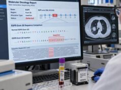
- Small insertions and deletions, such as EGFR exon 19 deletions or exon 20 insertions.
- Gene fusions or rearrangements, such as ALK, ROS1, RET, or NTRK fusions.
- Splice-related alterations, including variants that lead to MET exon 14 skipping.
- Copy-number changes, such as amplification of MET or other genes.
- Complex genomic biomarkers, depending on the platform, such as microsatellite instability (MSI) or tumor mutational burden (TMB).
Some panels analyze DNA only. Others combine DNA and RNA. That distinction matters because RNA-based methods can improve detection of expressed gene fusions and splice alterations. A panel advertised as “large” is not automatically better if it lacks reliable coverage for the alteration types that matter in NSCLC.
Broad NGS is increasingly favored over sequential single-gene testing because lung tumors may contain one of many mutually exclusive oncogenic drivers. Testing genes one at a time can consume tissue, delay results, and leave less material for later studies. A well-designed NSCLC biomarker workup combines genomic testing with pathology and non-genomic biomarkers rather than treating them as separate competing steps.
Who Needs NGS Testing and When
Comprehensive molecular profiling is especially important in advanced or metastatic non-squamous NSCLC, where guideline-based treatment depends heavily on actionable genomic findings. Broad testing may also be appropriate in selected squamous cancers, particularly when clinical features or limited biopsy material raise concern for an adenocarcinoma component, and in other settings where actionable targets or tumor-agnostic markers could affect care.
The timing is critical. For newly diagnosed advanced NSCLC, molecular testing should ideally be initiated early enough that the results are available before choosing systemic therapy, provided the patient is clinically stable. Starting an empiric treatment before key driver results are known can complicate later treatment sequencing.
NGS may also be ordered:
- after surgery or chemoradiation when a specific biomarker affects adjuvant or consolidation treatment;
- at recurrence if prior molecular testing was incomplete or outdated;
- at progression on targeted therapy to search for acquired resistance;
- when only a tiny biopsy is available and many biomarkers must be assessed efficiently; or
- when a patient is being considered for a molecularly matched clinical trial.
The exact test should match the clinical question. A newly diagnosed tumor may need broad driver profiling, whereas a patient progressing on a targeted drug may need a panel designed to detect both the original driver and secondary resistance mechanisms.
PD-L1 testing is usually performed separately by immunohistochemistry. A genomic NGS panel does not replace a PD-L1 test unless the treatment question specifically concerns a genomic biomarker rather than protein expression.
Tissue NGS vs Blood-Based NGS
Tissue and plasma NGS answer overlapping questions but have different strengths. Tissue is obtained from a biopsy or surgical specimen and directly samples the tumor. Plasma NGS analyzes circulating tumor DNA (ctDNA) released into blood.
| Feature | Tissue NGS | Plasma ctDNA NGS |
|---|---|---|
| Specimen | Tumor biopsy or resection tissue | Blood plasma |
| Main advantage | Direct tumor material; can pair with histology and other tissue tests | Minimally invasive, fast to obtain, can sample DNA from multiple tumor sites |
| Main limitation | May require an invasive procedure and can be limited by small samples | False negatives occur when little ctDNA is released into blood |
| Useful at progression | Can show resistance plus histologic transformation | Can detect multiple circulating resistance clones without a new invasive biopsy |
A lung cancer liquid biopsy can be particularly valuable when tissue is insufficient, difficult to obtain, or needed urgently for other pathology tests. A positive plasma finding for a well-validated actionable driver may be enough to guide targeted treatment in the appropriate setting.
A negative plasma test is different. It may reflect low ctDNA shedding rather than a genuinely mutation-negative tumor. When plasma is negative or non-informative, tissue molecular testing should generally be pursued if feasible. Some clinicians order tissue and plasma in parallel because the methods can be complementary rather than mutually exclusive.
Specimen quality also matters for tissue. The pathology laboratory estimates tumor cellularity and may mark an area enriched for tumor. Decalcified bone samples, very small biopsies, old blocks, or heavily necrotic tissue can sometimes reduce DNA/RNA quality. The report may state that the specimen failed quality-control thresholds or that certain alteration types could not be assessed reliably.
How to Read an NGS Report
An NGS report can look intimidating because it may include dozens or hundreds of genes. The most useful approach is to focus first on the clinical classification of each result rather than the raw number of findings.
Pathogenic or likely pathogenic actionable alterations
These are the highest-priority findings. They are alterations known or strongly suspected to drive cancer and may be linked to an approved targeted therapy, a guideline-supported treatment, or a relevant trial. The report may place them in tiers based on level of evidence.
The exact alteration matters. “EGFR positive” is less informative than “EGFR exon 19 deletion” or “EGFR L858R,” because different EGFR alterations can have different treatment implications. The same principle applies across other genes.
Variants of uncertain significance
A variant of uncertain significance (VUS) is a genetic change whose role is not established. VUS findings are common on large panels. They should not be treated as equivalent to an actionable driver and generally should not determine therapy by themselves.
Allele fraction and tumor content
Reports may provide a variant allele fraction (VAF), which is the percentage of sequenced DNA molecules carrying the variant at a specific location. VAF does not directly equal the percentage of tumor cells. It can be affected by tumor purity, copy-number changes, normal-cell contamination, tumor heterogeneity, and whether the sample is tissue or plasma.
Negative results and technical comments
A “negative” report should be checked for what was actually tested. Important questions include whether the panel covered fusions, whether RNA sequencing succeeded, whether tumor content was adequate, and whether the assay could evaluate copy-number changes. A negative limited DNA panel is not equivalent to a complete DNA-plus-RNA profile.
Some reports also list MSI or TMB. These biomarkers can have clinical relevance in selected settings, but they should not distract from established lung cancer drivers. A dedicated TMB test for lung cancer or MSI assessment may be interpreted differently from a driver-gene result.
Driver Mutations, Fusions, and Other Key Findings
A driver alteration gives cancer cells a growth advantage and can become a therapeutic target. Modern NSCLC treatment relies on identifying specific drivers rather than treating all tumors as molecularly identical.
Commonly evaluated actionable categories include:
- EGFR: activating mutations such as exon 19 deletions and L858R, plus less common sensitizing or resistance-associated variants. See the focused EGFR mutation test for alteration-specific interpretation.
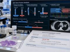
- ALK: rearrangements create oncogenic fusions that can respond to ALK inhibitors. An ALK fusion result is fundamentally different from a nonspecific ALK sequence variant.
- ROS1 and RET: actionable fusions can be found by appropriately designed DNA or RNA assays.
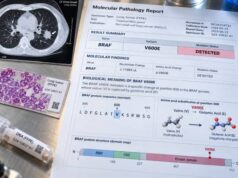
- BRAF: BRAF V600E is a well-established actionable driver, while non-V600 BRAF variants have different implications.
- KRAS: KRAS G12C has specific targeted-treatment relevance; other KRAS variants do not automatically carry the same treatment options.
- MET: clinically important alterations include MET exon 14 skipping and, in selected contexts, MET amplification. A MET exon 14 skipping test may require careful evaluation of splice-site variants and RNA evidence.
- NTRK: NTRK1/2/3 fusions are rare in lung cancer but can be actionable under tumor-agnostic treatment indications.
- ERBB2/HER2: activating ERBB2 mutations, especially in the kinase domain, can have treatment implications and should be distinguished from HER2 protein expression or gene amplification.
The panel may also identify co-mutations in genes such as TP53, STK11, or KEAP1. These findings can provide prognostic or biologic context and may influence clinical-trial interpretation, but they do not necessarily function as direct drug targets in routine NSCLC care.
The key question is not “How many mutations were found?” It is “Which alterations are valid, clinically actionable, and relevant to this patient’s treatment setting?” A tumor with one strong driver can have a much clearer treatment path than a tumor with several non-actionable variants.
NGS for Resistance Testing
Cancer genomes change during therapy. Targeted drugs suppress sensitive tumor cells, but resistant clones can emerge. Repeat NGS at progression can identify how the tumor escaped treatment and whether another targeted strategy is possible.
Resistance mechanisms include:
- a second mutation in the original drug target;
- amplification or activation of a bypass pathway;
- a new fusion or other acquired genomic change;
- loss of the original detectable clone; or
- histologic transformation that may not be identifiable by genomic testing alone.
Examples include secondary EGFR resistance mutations, ALK kinase-domain substitutions, and MET amplification after EGFR-directed therapy. A broad lung cancer resistance mutation panel can be useful when progression occurs after a targeted drug.
Tissue and plasma have complementary roles here. Plasma may capture DNA from several metastatic sites and reveal multiple resistant subclones. Tissue can show morphology, including transformation to small cell carcinoma or another phenotype, which a DNA report cannot diagnose by itself.
The timing of repeat testing should be clinically purposeful. Routine serial NGS without a treatment decision in mind can generate ambiguous data. In contrast, testing at confirmed progression may directly inform the next therapy or trial.
A practical detail is whether the panel uses DNA, RNA, or both. DNA sequencing is excellent for many substitutions and small insertions or deletions, and it can often detect rearrangements when the assay covers the relevant introns. RNA sequencing looks at gene transcripts after splicing has occurred. That can make it especially useful for confirming that a rearrangement produces an expressed fusion or that a splice alteration actually results in an abnormal transcript. In a small lung biopsy, a combined strategy can reduce the chance that an important fusion is missed because the DNA breakpoint lies in a difficult region.
The number of genes on a panel should therefore not be the only quality measure. A 500-gene DNA-only assay may be less useful for a specific fusion question than a smaller, well-validated DNA-plus-RNA assay. Coverage depth, tumor-content requirements, fusion design, copy-number validation, and reporting standards all affect clinical performance.
NGS can also report broad genomic signatures. TMB estimates the number of somatic mutations across a defined amount of sequenced DNA, while MSI reflects defects in DNA mismatch repair that create instability in repetitive sequences. These are conceptually different from oncogenic drivers. A high TMB or MSI-high result does not identify one mutated protein to inhibit in the same way that an EGFR or ALK driver does. In lung cancer, such signatures should be interpreted in the treatment context and alongside established biomarkers rather than used as shortcuts around complete molecular profiling.
Limitations and Next Steps After Testing
NGS is powerful, but it does not guarantee that an actionable target will be found. It also does not replace the clinical judgment needed to decide whether a detected alteration is truly driving the tumor.
Important limitations include:
- Insufficient tumor material: Small biopsies may not contain enough viable tumor for high-quality sequencing.
- Incomplete assay design: Some panels have strong mutation coverage but weak fusion or copy-number detection.
- False-negative plasma results: Low-shedding disease may produce little ctDNA.
- VUS overinterpretation: Uncertain variants can be mistaken for actionable mutations.
- Changing evidence: The significance of a rare alteration may evolve as new trials and approvals emerge.
- Germline implications: Some tumor findings can suggest an inherited variant, but tumor-only NGS cannot always distinguish somatic from germline origin. Confirmatory germline testing may be needed when clinically appropriate.
- Turnaround time: Results can take roughly one to several weeks depending on the specimen, assay, need for repeat extraction, and laboratory workflow.
After receiving the report, the oncology team should confirm whether all major clinically relevant alteration classes were adequately tested. If an actionable driver is found, the result should be matched to the patient’s stage, line of therapy, comorbidities, and treatment history. If no driver is found, the team should decide whether the result is truly comprehensive or whether additional tissue, RNA, fusion, or plasma testing is justified.
Complex reports may benefit from review by a molecular tumor board, especially when the finding is rare, the evidence is emerging, or multiple possible therapies compete. The goal of NGS is not simply to produce a long genomic report. It is to turn a limited cancer specimen into precise information that helps choose the safest and most effective next step.
References
- Recommendations for the use of next-generation sequencing (NGS) for patients with advanced cancer in 2024: a report from the ESMO Precision Medicine Working Group 2024 (Guideline)
- Recommendations for reporting tissue and circulating tumour (ct)DNA next-generation sequencing results in non-small cell lung cancer 2024 (Position Statement)
- Clinical Practice Recommendations for the Use of Next-Generation Sequencing in Patients with Solid Cancer: A Joint Report from KSMO and KSP 2024 (Guideline)
- A Paradigm Shift in Non-Small-Cell Lung Cancer (NSCLC) Diagnostics: From Single Gene Tests to Comprehensive Genomic Profiling 2024 (Review)
- Guidance for clinicians and patients with non-small cell lung cancer in the time of precision medicine 2023 (Review)
Disclaimer
This article provides general educational information and is not a substitute for individualized cancer care. NGS results should be interpreted by qualified oncology and pathology professionals using the exact assay, specimen quality, cancer subtype, stage, and treatment history. Do not change treatment based on a genomic report without discussing it with the treating team.