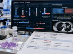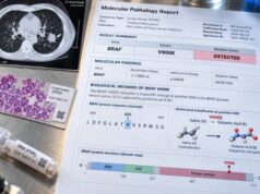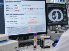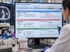
A lung cancer resistance mutation panel is a molecular test used after targeted therapy stops controlling the cancer or when resistance is strongly suspected. It looks for new genomic changes that may explain why a previously effective drug is no longer working. In non-small cell lung cancer (NSCLC), resistance can develop through a new mutation in the original driver gene, activation of a bypass pathway such as MET, a new gene fusion, amplification of another oncogene, or a change in tumor biology that genomic testing alone may not capture. The panel may be performed on a new tissue biopsy, a blood-based circulating tumor DNA (ctDNA) sample, or both. The most useful result is one that identifies a clinically meaningful resistance mechanism and changes the next treatment decision. A negative result does not prove that resistance is absent, because low tumor DNA, incomplete assay coverage, tumor heterogeneity, or non-genetic mechanisms can leave the cause unexplained.
- What it measures: Resistance panels may evaluate secondary EGFR, ALK, and ROS1 mutations, MET amplification or activation, new fusions, and other bypass-pathway alterations that emerge during targeted therapy.
- When it is ordered: Testing is most useful at confirmed progression on a targeted drug, when the result could guide a new therapy, combination strategy, or clinical trial.
- What a positive result means: A detected resistance alteration may explain treatment failure and can sometimes identify a specific next-line targeted option.
- What a negative result means: No detected resistance marker does not rule out resistance; tissue biopsy may still be needed, especially if plasma ctDNA is negative or histologic transformation is possible.
- Blood vs tissue: Plasma can sample DNA from several tumor sites at once, while tissue can reveal tumor-cell morphology and transformations that a blood genomic test cannot diagnose.
Table of Contents
- What Resistance Mutation Testing Means
- When a Resistance Panel Is Useful
- EGFR Resistance Markers
- ALK, ROS1, and MET Resistance
- Tissue vs Plasma Resistance Testing
- How to Read Resistance Panel Results
- Limitations and Next Steps
What Resistance Mutation Testing Means
Targeted therapy works by blocking a molecular feature that the cancer depends on. Examples include EGFR mutations, ALK fusions, ROS1 fusions, and MET alterations. These treatments can produce deep and durable responses, but advanced cancers often evolve under drug pressure. Cells that can grow despite treatment become more prominent, producing acquired resistance.
A resistance mutation panel is designed to investigate that new tumor state. It is different from an initial diagnostic biomarker panel because the question has changed from “What drives this cancer?” to “How has this cancer escaped the drug?”
Resistance mechanisms fall into several broad groups:
- On-target resistance: the original drug target changes so the drug binds less effectively or no longer suppresses signaling.
- Bypass signaling: another pathway becomes active and allows the cancer to grow even though the original target remains inhibited.
- Gene amplification: extra copies of a growth-promoting gene can create enough signaling to overcome treatment.
- New oncogenic fusions or mutations: an alternate driver can emerge during therapy.
- Phenotypic or histologic transformation: the tumor changes cell state, such as transformation from adenocarcinoma to small cell lung cancer.
- Non-genetic resistance: epigenetic changes, drug-tolerant persister states, and interactions with the tumor microenvironment can contribute even when no clear DNA resistance alteration is found.
A broad lung cancer NGS panel may detect many of these genomic mechanisms if its design includes the relevant genes and alteration types. However, a panel cannot diagnose every form of resistance, especially changes that depend on tumor morphology.
When a Resistance Panel Is Useful
Resistance testing is most useful when there is objective disease progression on targeted therapy and the result could change management. Progression may be seen on imaging, inferred from new metastatic disease, or supported by worsening symptoms. A rising tumor marker alone is generally not enough to define a molecular resistance mechanism.
The timing should match a clinical decision. Examples include:
- progression on an EGFR tyrosine kinase inhibitor (TKI);
- progression on an ALK- or ROS1-directed TKI;
- progression after therapy for MET exon 14–altered disease;
- mixed progression, where some lesions remain controlled while others grow;
- recurrence after a prior targeted treatment; or
- consideration of a trial that requires proof of a specific acquired alteration.
Testing is less useful when the next treatment would be the same regardless of the result. It can also be misleading if ordered too early during a brief radiographic fluctuation that has not been confirmed as true progression.
Resistance testing should start with a clear record of the original driver alteration, the drugs already used, how long they worked, and the pattern of progression. The same secondary mutation can have different implications depending on which drug selected for it. The patient’s treatment history is therefore part of the molecular interpretation, not just background information.
EGFR Resistance Markers
EGFR-mutant NSCLC provides the clearest example of how resistance evolves over successive generations of targeted therapy. The specific mechanism depends strongly on which EGFR inhibitor was used.
EGFR T790M
EGFR T790M became a classic resistance marker after first- and second-generation EGFR TKIs. It changes the kinase domain and reduces the effectiveness of earlier drugs. The emergence of third-generation EGFR inhibitors changed the treatment landscape because these drugs were designed to inhibit sensitizing EGFR mutations together with T790M.
Today, the importance of a T790M resistance test depends on the prior therapy and clinical setting. It remains an important example of an on-target resistance mutation, but resistance after first-line third-generation therapy is more heterogeneous.
EGFR C797S and other on-target changes
After osimertinib and related third-generation therapy, EGFR C797S is one recognized on-target resistance mechanism. The C797 residue is involved in covalent binding of these inhibitors, so alteration at that site can reduce drug activity. Other less common EGFR kinase-domain substitutions may also emerge.
The exact configuration of resistance mutations can matter. For example, whether two EGFR mutations occur on the same DNA copy or on different copies can influence theoretical sensitivity to combinations of inhibitors. This level of interpretation requires an experienced molecular oncology team and is not captured by a simple “positive/negative” label.
EGFR-independent resistance
Many cancers progressing after an EGFR inhibitor do not acquire another EGFR mutation. Instead, they activate alternative signaling. MET amplification is one of the best-known bypass mechanisms. HER2 amplification, alterations in RAS-MAPK or PI3K pathways, new oncogenic fusions, and cell-cycle changes have also been reported.
This is why testing only the EGFR gene at progression can miss clinically important biology. A broad resistance panel may be more informative than a single-gene assay when the resistance mechanism is unknown.
Histologic transformation is another major category. Some EGFR-mutant adenocarcinomas transform into small cell lung cancer. That diagnosis requires tissue morphology and immunophenotyping; a plasma panel cannot confirm it.
ALK, ROS1, and MET Resistance
Resistance in fusion-driven lung cancers often follows the same broad principles as EGFR resistance: the kinase target itself can mutate, bypass pathways can activate, and tumor heterogeneity can generate several resistant clones at once.
ALK resistance mutations
ALK-positive NSCLC is driven by a fusion that activates the ALK kinase. Successive ALK inhibitors have different activity against secondary ALK mutations. Important reported resistance substitutions include G1202R, a solvent-front mutation, and L1196M, a gatekeeper mutation, among others.
The significance of an ALK mutation depends on the drug used previously and the sensitivity profile of possible next-generation inhibitors. A resistance report should therefore name the exact amino-acid substitution rather than simply state “ALK mutation detected.”
Bypass mechanisms such as MET amplification can also occur in ALK-driven disease, so an ALK fusion should not be interpreted in isolation when the cancer has already progressed on an ALK TKI.
ROS1 resistance mutations
ROS1 fusion-positive NSCLC can develop kinase-domain resistance mutations after ROS1-directed therapy. ROS1 G2032R is a well-described solvent-front mutation; other substitutions such as D2033N, L2026M, and additional kinase-domain changes have also been reported.
The exact resistance spectrum varies by prior inhibitor. Because some next-generation ROS1 inhibitors were designed to retain activity against particular resistant kinase variants, precise molecular identification can matter for treatment sequencing and clinical-trial selection.
A dedicated ROS1 fusion test confirms the original driver, but progression often requires broader testing that can detect secondary ROS1 mutations and alternative pathways.
MET as a driver and as a bypass resistance pathway
MET can be relevant in more than one way. MET exon 14 skipping is a primary oncogenic driver in a subset of NSCLC, while MET amplification can function either as a primary genomic event or as an acquired bypass mechanism after therapy directed at another driver.
This distinction matters clinically. A MET exon 14 skipping result is not the same as increased MET copy number. Likewise, a MET amplification test must be interpreted according to the assay method, copy-number threshold, tumor fraction, and treatment context.
Resistance to MET-directed therapy can itself involve secondary MET kinase-domain mutations or activation of alternative pathways. This illustrates why resistance testing is an evolving snapshot rather than a permanent molecular label.
Tissue vs Plasma Resistance Testing
Both tissue and blood-based ctDNA testing can be useful at progression, but they do not provide identical information.
| Question | Plasma ctDNA | Tissue biopsy |
|---|---|---|
| Can it detect genomic resistance mutations? | Yes, if enough ctDNA is present and the assay covers the alteration | Yes, if the sampled lesion contains the resistant clone |
| Can it sample multiple tumor sites? | Potentially, because DNA may come from several lesions | No; it directly samples one biopsy site |
| Can it diagnose small cell transformation? | No | Yes, through histopathology |
| Can a negative result miss resistance? | Yes, especially with low ctDNA shedding | Yes, especially if resistance differs across tumor sites or tissue quality is poor |
A liquid biopsy is attractive because it is minimally invasive and can be repeated. It may reveal more than one resistance clone circulating from different metastatic deposits. This is particularly useful in genetically heterogeneous progression.
Its main weakness is sensitivity. If plasma does not contain enough tumor DNA, the test can be falsely negative. In that situation, a tissue biopsy should be considered when feasible. Tissue is also preferred when transformation is suspected because only tissue can show the tumor’s cell type and architecture.
The best strategy is often complementary: use plasma for fast broad genomic information and tissue when morphology, deeper molecular characterization, or confirmation is needed.
How to Read Resistance Panel Results
A resistance report should be interpreted in three steps: confirm the original driver, identify the new alteration, and ask whether the new finding has a validated therapeutic implication.
A result may fall into one of four practical categories:
- A recognized actionable resistance mechanism is detected. This is the most useful outcome because it can support a specific next-line treatment or trial.
- A resistance alteration is detected, but there is no established standard therapy for it. The finding may still help with trial selection or explain why the prior drug failed.
- Only the original driver is detected. The cancer is still shedding tumor DNA, but the assay has not identified a clear genomic escape mechanism.
- No informative tumor DNA is detected. This is not the same as “no resistance.” It may indicate low shedding or inadequate assay sensitivity and can justify tissue testing.
The variant allele fraction (VAF) can provide context but should not be interpreted as the percentage of cancer cells carrying the mutation. It depends on the amount of tumor DNA in the specimen, copy number, sampling, and assay characteristics.
Copy-number findings require particular care. A report of MET amplification may use different thresholds depending on whether the test is tissue NGS, fluorescence in situ hybridization (FISH), or plasma sequencing. “Amplified” is therefore not a perfectly interchangeable term across laboratories.
Variants of uncertain significance should not be treated as proven resistance drivers. A new rare mutation that appears during progression may be biologically interesting, but treatment should not be based on it unless evidence supports its role.
Resistance testing also has to be interpreted alongside the pattern of progression. A patient with growth in one or two isolated lesions while the rest of the cancer remains controlled may have a resistant subclone confined to those sites. In selected cases, clinicians may treat the growing lesions locally and continue the same targeted drug rather than switch all systemic therapy immediately. By contrast, widespread progression across several organs suggests that a resistant population has become more dominant and may make a systemic treatment change more urgent. Molecular results do not make this decision by themselves, but they can help explain the pattern.
Brain progression deserves special attention. Some targeted drugs penetrate the central nervous system better than others, and plasma ctDNA can underrepresent disease confined to the brain because tumor DNA from the central nervous system may not reach peripheral blood efficiently. A negative plasma resistance panel in a patient with brain-only progression is therefore especially weak evidence against a resistance mechanism. Imaging, cerebrospinal fluid testing in selected circumstances, or tissue from an accessible progressing lesion may provide more useful information.
Finally, resistance is not always a single-step event. A tumor can contain several coexisting resistant populations, and those populations can change again after the next therapy. A resistance panel is best viewed as a time-specific molecular snapshot. The most clinically useful reports clearly distinguish the original driver, acquired alterations, uncertain variants, assay limitations, and whether tumor DNA was present at a level that makes a negative result interpretable.
Limitations and Next Steps
Resistance testing is valuable because it can turn progression into a new molecular treatment question, but it has limits.
The main limitations are:
- resistant clones can differ between tumor sites;
- some cancers release little ctDNA into blood;
- a panel may not cover all relevant fusions, copy-number changes, or rare kinase-domain mutations;
- non-genetic resistance may not appear on DNA sequencing;
- histologic transformation requires tissue;
- prior treatment changes which resistance mechanisms are plausible; and
- a molecular finding may be scientifically valid without having an approved treatment attached to it.
After results are available, the oncology team should ask whether the finding is actionable now, whether another specimen is needed, and whether the tumor should be reviewed in a molecular tumor board. If plasma is negative and disease is clearly progressing, tissue is often the next logical step when biopsy is safe. If tissue shows transformation, treatment may change even when the genomic driver remains detectable.
It is also important not to assume that every new mutation is the reason for resistance. Cancer progression can involve several mechanisms at once. A small subclone may appear on sequencing without being the dominant cause of treatment failure. The pattern of progression, relative allele fractions, pathology, and response to prior drugs all help determine whether a finding is clinically credible.
The best resistance panel is therefore not simply the assay with the most genes. It is the assay that answers the patient’s specific progression question, includes the relevant alteration types, uses an appropriate specimen, and produces a result that can be integrated with pathology and treatment history.
References
- Research progress and challenges in the treatment of oncogene-addicted non-small cell lung cancer 2025 (Review)
- Understanding the treatment response and resistance to targeted therapies in non-small cell lung cancer: clinical insights and perspectives 2024 (Review)
- Mechanisms of resistance to targeted therapy and immunotherapy in non-small cell lung cancer: promising strategies to overcoming challenges 2024 (Review)
- Acquired resistance mechanisms to osimertinib: The constant battle 2023 (Review)
- Mechanisms of Acquired Resistance and Tolerance to EGFR Targeted Therapy in Non-Small Cell Lung Cancer 2023 (Review)
Disclaimer
This article is for general education and does not replace individualized oncology care. Resistance-test results must be interpreted with the exact prior therapy, pathology, imaging, specimen type, and assay design. Do not change or combine cancer treatments based on a molecular result without guidance from the treating oncology team.