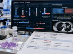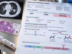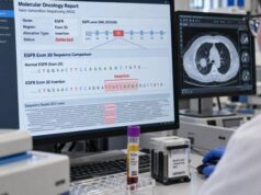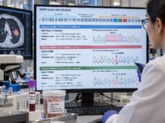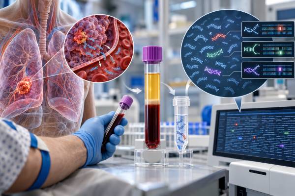
A lung cancer liquid biopsy is a blood-based molecular test that looks for tumor-derived material, most often circulating tumor DNA (ctDNA). It can identify actionable gene changes when tissue is limited, speed treatment decisions in advanced non-small cell lung cancer (NSCLC), and sometimes reveal resistance changes after targeted therapy stops working. The test is convenient because it usually requires only a blood draw, but it is not a complete replacement for tumor biopsy. Some cancers release little DNA into the bloodstream, so a negative plasma result can miss a mutation that is present in the tumor. Liquid biopsy is most useful when its strengths and limits are interpreted together with tissue pathology, imaging, cancer stage, treatment history, and the specific assay used. Results may include mutations, gene fusions, copy-number changes, and other genomic findings, but what can be detected depends on the panel and the amount of ctDNA in the sample.
- What it measures: Most lung cancer liquid biopsies analyze plasma ctDNA for tumor-related genetic alterations such as EGFR, KRAS, BRAF, MET, ALK, ROS1, RET, NTRK, and ERBB2/HER2 changes.
- What a positive result means: A clearly detected actionable alteration can often guide targeted treatment without waiting for another tissue biopsy, if the finding fits the diagnosis and assay requirements.
- What a negative result means: A negative plasma test does not rule out a tumor mutation; low tumor shedding can cause false-negative results, so tissue testing is often needed when feasible.
- Preparation and timing: Fasting is usually unnecessary. The test uses a routine blood draw, and comprehensive NGS results commonly take several days to about two weeks, depending on the laboratory.
- When it is especially useful: Liquid biopsy can help when tissue is unavailable or unsafe to obtain, when rapid molecular information is needed, or when cancer progresses and acquired resistance is suspected.
Table of Contents
- What a Lung Cancer Liquid Biopsy Measures
- When Liquid Biopsy Is Used
- How the Test Is Done and What Affects Accuracy
- How to Read Liquid Biopsy Results
- Resistance Markers and Treatment Monitoring
- Limitations, False-Negative Results, and Other Pitfalls
- What to Ask After You Get the Results
What a Lung Cancer Liquid Biopsy Measures
A lung cancer liquid biopsy usually examines cell-free DNA (cfDNA) in plasma. cfDNA is made of small DNA fragments released into the bloodstream from many cells in the body. The portion that comes from cancer cells is called circulating tumor DNA, or ctDNA. Modern assays use sensitive polymerase chain reaction (PCR) methods or, more commonly for broad testing, next-generation sequencing (NGS) to identify tumor-associated genetic changes in that DNA.
The main goal in advanced NSCLC is to find molecular alterations that may influence treatment. Depending on the assay, a plasma panel may evaluate:
- single-nucleotide variants, such as some EGFR, KRAS, BRAF, or ERBB2 mutations;
- small insertions or deletions, including EGFR exon 19 deletions and some exon 20 insertions;
- splice-related alterations such as MET exon 14 skipping-associated variants;
- gene fusions involving ALK, ROS1, RET, or NTRK genes;
- copy-number gains, such as MET amplification; and
- selected resistance alterations that appear during targeted treatment.
A broad plasma assay can therefore overlap with a tissue lung cancer NGS panel. The important difference is the specimen. Tissue testing analyzes DNA or RNA taken directly from a tumor sample, while liquid biopsy analyzes tumor-derived material circulating in blood.
Not every liquid biopsy includes the same genes or alteration types. Two tests both called “comprehensive” can differ in gene count, fusion coverage, copy-number performance, detection threshold, and whether they report tumor fraction. The report should be interpreted according to the exact validated capabilities of that assay rather than by the general label “liquid biopsy.”
Liquid biopsy also does not replace all biomarker testing. For example, PD-L1 is usually assessed by immunohistochemistry on tumor tissue, so a plasma genomic panel does not substitute for a PD-L1 lung cancer test when PD-L1 expression is needed for an immunotherapy decision.
When Liquid Biopsy Is Used
Liquid biopsy is most established in advanced NSCLC, where timely molecular profiling can determine whether a patient is eligible for targeted therapy. It is particularly useful when tissue is too small, exhausted by prior testing, difficult to obtain, or unsafe to biopsy.
A clinician may order plasma ctDNA testing at several points in care.
At the time of advanced or metastatic diagnosis
When a new diagnosis of advanced NSCLC is made, comprehensive biomarker testing is important before treatment whenever the clinical situation allows. Plasma testing may be ordered at the same time as tissue testing or while a tissue biopsy is being arranged. A positive, clearly actionable plasma result can sometimes shorten the time to a treatment decision.
However, a plasma test cannot establish the histologic diagnosis of lung cancer. Tissue remains important for determining whether a tumor is adenocarcinoma, squamous cell carcinoma, small cell lung cancer, or another subtype, and for performing immunohistochemical tests.
When tissue cannot be obtained
Some lung tumors are hard to reach safely, and some patients cannot undergo an invasive biopsy because of medical risk. In that setting, plasma ctDNA can provide valuable molecular information without a new tumor procedure. The tradeoff is that a negative blood result is less reassuring than a negative high-quality tissue result because not all tumors shed enough ctDNA to be detected.
When cancer progresses on targeted therapy
Cancer can evolve under treatment pressure. A tumor that initially responds to a targeted drug may later develop a new alteration that allows cancer cells to grow again. A blood test can sample DNA released from multiple tumor sites at once, which may help reveal heterogeneous resistance that a biopsy of one lesion could miss.
For this reason, clinicians may use a lung cancer resistance mutation panel or a broad ctDNA assay at progression. Tissue biopsy may still be needed if plasma is negative, if the resistance mechanism is unclear, or if the cancer may have changed histologically.
After treatment for earlier-stage disease
Research increasingly uses serial ctDNA to detect molecular residual disease (MRD), meaning tiny amounts of tumor DNA remaining after treatment even when scans show no visible cancer. A positive result after curative-intent treatment can be associated with higher recurrence risk. However, MRD-guided treatment in lung cancer remains an evolving area, and whether changing therapy solely because of a ctDNA result improves outcomes depends on the clinical setting and evidence. A dedicated lung cancer MRD ctDNA test should therefore be distinguished from routine metastatic plasma genotyping.
How the Test Is Done and What Affects Accuracy
The patient usually has blood drawn into special tubes designed to preserve cell-free DNA. Fasting is generally not required. The sample is processed to separate plasma, DNA is extracted, and the laboratory applies a validated molecular method such as targeted NGS or digital PCR.
For broad NGS, the report may take roughly 5 to 14 days, although turnaround varies. Focused tests for one mutation can sometimes be faster. The practical details matter because ctDNA is present at low concentrations and can be diluted by DNA from normal blood cells.
Several factors affect the chance of detecting a true tumor alteration:
- Tumor burden: Larger or more widespread disease often releases more ctDNA than small-volume disease.
- Site of disease: Some sites shed less DNA into peripheral blood. Isolated central nervous system disease can be especially difficult to detect in plasma.
- Recent treatment: Effective therapy may reduce ctDNA quickly, making some variants harder to detect.
- Tumor biology: Two cancers of similar size may release very different amounts of DNA.
- Assay sensitivity: Different laboratories have different limits of detection and different performance for mutations, fusions, and copy-number changes.
- Sample handling: Delayed processing or poor specimen quality can increase background DNA and reduce useful signal.
Many reports give a variant allele fraction (VAF), which is the proportion of DNA molecules carrying a particular variant among the molecules assessed at that location. A VAF of 1%, for example, does not mean that 1% of the body is cancer or that 1% of tumor cells contain the mutation. It is a measurement in the DNA sample and is influenced by tumor fraction, tumor genetics, normal cfDNA, treatment, and assay design.
A very low VAF can still be clinically meaningful if the alteration is real and well validated. Conversely, the numerical VAF alone should not determine whether a targeted drug will work.
How to Read Liquid Biopsy Results
The most useful way to read a plasma report is to separate findings into actionable tumor alterations, other tumor-associated findings, and results that are negative or uncertain.
Actionable positive findings
An actionable finding is a molecular alteration linked to an approved therapy, guideline-supported treatment strategy, or potentially relevant clinical trial in the patient’s cancer type and setting. Examples in NSCLC can include activating EGFR mutations, KRAS G12C, BRAF V600E, MET exon 14 skipping, ERBB2/HER2 mutations, and ALK, ROS1, RET, or NTRK fusions.
If plasma detects a well-established actionable driver, clinicians may be able to make a treatment decision from the blood result, assuming the result is technically valid and consistent with the diagnosis. For example, an activating alteration found on an EGFR mutation test can have direct treatment implications in NSCLC.
A positive result is not the same as a prognosis. The effect depends on the specific mutation, cancer stage, available treatment, previous therapies, co-mutations, and other clinical factors.
Negative or non-informative results
A negative liquid biopsy means the assay did not detect a reportable alteration above its detection threshold. It does not prove the tumor lacks actionable mutations.
This is one of the most important principles in ctDNA testing. A tumor may carry an actionable alteration yet release too little ctDNA for detection. Meta-analytic data in advanced NSCLC show very high specificity for plasma detection of mutations but lower overall sensitivity, with sensitivity varying substantially by alteration type. Therefore, when plasma testing is negative or non-informative and tissue is available, reflex tissue testing is usually appropriate.
The report may also state that no ctDNA was detected, that tumor fraction is low, or that the sample is inadequate. Those phrases strengthen the reason not to overinterpret a negative result.
Variants of uncertain significance
A variant of uncertain significance (VUS) is a DNA change whose clinical importance is not established. A VUS should not automatically be treated as a driver mutation or used as the sole reason to choose a targeted drug. Its classification may change as evidence develops.
Some reports also list alterations that are biologically interesting but not currently actionable in lung cancer. These can still help describe tumor biology or trial eligibility, but they should be separated from findings with established treatment implications.
Resistance Markers and Treatment Monitoring
One of liquid biopsy’s strongest advantages is that it can be repeated with much less burden than serial tumor biopsies. That makes it useful for investigating acquired resistance when disease progresses during targeted therapy.
Resistance can occur through several mechanisms. The original drug target may change, a bypass pathway may become activated, or the cancer may develop broader genomic or histologic changes. Examples include secondary EGFR mutations, kinase-domain mutations after ALK-targeted therapy, and increased MET signaling. MET amplification, for instance, can function as a bypass resistance mechanism in some EGFR-driven cancers.
Liquid biopsy can help because plasma contains DNA released from multiple tumor deposits. If one metastatic site develops one resistance mechanism and another site develops a different one, a blood sample may capture both. A single tissue biopsy samples only one location.
Still, plasma does not reveal everything. Gene fusions and copy-number changes can be harder to detect than some point mutations, and a transformation in tumor type cannot be diagnosed from ctDNA alone. For example, an EGFR-mutant adenocarcinoma can occasionally transform into small cell lung cancer after treatment. That kind of histologic change requires tissue evaluation.
Serial ctDNA levels can also fall when treatment is effective and rise as disease progresses. This has strong research value and may offer prognostic information, but routine clinical decisions should not be made from a change in VAF alone unless the test and use are validated for that purpose. Imaging, symptoms, treatment history, and the complete molecular report remain essential.
Limitations, False-Negative Results, and Other Pitfalls
The biggest practical limitation of plasma ctDNA testing is false-negative risk. Low-shedding tumors, small tumor volume, treatment response, and certain metastatic patterns can leave too little tumor DNA in blood. This is why a negative plasma test often needs tissue follow-up.
Another important issue is clonal hematopoiesis. As people age, blood-forming stem cells can acquire mutations and expand into small clones. DNA from these blood cells enters plasma and can create variants that are not from the lung tumor. Depending on the gene and assay, this can complicate interpretation. Some laboratories use paired white-blood-cell sequencing or other bioinformatic approaches to reduce this risk.
Copy-number gains deserve special caution. A plasma finding suggesting amplification may be influenced by tumor fraction and assay methodology. Conversely, a true MET copy-number gain may be missed when ctDNA levels are low. Tissue-based methods can be useful when a copy-number result will change treatment.
Other common interpretation mistakes include:
- assuming that every mutation is a drug target;
- assuming a negative result means “no mutations”;
- treating VAF as a direct measure of cancer percentage;
- ignoring whether the assay can reliably detect fusions or amplifications;
- overlooking the possibility of clonal hematopoiesis;
- using plasma genomics as a substitute for pathologic diagnosis; and
- assuming MRD testing and metastatic genotyping are interchangeable.
A good liquid biopsy report should be read in context. The same genomic finding can have different meaning at diagnosis, during response, or after progression.
What to Ask After You Get the Results
A liquid biopsy result is most useful when it leads to a specific next step. Patients and caregivers can ask the oncology team:
- Was enough ctDNA present for the result to be reliable? A report with very low or undetectable tumor fraction deserves more caution, especially if no actionable mutation was found.
- Did the assay test all alteration types that matter for this cancer? Confirm whether it included relevant mutations, fusions, and copy-number changes rather than only a small mutation panel.
- Is any detected alteration clearly actionable in lung cancer? Ask whether the evidence applies to this tumor type, disease stage, and treatment line.
- If the test was negative, should tissue testing be done? In many advanced NSCLC cases, the answer is yes if adequate tissue can safely be obtained.
- Could a result come from blood cells rather than the tumor? This is especially relevant for genes associated with clonal hematopoiesis.
- Does progression require a new tissue biopsy? A tissue sample may be important if transformation is suspected or plasma does not identify a resistance mechanism.
- Will this result change treatment now, or is it mainly useful for a clinical trial or future planning? Not every reported variant has immediate clinical value.
For newly diagnosed advanced NSCLC, comprehensive molecular profiling should be viewed as a coordinated process rather than a contest between blood and tissue. Plasma can provide speed, convenience, and a broad view of tumor DNA, while tissue provides histology, protein markers, and a direct tumor specimen. Used together, they can reduce missed opportunities for biomarker-matched treatment.
References
- Liquid biopsy in lung cancer 2025 (Review)
- Liquid biopsy techniques and lung cancer: diagnosis, monitoring and evaluation 2024 (Review)
- Clinical Validity and Utility of Circulating Tumor DNA (ctDNA) Testing in Advanced Non-small Cell Lung Cancer (aNSCLC): A Systematic Literature Review and Meta-analysis 2024 (Systematic Review)
- Recommendations for reporting tissue and circulating tumour (ct)DNA next-generation sequencing results in non-small cell lung cancer 2024 (Position Statement)
- ESMO recommendations on the use of circulating tumour DNA assays for patients with cancer: a report from the ESMO Precision Medicine Working Group 2022 (Guideline)
Disclaimer
This article is for general education and does not replace personalized medical advice. Liquid biopsy results should be interpreted by an oncology team together with tissue pathology, imaging, treatment history, and the exact assay used. Do not start, stop, or change cancer treatment based on a molecular result without discussing it with the treating clinician.