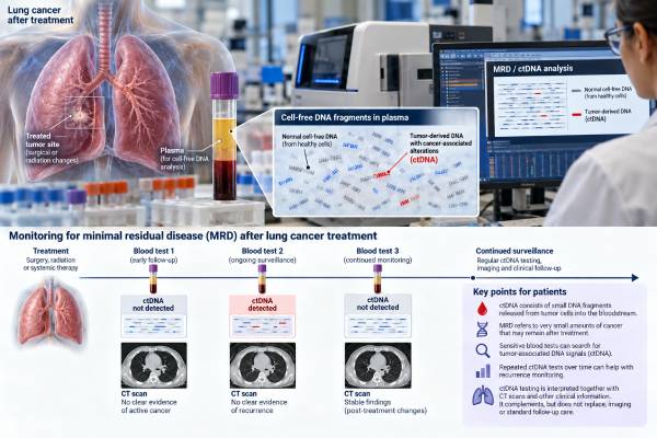
A minimal residual disease (MRD) ctDNA test looks for tiny amounts of lung cancer DNA that may remain in the bloodstream after treatment intended to eliminate visible disease. The test is most often studied after surgery, chemoradiation, or other curative-intent treatment for non-small cell lung cancer (NSCLC). A positive result can indicate molecular evidence of residual cancer before recurrence is visible on scans, and repeated positive results are associated with a higher risk of relapse. A negative result is reassuring but cannot guarantee that cancer is gone because early-stage lung cancers may release very little circulating tumor DNA (ctDNA). MRD testing can be tumor-informed, using mutations identified from an individual patient’s tumor, or tumor-naive/tumor-agnostic, using a predefined genomic or epigenomic approach. Although the prognostic value of ctDNA MRD is strong, using MRD results alone to start, stop, intensify, or de-escalate treatment remains an evolving area and should follow current clinical evidence and specialist guidance.
- What it measures: MRD testing searches blood for very low levels of tumor-derived DNA after treatment, when imaging may show no measurable cancer.
- What a positive result usually means: Detectable ctDNA after curative-intent therapy is associated with a substantially higher risk of recurrence and can precede radiographic relapse.
- What a negative result means: No ctDNA detected lowers the estimated recurrence risk but does not prove that every cancer cell has been eliminated.
- Why repeat testing may help: Serial surveillance generally detects more relapses than a single post-treatment “landmark” blood draw because ctDNA can become detectable later.
- Clinical caution: MRD is a powerful prognostic biomarker, but treatment changes based only on MRD are not universally established outside validated clinical pathways or trials.
Table of Contents
- What MRD ctDNA Means in Lung Cancer
- When MRD Testing Is Done
- Tumor-Informed vs Tumor-Agnostic MRD Tests
- How to Read MRD Results
- How MRD Relates to Recurrence Monitoring
- Can MRD Guide Treatment Decisions?
- Limitations and Next Steps
What MRD ctDNA Means in Lung Cancer
Minimal residual disease, also called molecular residual disease in this context, means cancer may persist at a level too small to see with standard imaging. After a lung tumor is removed or treated definitively, microscopic cancer cells can remain locally or elsewhere in the body. If those cells release fragments of tumor DNA into the bloodstream, highly sensitive assays may detect that material as ctDNA.
MRD is therefore not the same as a visible tumor mass. A CT scan may be clear while ctDNA is detectable. Conversely, an MRD test may be negative even when microscopic cancer remains because not every tumor sheds enough DNA into plasma to cross the assay’s detection threshold.
This creates two distinct concepts:
- Clinical or radiographic recurrence: cancer becomes visible on scans, causes symptoms, or is confirmed pathologically.
- Molecular recurrence: tumor-derived DNA becomes detectable before conventional recurrence is evident.
The goal of MRD testing is to identify molecular recurrence earlier and improve risk stratification. In studies of lung cancer after definitive therapy, ctDNA positivity has consistently been associated with poorer recurrence-free outcomes. A 2023 meta-analysis found high specificity but only moderate sensitivity, meaning a positive result is often meaningful while a negative result cannot rule out residual disease with certainty.
MRD testing is different from a routine lung cancer liquid biopsy used to find actionable mutations in advanced disease. Both use ctDNA, but the analytical challenge is much harder for MRD because the amount of tumor DNA after treatment can be extremely small.
When MRD Testing Is Done
MRD can be assessed at different time points, and timing changes what the result means.
Before definitive treatment
A pre-treatment blood sample can show whether the tumor sheds detectable ctDNA at baseline. This information can help with assay design in tumor-informed testing and gives context for interpreting later negative results. A tumor that never shed detectable ctDNA before surgery may be difficult to monitor reliably with plasma afterward.
After surgery or definitive local therapy
A landmark MRD test is taken at a predefined interval after treatment. The purpose is to ask whether tumor DNA remains once the main tumor burden has been removed or treated.
The exact timing varies by study and platform. Drawing blood too soon after surgery can be problematic because tissue injury releases large amounts of normal cell-free DNA, which can dilute tumor signal. Many research protocols therefore use a postoperative interval rather than testing immediately after the operation.
During adjuvant treatment
Serial ctDNA may be measured during chemotherapy, targeted therapy, or immunotherapy after surgery. Clearance of previously detectable ctDNA can suggest molecular response, while persistent or re-emerging ctDNA can indicate higher residual risk. Whether therapy should be changed solely because of these dynamics is still being tested prospectively.
During surveillance
Repeated blood draws after treatment can detect molecular relapse. A surveillance strategy is generally more sensitive than one landmark test because a patient who is negative soon after treatment may later become positive as residual cancer grows and sheds more DNA.
MRD testing does not replace guideline-based imaging follow-up. It is an additional molecular signal, not a substitute for CT scans, clinical review, or diagnostic evaluation of new symptoms.
Tumor-Informed vs Tumor-Agnostic MRD Tests
MRD assays use different strategies to find tiny amounts of cancer-derived material. The two broad categories are tumor-informed and tumor-agnostic approaches.
| Approach | How it works | Main strength | Main limitation |
|---|---|---|---|
| Tumor-informed | Sequences the patient’s tumor first, then tracks selected tumor-specific variants in plasma | Can achieve high specificity and personalized tracking | Requires adequate tumor tissue and extra setup time |
| Tumor-agnostic | Uses a predefined panel or other signal without building a personalized assay from the tumor | Can be faster and does not always require tumor tissue | May face more background noise or lower sensitivity depending on design |
Tumor-informed tests often begin with NGS of the resected tumor. The laboratory selects mutations that appear specific to that cancer and then looks for those exact variants in follow-up blood samples. Tracking several mutations at once can improve confidence that a detected signal truly comes from the tumor.
Tumor-agnostic methods may use broad mutation panels, methylation patterns, fragment characteristics, or combinations of signals. They avoid the need to customize an assay for each patient, but performance varies widely by platform.
Neither strategy is automatically superior in every setting. Important performance features include limit of detection, false-positive rate, number of variants tracked, background suppression, sample volume, reproducibility, and how clonal hematopoiesis is handled.
Clonal hematopoiesis deserves special attention because age-related blood-cell clones can release mutated DNA into plasma. If a test mistakes that DNA for tumor DNA, it can create a false-positive result. Some assays sequence white blood cells or use other filtering methods to reduce this risk.
How to Read MRD Results
Most MRD reports are interpreted as detected/positive, not detected/negative, or occasionally indeterminate/not evaluable. The meaning depends on timing and assay quality.
Positive or detected
A positive post-treatment ctDNA result means the assay found tumor-associated DNA above its validated threshold. In early-stage or locally advanced NSCLC after definitive therapy, this is a strong adverse prognostic sign. Across studies, ctDNA positivity is associated with a much higher likelihood of later recurrence.
A positive result does not tell exactly where residual cancer is located, how large it is, or when it will become visible on imaging. It should trigger careful clinical interpretation rather than an assumption that a scan must already show disease.
Negative or not detected
A negative result means the assay did not find ctDNA above its detection limit in that blood sample. It is generally favorable, but it is not equivalent to “cured.”
Reasons a patient with residual cancer may still test negative include:
- very small tumor burden;
- low-shedding tumor biology;
- disease limited to sites that release little DNA into peripheral blood;
- an assay tracking variants no longer present in the dominant clone;
- inadequate plasma volume or pre-analytic problems; and
- testing at a time when the ctDNA level is temporarily below detection.
The meaning of a negative result is stronger when ctDNA was clearly detectable before treatment and then becomes repeatedly undetectable afterward.
Quantitative changes
Some tests report a numerical ctDNA level, mean tumor molecules, variant copies, or another proprietary metric. The trend can be useful, but numbers from different platforms are not directly interchangeable. A doubling or decline should be interpreted using that assay’s validated framework rather than a universal threshold.
How MRD Relates to Recurrence Monitoring
One of the most promising uses of MRD is detecting recurrence earlier than imaging. In a systematic review of resected NSCLC, postoperative ctDNA detection often preceded radiographic or clinical relapse, with a reported mean lead time of roughly 5.5 months across included studies. Lead time varies substantially by assay, sampling frequency, tumor biology, and scan schedule.
This early signal can be clinically valuable, but it also creates a difficult question: what should be done during the interval between molecular recurrence and visible disease?
More intensive imaging may be reasonable in some contexts, but repeated scans can also detect indeterminate findings and increase radiation exposure. Starting systemic therapy before radiographic recurrence may help if the molecular signal accurately identifies disease that is destined to recur, but that benefit must be demonstrated in prospective trials rather than assumed.
Serial testing can also help distinguish persistent MRD from a one-time borderline result. A patient who remains ctDNA-positive on repeated samples has a different risk profile from someone with a single low-level signal that is not reproduced.
MRD should be integrated with stage, pathology, lymph-node involvement, surgical margins, driver mutations, treatment received, and imaging. It adds a molecular layer to recurrence risk but does not erase traditional prognostic factors.
For advanced NSCLC, ctDNA monitoring serves a different purpose. It may track treatment response or emerging resistance, whereas true MRD testing is most often discussed after treatment with curative intent when no measurable disease remains.
The timing and frequency of blood collection can strongly affect apparent performance. A single sample taken soon after treatment provides a snapshot. Serial sampling provides multiple opportunities to detect a low-level clone as it grows, which is one reason surveillance strategies tend to improve sensitivity. However, more testing also creates more chances for borderline or technically ambiguous findings. A useful surveillance plan should therefore define in advance when blood is drawn, how a new positive result is confirmed, and what action follows.
The biology of ctDNA also helps explain why MRD is difficult in lung cancer. Tumor DNA represents only a tiny fraction of all cell-free DNA in plasma after successful treatment. Normal tissue turnover, inflammation, recent surgery, infection, and other processes can increase background cfDNA without increasing tumor DNA. The assay must detect a few tumor-specific molecules against that much larger background while avoiding sequencing errors. This is why MRD platforms use very deep sequencing, unique molecular identifiers, personalized mutation tracking, or other error-suppression methods.
An MRD result should also be separated from a routine tumor-marker blood test. ctDNA is not a protein concentration such as CEA. It is molecular evidence that specific tumor-associated DNA fragments are present. There is therefore no universal “normal range” in ng/mL that applies across platforms. Each assay has its own analytical threshold and reporting system.
Finally, molecular lead time is not automatically the same as extra survival time. Detecting recurrence five months earlier may provide a valuable window for research or intervention, but only a clinical trial can show whether acting during that window improves outcome compared with waiting for standard radiographic detection. This distinction prevents overpromising what an early positive blood test can accomplish today.
Can MRD Guide Treatment Decisions?
MRD has strong clinical validity as a prognostic marker: positive ctDNA after definitive treatment predicts a higher likelihood of recurrence. The harder question is clinical utility: does changing treatment because of the result improve survival or cure rates?
That distinction is essential. A biomarker can accurately predict relapse without proving that acting on it early improves outcomes.
Potential future uses include:
- Escalation: giving additional or more intensive treatment to MRD-positive patients who have a high residual risk.
- De-escalation: reducing treatment in repeatedly MRD-negative patients to avoid unnecessary toxicity.
- Switching therapy: changing an adjuvant drug if ctDNA persists or reappears.
- Earlier salvage treatment: starting treatment at molecular recurrence before imaging confirms relapse.
These strategies are being studied, but they require prospective evidence. The 2022 ESMO ctDNA recommendations recognized the high clinical validity of MRD detection while stating that MRD-directed treatment could not yet be recommended routinely because clinical utility had not been established. More recent expert consensus continues to emphasize assay standardization and cautions against using variable-sensitivity MRD tests for treatment de-escalation outside appropriate evidence-based settings.
A patient should therefore ask not only “Is my MRD positive?” but also “What evidence shows that changing treatment because of this result helps people in my exact situation?”
Limitations and Next Steps
The biggest limitation of lung cancer MRD is sensitivity. Early-stage tumors often release little ctDNA, so a negative test can miss microscopic disease. Meta-analysis suggests specificity is generally high while sensitivity is only moderate, particularly with a single landmark sample.
Other challenges include:
- no universal standard for assay design or reporting;
- different blood-draw schedules across studies;
- variable definitions of MRD positivity;
- clonal hematopoiesis and other background variants;
- limited ctDNA shedding from some tumor sites;
- inability of ctDNA to show the anatomic location of recurrence; and
- uncertainty about the best intervention after molecular relapse.
Before using an MRD result to make a major decision, useful questions include:
- Was the test tumor-informed or tumor-agnostic?
- Was ctDNA detectable before treatment?
- How long after surgery, radiation, or chemoradiation was the blood drawn?
- Is the result a one-time measurement or part of a serial trend?
- What is the assay’s validated sensitivity and specificity in lung cancer?
- Does the proposed treatment change have evidence for MRD-positive patients in this exact setting?
- Will standard imaging surveillance continue regardless of the result?
MRD ctDNA is one of the most promising tools for making recurrence risk more precise, but its strength is currently greatest as a prognostic and research biomarker. The safest interpretation uses MRD alongside pathology, imaging, stage, molecular profile, and evidence-based treatment standards rather than allowing one blood result to replace them.
Sampling timing and serial trends also matter when a result is close to the assay’s detection limit.
References
- Consensus Statement on ctDNA Minimal Residual Disease Testing in Early Stage NSCLC: A Delphi Study by the Asian Thoracic Oncology Research Group 2026 (Position Statement)
- Circulating tumour DNA in early stage and locally advanced NSCLC: ready for clinical implementation? 2025 (Review)
- Clinical application of minimal residual disease detection by ctDNA testing in non-small cell lung cancer: a narrative review 2025 (Review)
- Postoperative ctDNA in indicating the recurrence risk and monitoring the effect of adjuvant therapy in surgical non-small cell lung cancer 2024
- Accuracy of minimal residual disease detection by circulating tumor DNA profiling in lung cancer: a meta-analysis. 2023 (Systematic Review)
- ESMO recommendations on the use of circulating tumour DNA assays for patients with cancer: a report from the ESMO Precision Medicine Working Group 2022 (Guideline)
Disclaimer
This article is for general education and does not replace individualized medical advice. Lung cancer MRD results should be interpreted with the assay used, timing of the blood draw, pathology, stage, imaging, and treatment history. Do not start, stop, or reduce cancer treatment based on an MRD result without guidance from the treating oncology team.