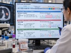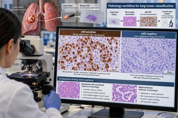
The p40 immunohistochemistry test is a tissue stain used to identify squamous differentiation in lung cancer. p40 is an isoform of the TP63 protein, known as ΔNp63, and it is usually seen as nuclear staining in squamous epithelial cells and squamous cell carcinomas. In a poorly differentiated non-small cell lung cancer, strong, diffuse p40 positivity strongly supports squamous cell carcinoma, especially when adenocarcinoma markers such as TTF-1 are negative. p40 is more specific for squamous differentiation than the broader p63 stain, which can be positive in a substantial minority of lung adenocarcinomas. However, p40 does not prove that a tumor started in the lung because squamous cancers from other organs can also be p40 positive. The final diagnosis combines p40 with tumor morphology, clinical history, imaging, and complementary immunostains. A negative p40 result also does not completely exclude squamous cell carcinoma because no immunohistochemical marker has perfect sensitivity.
- What p40 detects: p40 immunohistochemistry detects the ΔNp63 isoform of TP63 in tumor-cell nuclei and is a highly specific marker of squamous differentiation.
- What positive staining usually means: Strong, diffuse nuclear staining supports squamous cell carcinoma when the morphology and other markers fit.
- How accurate it is: A 2022 meta-analysis estimated p40 sensitivity near 92% and specificity near 94% for distinguishing lung squamous carcinoma from non-squamous NSCLC.
- Why p40 is preferred over p63: p63 is sensitive but less specific because lung adenocarcinomas can show p63 staining; p40 reduces that diagnostic pitfall.
- Important limitation: p40 identifies squamous phenotype, not lung origin, so metastatic squamous cancers can also be positive.
Table of Contents
- What p40 Is and How the Stain Works
- What Positive p40 Staining Means
- What Negative p40 Staining Means
- p40 Versus p63, TTF-1, and Other Markers
- Using p40 on Small Biopsies and Cytology
- Limits and Diagnostic Pitfalls
- What Happens After the p40 Result
What p40 Is and How the Stain Works
p40 is the common pathology name for ΔNp63, a set of protein isoforms produced from the TP63 gene. TP63 is important in the development and maintenance of basal and squamous epithelial cells. Squamous cell carcinomas often preserve strong expression of the ΔNp63 isoform, which makes p40 useful as an immunohistochemical marker.
The test is performed on tumor tissue rather than blood. A pathology laboratory can use a formalin-fixed, paraffin-embedded biopsy, surgical specimen, or an adequately prepared cytology cell block. An antibody binds p40 in the nuclei of cells, and a staining system makes the binding visible under the microscope.
True p40 positivity is a nuclear pattern. The pathologist evaluates how many tumor cells stain, how intense the staining is, and whether the pattern is diffuse or focal. Strong staining in most tumor nuclei provides much stronger evidence of squamous differentiation than rare isolated positive cells.
There is no universal treatment score such as a PD-L1 TPS. Pathology reports may say “p40 positive,” “diffusely positive,” “strong nuclear staining,” or describe a percentage. The significance comes from the pattern and the diagnostic context rather than from a single standardized numeric threshold.
p40 became especially important as lung cancer diagnosis moved toward precise NSCLC subtyping on very small specimens. Distinguishing squamous cell carcinoma from adenocarcinoma matters because the two groups have different molecular-testing probabilities, treatment considerations, and tissue-triage needs.
A p40 stain is therefore a classification tool. It tells the pathologist about cell differentiation. It does not identify a driver mutation, measure tumor burden, or show how far the cancer has spread.
This separation between classification and prediction is important for patients reading a report. A “p40 positive” result describes what the tumor cells resemble biologically; it does not mean the cancer is more advanced, more aggressive, or more responsive to a particular drug. Those questions require stage, molecular biomarkers, PD-L1, and other clinical information interpreted together carefully by the treating oncology team.
What Positive p40 Staining Means
Strong, diffuse nuclear p40 staining in a non-small cell lung carcinoma strongly supports squamous differentiation. In the right morphologic setting, that pattern can support a final diagnosis of lung squamous cell carcinoma.
The marker performs well because most lung squamous cell carcinomas express p40, while most lung adenocarcinomas do not. A 2022 diagnostic meta-analysis of 85 studies found summary sensitivity of about 92% and specificity of about 94% for p40 in distinguishing lung squamous carcinoma from non-squamous NSCLC.
That performance does not mean every positive cell proves squamous carcinoma. The extent of staining matters. Rare lung adenocarcinomas can show focal p40 positivity, often in only a small proportion of cells. A pathologist is much more cautious with focal weak staining than with a classic diffuse strong pattern.
Positive p40 can be particularly helpful when morphology is poorly differentiated. A tiny biopsy may show sheets of malignant cells without obvious keratinization or gland formation. If those cells are diffusely p40 positive and TTF-1 negative, the immunophenotype strongly favors squamous cell carcinoma.
| p40 | TTF-1 | Typical interpretation |
|---|---|---|
| Diffuse positive | Negative | Strongly supports squamous differentiation |
| Negative | Positive | Supports adenocarcinoma or another TTF-1-positive pulmonary tumor in the right context |
| Negative | Negative | May remain NSCLC not otherwise specified or require additional markers |
| Both diffusely positive | Positive | Unusual pattern requiring careful review and possible broader workup |
The rare tumor that diffusely co-expresses p40 and TTF-1 deserves special attention. Recent work suggests this can represent an unusual biphenotypic NSCLC rather than a routine squamous carcinoma or adenocarcinoma. The pathologist may use additional stains and molecular testing rather than forcing the case into a conventional category.
What Negative p40 Staining Means
A negative p40 stain means convincing nuclear expression was not detected in the sampled tumor cells. It makes classic squamous differentiation less likely but does not completely exclude squamous cell carcinoma.
The 2022 meta-analysis estimated sensitivity around 92%, which means a minority of true lung squamous carcinomas can be p40 negative. Technical issues and tumor biology both contribute. Poor fixation, scant viable tumor, decalcification, crush artifact, necrosis, and very poorly differentiated morphology can reduce staining reliability.
When p40 is negative, the pathologist looks at the full microscopic pattern and other markers. A tumor with gland formation, mucin, TTF-1 positivity, or Napsin A positivity may be classified as adenocarcinoma. A tumor negative for both squamous and glandular markers may require a broader differential diagnosis.
The TTF-1 IHC test is commonly paired with p40 because it provides complementary lineage information. This minimalist combination can classify many poorly differentiated NSCLCs while preserving precious biopsy material for molecular studies.
Negative p40 also does not mean a tumor is necessarily adenocarcinoma. Neuroendocrine carcinomas, mesothelioma, metastatic tumors, sarcomatoid carcinomas, and other entities can be p40 negative. The next marker is chosen based on morphology and clinical possibilities, not by a fixed automatic sequence.
If the tissue is extremely limited, the pathologist may choose not to perform many additional stains. Conserving tissue for predictive biomarkers can be more valuable than achieving unnecessary immunohistochemical detail once a clinically useful classification has been reached.
p40 Versus p63, TTF-1, and Other Markers
p40 and p63 are related but not identical stains. The commonly used p63 antibody recognizes multiple TP63 isoforms, while p40 is directed toward ΔNp63. That narrower target gives p40 greater specificity for squamous differentiation.
Classic studies showed that p63 can stain a meaningful minority of lung adenocarcinomas, sometimes extensively. That creates a risk of misclassifying a poorly differentiated adenocarcinoma as squamous cell carcinoma if p63 is used alone. p40 greatly reduces this problem while retaining high sensitivity for squamous tumors.
The 2022 meta-analysis compared p40, p63, CK5/6, and desmocollin-3. p40 had the best overall diagnostic odds ratio, with both high sensitivity and high specificity. CK5/6 is also a useful squamous marker but shows cytoplasmic/membranous staining rather than the crisp nuclear pattern of p40.
TTF-1 works in the opposite direction. It is primarily an adenocarcinoma and pulmonary lineage marker, although it can also be expressed in many small cell lung cancers. The Napsin A IHC marker can further support lung adenocarcinoma when morphology and TTF-1 are compatible.
A useful way to think about the panel is:
- p40: strongest common marker for squamous differentiation;
- p63: sensitive squamous/basal marker but less specific;
- CK5/6: useful supporting squamous marker;
- TTF-1: supports pulmonary adenocarcinoma and some neuroendocrine tumors;
- Napsin A: supports adenocarcinoma, especially lung origin in the proper differential.
These markers are not treatment targets. Their job is to create an accurate histologic diagnosis so that the right predictive testing can follow.
Using p40 on Small Biopsies and Cytology
Most patients with advanced lung cancer are diagnosed from small biopsies or cytology rather than a large surgical resection. That makes tissue conservation a central part of pathology practice.
A core biopsy may contain only a few millimeters of tumor. The same block may be needed for H&E slides, lineage immunostains, PD-L1, DNA sequencing, and RNA fusion testing. Using a focused panel such as p40 plus TTF-1 can answer the subtype question with minimal tissue consumption.
p40 also performs well on many cytology preparations. A 2023 study using automated immunocytochemistry on ThinPrep material found p40 to have high specificity for lung squamous carcinoma and strong agreement with standard tissue immunohistochemistry. Laboratories still need to validate their own cytology methods because fixation and processing differ from routine tissue.
A practical workflow for an undifferentiated NSCLC might be:
- review morphology for glandular, squamous, or neuroendocrine features;
- use a small number of lineage stains if morphology is insufficient;
- interpret p40 and TTF-1 together rather than independently;
- classify the tumor at the level supported by evidence;
- preserve remaining material for molecular and PD-L1 testing.
This approach helps avoid “stain exhaustion,” where so many diagnostic antibodies are ordered that too little tumor remains for predictive biomarkers. In modern lung cancer care, saving tissue can directly affect treatment options.
The size of the biopsy can also affect how confidently the pathologist names the subtype. A resection specimen may show keratin pearls, intercellular bridges, gland formation, or separate histologic components that are absent from a needle core. On a tiny sample, immunostains sometimes supply the decisive evidence that morphology cannot provide. Even then, the report may appropriately use qualifying language such as “favor squamous cell carcinoma” when the available tissue does not support absolute certainty.
Cytology creates similar challenges. Tumor cells in pleural fluid, bronchial brushings, or fine-needle aspirates may appear as isolated cells rather than the architectural groups seen in a tissue section. A validated p40 stain can help recover lineage information, but the pathologist still needs suitable controls and enough malignant cells. Benign mesothelial cells, inflammatory cells, and bronchial basal cells can be present in the same preparation and must be separated morphologically from the tumor population.
The test can also be useful when only a metastatic site is sampled. For example, a lymph-node biopsy may show a poorly differentiated carcinoma without obvious keratinization. Diffuse p40 positivity can establish squamous differentiation, but the question of where that squamous carcinoma began remains open until the clinical and radiologic evidence is reviewed. This distinction is particularly important in patients with prior head-and-neck, esophageal, cervical, or cutaneous squamous cancers.
Limits and Diagnostic Pitfalls
The biggest limitation of p40 is that it identifies squamous differentiation, not organ of origin. A metastatic squamous carcinoma from the head and neck, esophagus, cervix, skin, or another site can also be p40 positive.
Therefore, a p40-positive lesion in the lung is not automatically a primary lung squamous carcinoma. Clinical history and imaging are essential. If a patient has a known p40-positive cancer elsewhere, the pathologist may need additional site-specific markers and morphologic comparison with the prior tumor.
Other pitfalls include:
- Focal positivity: Rare adenocarcinomas and unusual tumors can show limited p40 staining; diffuse strong staining is more convincing.
- Basal or reserve cells: Benign bronchial basal cells are normally p40 positive and should not be mistaken for malignant staining.
- Adenosquamous carcinoma: Small biopsies may sample only one component, so a p40-positive biopsy may not reveal a separate adenocarcinoma component elsewhere in the tumor.
- Rare biphenotypic tumors: Diffuse p40 and TTF-1 co-expression is unusual and warrants expert review.
- Technical failure: Poor fixation or tissue damage can weaken a truly positive tumor.
- Squamous morphology without p40: Morphology still matters; a negative stain cannot overrule convincing keratinization and intercellular bridges without further review.
Another common misunderstanding is that p40 positivity determines whether molecular testing should be performed. Histology influences testing strategy, but current molecular practice is broader than older algorithms. Selected squamous NSCLCs—especially in never-smokers, younger patients, small biopsies where adenocarcinoma cannot be completely excluded, or according to local guidelines—may still need genomic profiling.
What Happens After the p40 Result
After p40 is interpreted, the pathologist integrates it into the final diagnosis. A typical report may read “non-small cell carcinoma, favor squamous cell carcinoma” or “squamous cell carcinoma,” depending on morphology, sample quality, and the complete immunophenotype.
That classification then guides the next layer of biomarker work. The NSCLC biomarker workup may include PD-L1 and molecular profiling according to histology, stage, smoking history, treatment setting, and current guidelines.
Patients reading the pathology report can ask several useful questions:
- Was p40 staining strong and diffuse or only focal?
- Were TTF-1 or Napsin A also tested?
- Does the overall profile support a primary lung cancer or could it be metastatic squamous carcinoma?
- Is the sample large enough for PD-L1 and molecular testing?
- If the biopsy is small, is the diagnosis definitive or qualified as “favor squamous”?
p40 itself does not need to be monitored over time. It is not a serum tumor marker and there is no reason to repeat p40 on serial blood draws. Repeat immunohistochemistry may be performed on a new biopsy if the tumor changes, if a second primary cancer is suspected, or if the original diagnosis needs reevaluation.
The most practical interpretation is simple: diffuse nuclear p40 staining is strong evidence that a carcinoma has squamous differentiation. That evidence becomes a lung squamous cell carcinoma diagnosis only when morphology and clinical context support lung origin. Used with TTF-1 and a carefully chosen panel, p40 allows accurate classification while preserving tissue for the molecular tests that increasingly shape treatment.
References
- Non-small cell lung carcinomas with diffuse co-expression of TTF-1 and p40: Clinical, pathological and molecular characterization of a tumor subtype 2026
- Unravelling the diagnostic pathology and molecular biomarkers in lung cancer 2024 (Review)
- Immunocytochemical detection by a fully automated immunostainer for assisting subclassification of pulmonary tumors on ThinPrep slides in real-life clinical samples: A single-institution experience 2023
- Accuracy of Classifying Lung Carcinoma Using Immunohistochemical Markers on Limited Biopsy Material: A Two-Center Study 2023
- Immunohistochemical markers to diagnose primary squamous cell carcinoma of the lung: a meta-analysis of diagnostic test accuracy 2022 (Meta-analysis)
- Immunohistochemical Detection of p40 Expression in Lung Cancer Clinical Samples 2021
Disclaimer
p40 immunohistochemistry is a pathology classification tool and cannot establish lung origin, stage, or treatment by itself. Results must be interpreted by a qualified pathologist with morphology, other immunostains, clinical history, imaging, and molecular tests when appropriate. Treatment decisions should not be based on p40 staining alone.