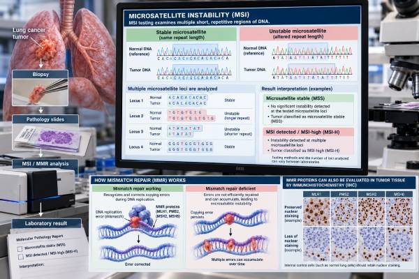
An MSI test for lung cancer checks whether tumor DNA shows microsatellite instability, a pattern caused by defective DNA mismatch repair. Microsatellites are short repeated DNA sequences that are especially prone to copying errors. Normally, mismatch repair proteins correct those errors. When that repair system fails, the repeats become unstable and mutations accumulate across the genome. A tumor with high-level microsatellite instability is called MSI-high (MSI-H); a tumor without that pattern is usually called microsatellite stable (MSS). MSI-H and mismatch repair deficiency (dMMR) are uncommon in lung cancer, but they can matter because they identify a biologically distinct, often highly mutated tumor subtype and may have immunotherapy implications. Testing may be performed with next-generation sequencing (NGS), PCR-based MSI analysis, or immunohistochemistry for the mismatch repair proteins MLH1, PMS2, MSH2, and MSH6. Results should be interpreted with the testing method, tumor type, PD-L1, tumor mutational burden, molecular profile, and clinical setting.
- What it measures: MSI testing looks for instability in repetitive DNA sequences; MMR immunohistochemistry checks whether key mismatch repair proteins are retained or lost.
- How common MSI-H is in lung cancer: Large genomic series have found MSI-H in roughly 0.4%–0.5% of NSCLC, making it a rare biomarker.
- What MSI-H means: MSI-H strongly suggests defective mismatch repair and a hypermutated tumor state, but it does not guarantee response to immunotherapy.
- What MSS means: Microsatellite-stable lung cancer does not show the characteristic MSI pattern; most lung cancers are MSS.
- Inherited cancer caution: Most MSI-H lung cancers are not automatically hereditary, but an MMR-gene pattern can occasionally raise concern for Lynch syndrome and justify germline evaluation.
Table of Contents
- What MSI and Mismatch Repair Mean
- How Common MSI-H Is in Lung Cancer
- How MSI Is Tested
- How to Read MSI and MMR Results
- MSI-H, TMB, PD-L1, and Immunotherapy
- When MSI or dMMR Raises Hereditary Cancer Questions
- Limitations and Next Steps
What MSI and Mismatch Repair Mean
DNA copying is not perfectly error-free. The mismatch repair (MMR) system acts like a proofreading mechanism that corrects base mismatches and small insertion/deletion errors produced during DNA replication. Four proteins are especially important in routine cancer testing: MLH1, PMS2, MSH2, and MSH6.
Microsatellites are short repetitive DNA sequences. Because the same sequence is repeated many times, the replication machinery can slip while copying them. An intact MMR system repairs most of those mistakes. When MMR is defective, the length of microsatellite sequences begins to vary across tumor cells. That molecular pattern is microsatellite instability.
The terminology is related but not identical:
- MSI-H: high-level microsatellite instability is detected by a molecular assay.
- MSS: microsatellite stable; the assay does not show the MSI-H pattern.
- dMMR: deficient mismatch repair, usually demonstrated by abnormal loss of one or more MMR proteins on immunohistochemistry or by other evidence of MMR dysfunction.
- pMMR: proficient mismatch repair; the expected MMR proteins are retained.
MSI-H and dMMR often occur together because loss of mismatch repair produces microsatellite instability. However, discordant results can occur, especially outside colorectal and endometrial cancers. A borderline or unusual result may therefore need review or confirmation by another method.
MSI is also different from tumor mutational burden. MSI-H tumors often have high TMB because defective repair allows mutations to accumulate, but TMB and MSI measure different genomic features.
How Common MSI-H Is in Lung Cancer
MSI-H is rare in lung cancer. That is one of the most important facts for interpreting the test.
A detailed genomic study of more than 5,000 NSCLC cases found MSI-H/MMR-deficient disease in about 0.41% of NSCLC and about 1.9% of small cell lung cancers in that cohort. Another large lung-cancer series reported MSI-H in approximately 0.5% of cases. These estimates vary with patient population, histology, assay, and definition, but they consistently show that MSI-H is uncommon compared with colorectal or endometrial cancer.
The rarity means MSI testing is usually not the first biomarker people think of in NSCLC. Established driver testing and PD-L1 remain central. However, MSI can appear on broad NGS panels, and identifying a genuine MSI-H tumor can still have meaningful treatment and hereditary implications.
The molecular profile of MSI-H lung cancer also appears distinctive. Because MSI-H is so uncommon, an unexpected result deserves the same quality check that would be applied to any rare biomarker: confirm specimen identity, review tumor percentage, make sure the assay was validated for this tumor type, and consider an orthogonal method if the finding will determine a major treatment decision. Rare does not mean impossible, but it raises the importance of technical confirmation when the result is discordant with the rest of the molecular profile. In one large study, MSI-H NSCLC was strongly associated with high TMB and frequent inactivation of MLH1. All NSCLC patients with MSI-H in that cohort had a smoking history, although that finding should not be used to decide who can or cannot have MSI testing.
Clinical characteristics are not accurate enough to diagnose MSI. A smoking history, age, or histologic subtype may change probability, but the diagnosis requires laboratory evidence.
How MSI Is Tested
Three main approaches can evaluate MSI/MMR biology: NGS-based MSI analysis, PCR-based MSI testing, and MMR immunohistochemistry.
NGS-based MSI testing
A broad lung cancer NGS panel can analyze many microsatellite loci and use a validated bioinformatic algorithm to classify the tumor as MSI-H or MSS. The same test may also report driver mutations, TMB, and other genomic features.
NGS is efficient when comprehensive profiling is already being performed. Its performance depends on the number and type of microsatellite loci assessed, sequencing quality, tumor content, and the laboratory’s validated algorithm. Not every NGS panel is designed to call MSI.
PCR-based MSI testing
PCR testing amplifies selected microsatellite markers and compares their lengths. Traditional methods may compare tumor DNA with normal DNA, while some mononucleotide-marker systems can classify instability using quasi-monomorphic reference ranges.
A tumor showing instability across a sufficient number of markers is classified as MSI-H. The exact criteria depend on the assay. PCR has a long history in colorectal cancer, but performance and interpretation can be more complex in non-colorectal tumors.
Mismatch repair immunohistochemistry
IHC uses antibodies to check for nuclear expression of MLH1, PMS2, MSH2, and MSH6 in tumor cells. Normal non-tumor cells within the slide act as internal positive controls.
The proteins function in pairs:
- MLH1 pairs with PMS2.
- MSH2 pairs with MSH6.
Loss patterns can therefore suggest which gene is affected. Loss of MLH1 often leads to loss of PMS2 as well, while loss of MSH2 commonly causes loss of MSH6. Isolated PMS2 or MSH6 loss can also occur.
IHC does not measure microsatellite length directly. It measures protein expression. That makes MMR IHC and MSI molecular testing complementary rather than perfectly interchangeable.
How to Read MSI and MMR Results
The report should first be classified into a clear practical category.
MSI-H and/or dMMR
An MSI-H result means the tumor has a high degree of microsatellite instability. A dMMR result means mismatch repair function is abnormal, often because one or more expected MMR proteins are lost.
When both tests are abnormal in a concordant pattern, confidence in true MMR deficiency is high. The report may then recommend correlation with the tumor’s genomic findings and, in selected cases, hereditary cancer assessment.
In lung cancer, an MSI-H result should be taken seriously despite its rarity. The result can help define immunotherapy eligibility in some settings and indicates a hypermutated tumor biology. However, MSI-H does not mean every immune checkpoint inhibitor will work or that immunotherapy is automatically the best first treatment.
MSS and/or pMMR
MSS means the molecular assay did not detect high-level microsatellite instability. pMMR means all expected mismatch repair proteins are retained. This is the typical result in lung cancer.
MSS does not mean a patient cannot benefit from immunotherapy. Most lung-cancer immunotherapy decisions are made using other factors, especially PD-L1 expression, stage, histology, driver status, and treatment setting.
Discordant or indeterminate findings
Examples include MSI-H with retained MMR protein expression, abnormal MMR IHC with an MSS PCR result, weak or patchy staining, or a technically inadequate specimen. These situations can arise from tumor heterogeneity, unusual biology, low tumor percentage, fixation problems, assay limitations, or interpretation errors.
A discordant result should not be forced into a simple positive/negative category without review. Repeat IHC, NGS, PCR, or testing of another tumor block may be appropriate depending on the clinical stakes.
A few technical details can prevent common misreadings. Older PCR systems sometimes reported MSI-low (MSI-L) when only a small number of markers were unstable. In current clinical practice, the major treatment-relevant distinction is usually MSI-H versus non-MSI-H/MSS, and the exact terminology depends on the validated assay. An MSI-L label should not automatically be treated as equivalent to MSI-H or dMMR.
MMR immunohistochemistry also requires a valid internal control. Normal stromal cells, lymphocytes, or non-neoplastic epithelium on the same slide should retain nuclear staining. If both tumor and internal-control cells are unstained, the test may have failed technically rather than showing true protein loss. Weak, patchy, or subclonal staining can also be difficult to classify. In those situations, repeating the stain on another block or using a molecular MSI method can be more informative than forcing a binary interpretation.
The protein-loss pattern can offer clues about mechanism. Because MLH1 stabilizes PMS2, loss of MLH1 often produces combined MLH1/PMS2 loss. MSH2 loss commonly causes accompanying MSH6 loss. By contrast, a pathogenic PMS2 or MSH6 alteration can produce isolated loss of that partner. These patterns do not diagnose the exact DNA event, but they can guide which genes deserve closer review.
Specimen quality matters for molecular testing as well. A tiny biopsy with only a small percentage of tumor can dilute the instability signal with normal DNA. Necrosis, poor fixation, or low DNA yield can make NGS or PCR less reliable. A report should therefore be checked for tumor percentage, quality-control comments, and whether the laboratory considered the sample adequate for MSI calling.
MSI can sometimes be reported from plasma ctDNA by comprehensive genomic profiling. This can be useful when tissue is limited, but low ctDNA shedding creates the same false-negative problem seen with other liquid-biopsy biomarkers. A negative plasma MSI result is weakest when the report shows little evidence that tumor DNA was present in the blood sample. In that situation, tissue testing remains more informative if a reliable MSI determination is clinically important.
MSI-H, TMB, PD-L1, and Immunotherapy
MSI-H tumors accumulate many mutations because their DNA proofreading system is impaired. Those mutations can generate abnormal proteins, or neoantigens, that make the cancer more visible to the immune system. This is the biological basis for the strong relationship between MSI-H/dMMR and response to immune checkpoint blockade across many tumor types.
Lung cancer is more complicated because immunotherapy already relies on several independent biomarkers and clinical factors. MSI-H, TMB, and PD-L1 expression are related to immune biology but are not the same test.
| Biomarker | What it measures | Typical result format |
|---|---|---|
| MSI | Instability in repetitive DNA caused by mismatch repair failure | MSI-H or MSS |
| MMR IHC | Presence or loss of MLH1, PMS2, MSH2, and MSH6 proteins | pMMR or dMMR with a loss pattern |
| TMB | Number of somatic mutations per amount of sequenced tumor DNA | Mutations per megabase or assay-specific category |
| PD-L1 | PD-L1 protein expression on tumor and/or immune cells, depending on assay | TPS or another assay-specific score |
Large lung-cancer datasets show that MSI-H tumors often have very high TMB, but PD-L1 expression does not necessarily rise in parallel. This reinforces the idea that the biomarkers provide overlapping but distinct information.
Importantly, MSI-H is not a guarantee of response. In one detailed lung-cancer cohort, some MSI-H patients had durable benefit from immune checkpoint inhibitors while others did not. Co-mutations in genes such as STK11, KEAP1, or JAK1 may influence immune response, although individual treatment decisions require broader evidence.
When MSI or dMMR Raises Hereditary Cancer Questions
MSI-H can result from somatic, tumor-only changes or from an inherited mismatch repair mutation such as Lynch syndrome. In lung cancer, most MSI-H cases should not automatically be assumed to represent Lynch syndrome because sporadic MMR inactivation can occur.
Still, certain findings justify closer review. A tumor with loss of MSH2/MSH6, isolated MSH6 or PMS2 loss, a pathogenic MMR-gene mutation, a strong personal or family history of Lynch-associated cancers, or unusually young age may prompt referral for genetic counseling and germline testing.
One large lung-cancer study identified a patient with an MSH2-altered MSI-H NSCLC who was subsequently confirmed to have Lynch syndrome. That example shows why hereditary implications should not be ignored simply because lung cancer is not a classic Lynch-associated tumor.
Tumor sequencing alone cannot always distinguish a somatic mutation from a germline variant. If an inherited condition is suspected, the appropriate test uses non-tumor DNA, usually blood or saliva, through a genetics service.
Limitations and Next Steps
The biggest interpretive challenge is that MSI-H is rare in lung cancer and testing methods were historically optimized in tumor types where MSI is much more common. Laboratories need validated methods and quality controls for non-colorectal tumors.
Other limitations include:
- low tumor content that reduces molecular sensitivity;
- poor tissue fixation that creates weak or artifactual IHC staining;
- discordance between IHC, PCR, and NGS;
- differences in MSI algorithms across NGS platforms;
- confusing high TMB with MSI-H;
- assuming an MSI-H result guarantees immunotherapy response; and
- assuming every dMMR tumor represents inherited Lynch syndrome.
After an abnormal result, useful questions include:
- Which method produced the result—NGS, PCR, IHC, or more than one?
- Was the tumor clearly MSI-H, or was the finding borderline or indeterminate?
- If IHC was abnormal, which proteins were lost?
- Is the result concordant with the tumor’s TMB and MMR-gene alterations?
- Does the finding change immunotherapy options in this exact lung-cancer setting?
- Is genetic counseling appropriate based on the MMR pattern and family history?
- If tests disagree, should another tumor block or another method be used?
For most patients with lung cancer, MSI will be MSS. When MSI-H is genuinely present, however, it is a meaningful molecular finding that deserves integrated interpretation rather than being treated as an incidental line on a large genomic report.
Because MSI-H is uncommon in lung cancer, an unexpected positive or borderline result deserves careful review of tumor purity, assay quality, and agreement between molecular and protein-based findings before it drives a major clinical decision. If the result is discordant, repeating the test with another validated method or tumor block can help distinguish a true rare biomarker from a technical artifact.
References
- Diagnostic Challenges and Clinical Implications of Microsatellite Instability/Mismatch Repair Deficiency in Solid Tumors 2026 (Review)
- Deficient Mismatch Repair and Microsatellite Instability in Solid Tumors. 2025 (Review)
- Microsatellite instability: A 2024 update 2024 (Review)
- Microsatellite Instability, Mismatch Repair, and Tumor Mutation Burden in Lung Cancer 2024 (Review)
- Microsatellite Instability and Mismatch Repair Deficiency Define a Distinct Subset of Lung Cancers Characterized by Smoking Exposure, High Tumor Mutational Burden, and Recurrent Somatic MLH1 Inactivation. 2024
Disclaimer
This article is for general education and does not replace individualized oncology or genetics advice. MSI and mismatch repair results should be interpreted using the exact assay, tumor type, pathology, genomic profile, treatment setting, and family history. Do not change cancer treatment or make hereditary-risk conclusions from an MSI result without qualified clinical review.