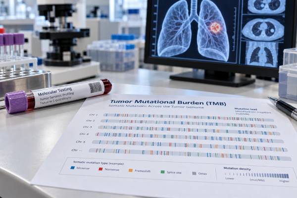
Tumor mutational burden (TMB) is a genomic score that estimates how many acquired DNA mutations are present in a tumor, usually reported as mutations per megabase (mut/Mb) of DNA analyzed. In lung cancer, TMB has been studied mainly as a biomarker of possible benefit from immune checkpoint inhibitors. The idea is that a tumor carrying more mutations may produce more abnormal proteins, or neoantigens, that the immune system can recognize. A high TMB can therefore be clinically relevant, but it does not guarantee that immunotherapy will work, and a low TMB does not prove that it will fail. Results also depend on the sequencing panel, the amount and quality of tumor DNA, the bioinformatics method, and the cutoff used. TMB should be read alongside lung cancer type, stage, driver mutations, PD-L1 expression, prior treatment, and the specific therapy being considered rather than as a stand-alone treatment decision.
- TMB is reported as mutations per megabase (mut/Mb): It estimates mutation density across a defined amount of tumor DNA, not the size or stage of the cancer.
- “TMB-high” is assay and indication specific: A cutoff such as 10 mut/Mb is important in one FDA tumor-agnostic setting but is not a universal lung-cancer threshold for every treatment.
- Higher TMB may increase the chance of immunotherapy benefit in some settings: It is a probabilistic biomarker, not a yes/no prediction of response.
- TMB and PD-L1 measure different biology: One cannot be substituted for the other, and both may be considered with other clinical and molecular findings.
- Testing method matters: Tissue TMB and blood TMB are not interchangeable, and different sequencing panels can produce different numerical estimates.
Table of Contents
- What tumor mutational burden measures
- How TMB is tested in lung cancer
- How to read a TMB score
- TMB and immunotherapy decisions
- TMB, PD-L1, and driver mutations
- Limitations and sources of variation
- Questions to ask about a TMB result
What tumor mutational burden measures
TMB measures the density of somatic mutations in cancer DNA. “Somatic” means the mutations were acquired by tumor cells rather than inherited in every cell of the body. Most clinical TMB assays count selected mutation types across a defined genomic region and then normalize the count to the number of megabases successfully analyzed.
A result of 10 mut/Mb, for example, means the assay estimated about 10 qualifying mutations for each megabase of evaluable tumor DNA under that test’s counting rules. It does not mean the tumor contains only 10 mutations total. The human coding genome spans far more DNA than the portion covered by a typical targeted panel.
The biological reason TMB can matter for immunotherapy is indirect. DNA mutations may alter protein sequences. Some altered proteins can be processed into neoantigens that immune cells recognize as foreign. In theory, a tumor with more mutations has more opportunities to create recognizable neoantigens. Immune checkpoint inhibitors can then remove inhibitory signals that were restraining antitumor T cells.
That chain is not automatic. Many mutations do not produce useful neoantigens, and tumors can evade immune attack through other mechanisms. Immune-cell infiltration, antigen presentation, oncogenic signaling, and the tumor microenvironment all influence response. TMB is therefore a probability marker, not a direct measurement of immune activity.
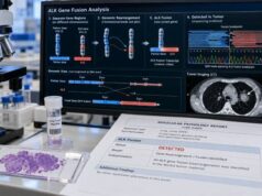
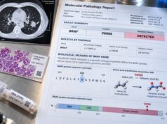
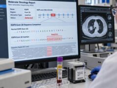
TMB is also different from a list of actionable mutations. A lung cancer NGS test may report EGFR, KRAS, BRAF, MET, RET, ALK, ROS1, and other alterations while also calculating TMB. The mutation list can identify specific treatment targets; the TMB score summarizes mutation density across the panel. One result should not be mistaken for the other.
Smoking-related lung cancers often carry more DNA damage and may have higher TMB than tumors arising in people with little or no tobacco exposure, but individual results vary widely. Histology and smoking history cannot substitute for an actual validated test when TMB is clinically needed.
How TMB is tested in lung cancer
TMB is usually calculated from next-generation sequencing (NGS). Whole-exome sequencing can count mutations across most protein-coding regions, but clinical laboratories more often estimate TMB from a large targeted gene panel because it requires less DNA, is faster, and can simultaneously report actionable alterations.
The basic process is:
- Obtain tumor DNA. A pathology laboratory selects tissue with enough viable tumor, usually from a biopsy, resection, or cytology-derived specimen that meets assay requirements.
- Sequence a defined genomic region. The panel reads hundreds of genes or another sufficiently large amount of DNA.
- Identify somatic variants. Bioinformatics filters remove sequencing artifacts and, depending on the method, known or likely germline variants.
- Apply the assay’s counting rules. The laboratory decides which mutation classes qualify for TMB calculation.
- Normalize by genomic territory. The qualifying count is divided by the number of megabases that were adequately assessed.
Different assays do not necessarily count exactly the same variants. Some include synonymous mutations; others focus on nonsynonymous changes. Panels also differ in gene content, sequence coverage, minimum variant allele frequency, germline filtering, and correction for technical artifacts. These differences can move the final score even when the same tumor is tested.
Panel size matters because a very small genomic region can give an unstable estimate. Harmonization studies have shown that larger panels and assay-specific calibration improve agreement with whole-exome TMB. This is why a clinically validated TMB result should come from a test with defined analytical performance rather than from manually counting mutations on a small sequencing report.
Tissue TMB versus blood TMB
Tissue TMB, sometimes written tTMB, is calculated from tumor tissue. Blood TMB, or bTMB, estimates mutation burden from circulating tumor DNA in plasma. Blood testing can be useful when tissue is limited, but low tumor DNA shedding can reduce sensitivity, and bTMB methods have their own thresholds and validation requirements.
A lung cancer liquid biopsy can detect mutations without another tissue procedure, but a blood-based TMB number should not be treated as numerically interchangeable with a tissue-based result unless the specific assay and clinical evidence support that use.
How to read a TMB score
The first step is to read the unit, assay name, specimen type, and laboratory interpretation together. A number without its testing context can be misleading.
Many reports classify TMB as low, intermediate, or high, while others provide only a numerical value and a defined threshold. The labels are not universally standardized. A result called “high” by one assay may not map perfectly to another platform because the panels and algorithms differ.
One widely recognized number is 10 mutations per megabase. In 2020, the U.S. Food and Drug Administration granted a tumor-agnostic accelerated approval for pembrolizumab in certain previously treated unresectable or metastatic solid tumors with TMB-high status defined as at least 10 mut/Mb by an FDA-approved test, when no satisfactory alternative treatment options exist. That regulatory threshold is important, but it should not be generalized into a rule that every lung cancer at 10 mut/Mb requires immunotherapy or that 9 mut/Mb means immunotherapy cannot help.
In lung cancer, treatment decisions can depend on disease stage, histology, PD-L1 level, actionable driver alterations, previous therapies, performance status, and the exact drug regimen. Trial-specific TMB thresholds have also varied.
A useful way to interpret a report is:
- Low TMB: The tumor has relatively fewer qualifying mutations under that assay. This may be associated with a lower chance of benefit from some checkpoint-inhibitor strategies, but it does not rule out benefit.
- High TMB: The tumor has relatively more qualifying mutations. This can be associated with greater immunotherapy benefit in some populations, but many TMB-high tumors still do not respond.
- Borderline result: A value close to the assay cutoff should be interpreted cautiously because technical and biological variation can affect classification.
- Not evaluable: Low DNA quantity, poor specimen quality, insufficient tumor content, or inadequate sequencing coverage may prevent a reliable calculation.
TMB is not a cancer screening test, a recurrence marker, or a direct measure of tumor burden. A TMB of 20 mut/Mb does not mean the cancer is twice as large or twice as aggressive as a tumor with 10 mut/Mb.
TMB and immunotherapy decisions
TMB became clinically interesting because multiple studies found that tumors with higher mutation burden were, on average, more likely to respond to immune checkpoint blockade. In advanced non-small cell lung cancer (NSCLC), randomized trials and pooled analyses have supported a relationship between higher TMB and benefit from certain immunotherapy-containing regimens.
However, the evidence is not simple enough to use TMB as an isolated switch. Different studies used different assays, cutoffs, drugs, treatment lines, and endpoints. Some trials showed a stronger relationship with progression-free survival or response than with overall survival. Combination treatment can also alter the value of a single predictive biomarker.
Current lung-cancer biomarker guidance therefore emphasizes validated testing and clinical context. TMB can provide additional information about the likelihood of immune responsiveness, but it does not replace biomarkers that are already required or strongly established for a specific treatment decision.
Several factors explain why TMB-high tumors can fail to respond:
- The mutations may not generate neoantigens that T cells can recognize.
- Tumor cells may lose antigen-presentation machinery.
- The tumor microenvironment may exclude or suppress immune cells.
- A strong oncogenic driver may shape an immune-resistant tumor state.
- The measured tissue sample may not represent every metastatic site.
- Prior therapy may change tumor clones and immune biology.
Likewise, some TMB-low tumors respond because mutation count is only one part of the immune response. This is why clinicians interpret the score as one dimension of a larger picture rather than a guarantee.
TMB can also be prognostic in some studies, but predictive and prognostic effects should not be confused. A predictive biomarker tells us whether a treatment is more likely to help relative to another option. A prognostic biomarker is associated with outcome regardless of treatment. The evidence for TMB is strongest in selected immunotherapy-prediction settings, and its pure prognostic meaning in untreated or early-stage NSCLC is less consistent.
TMB, PD-L1, and driver mutations
TMB and PD-L1 expression are related to immunotherapy but measure different biology. TMB estimates mutation density in DNA. The PD-L1 test for lung cancer uses immunohistochemistry to measure expression of a protein involved in suppressing T-cell activity.
A tumor can have high TMB and low PD-L1, low TMB and high PD-L1, both high, or both low. Neither result mathematically determines the other. Depending on the treatment regimen, PD-L1 may have a clearer validated role than TMB, and some therapies require a specific PD-L1 companion diagnostic or scoring system.
Driver mutations add another layer. In metastatic nonsquamous NSCLC, finding an actionable alteration can change first-line treatment more directly than TMB. EGFR mutations, ALK or ROS1 fusions, RET fusions, MET exon 14 skipping, BRAF V600E, and other oncogenic drivers have matched targeted therapies in appropriate settings. Some oncogene-driven tumors, especially classic EGFR-mutant cancers, may have lower TMB and less consistent benefit from single-agent checkpoint inhibition.
That is why comprehensive molecular testing should not be skipped simply because TMB or PD-L1 is high. A high immune biomarker does not erase a targetable driver. The NSCLC biomarker workup integrates histology, driver alterations, and immune biomarkers according to disease stage and treatment setting.
Microsatellite instability (MSI) and mismatch-repair deficiency are also distinct biomarkers. They can cause very high mutation burdens in some tumors, but MSI-high lung cancer is uncommon. A lung cancer MSI test evaluates a different biological mechanism and should not be inferred solely from the TMB number.
Limitations and sources of variation
TMB looks like a simple number, but several technical choices sit behind it. The largest interpretation problem is assuming that every mut/Mb value is measured the same way.
Panel design: Different gene panels cover different genomic territories. A panel enriched for genes that mutate frequently in cancer can overestimate mutation density unless the algorithm is calibrated appropriately.
Panel size: Smaller panels sample less DNA, so a few extra mutations can shift the estimated TMB substantially. Larger validated panels generally provide more stable estimates.
Variant filtering: Laboratories differ in how they exclude germline variants, sequencing artifacts, known driver mutations, and synonymous changes. Tumor-only sequencing must infer germline status without a matched normal sample, which can affect the final count.
Tumor purity: A specimen with few cancer cells may miss low-frequency mutations and underestimate TMB. Pathology review and assay-specific minimum tumor-content requirements help reduce this problem.
DNA quality: Formalin-fixed tissue can create artifacts, particularly in old or poorly processed specimens. Low DNA input and shallow sequencing depth can also reduce reliability.
Tumor heterogeneity: A biopsy samples one site at one time. Different regions or metastatic lesions can contain different clones. Treatment may also select new clones, so a historical TMB result may not perfectly describe current disease.
Blood-specific limitations: Plasma assays depend on enough circulating tumor DNA entering the bloodstream. Low-shedding tumors can produce an uninformative or falsely low blood estimate. Blood-derived clonal hematopoiesis can also introduce variants that require filtering.
Threshold effects: A cutoff creates a category from a continuous variable. A tumor at 9.8 mut/Mb is biologically unlikely to be completely different from one at 10.2 mut/Mb merely because they fall on opposite sides of a threshold. Near-cutoff values deserve especially careful assay-specific interpretation.
For these reasons, serial TMB testing is not commonly used as a routine disease-monitoring tool. TMB is usually assessed to characterize tumor biology and potential treatment relevance, while imaging, symptoms, and other clinical measures track response or progression.
Questions to ask about a TMB result
A TMB report is most useful when the patient and oncology team know exactly how it was generated and what decision it is supposed to inform. Useful questions include:
- Was TMB measured from tissue or blood?
- Which sequencing assay was used, and is it validated for TMB reporting?
- What is the numerical score in mut/Mb?
- What cutoff does this laboratory use, and is that cutoff linked to a specific drug indication or clinical trial?
- Was the sample adequate for reliable sequencing and tumor-content assessment?
- Are there actionable driver mutations on the same report that take priority for treatment selection?
- What is the PD-L1 result, and how does it affect the available immunotherapy options?
- Is the TMB value being used as a supportive biomarker or as part of a formally approved treatment indication?
- If the result is near the cutoff, would platform variability change the clinical interpretation?
Patients should also ask whether an old TMB result remains relevant after several lines of therapy. In many cases the original result still provides useful background, but repeat testing may be considered when new tissue is obtained for another clinical reason or when the molecular profile could affect a new treatment decision.
The clearest interpretation is not “high is good” or “low is bad.” A high TMB means the tested tumor sample carried a relatively high density of qualifying mutations according to that assay. It may strengthen the case for certain immunotherapy approaches, but treatment selection still depends on the full molecular and clinical context. A low TMB means fewer qualifying mutations were detected; it does not by itself exclude checkpoint inhibitor benefit. The score becomes clinically meaningful only when it is tied to a validated assay, a specific treatment setting, and the rest of the lung-cancer biomarker profile.
References
- Programmed Death Ligand-1 and Tumor Mutation Burden Testing of Patients With Lung Cancer for Selection of Immune Checkpoint Inhibitor Therapies: Guideline From the College of American Pathologists, Association for Molecular Pathology, International Association for the Study of Lung Cancer, Pulmonary Pathology Society, and LUNGevity Foundation 2024 (Guideline)
- Tumor mutational burden for the prediction of PD-(L)1 blockade efficacy in cancer: challenges and opportunities 2024 (Review)
- Tumor mutation burden in the prognosis and response of lung cancer patients to immune-checkpoint inhibition therapies 2023 (Review)
- Evaluating Tumour Mutational Burden as a Key Biomarker in Personalized Cancer Immunotherapy: A Pan-Cancer Systematic Review 2025 (Systematic Review)
- Aligning tumor mutational burden (TMB) quantification across diagnostic platforms: phase II of the Friends of Cancer Research TMB Harmonization Project 2021
- FDA approves pembrolizumab for adults and children with TMB-H solid tumors 2020
Disclaimer
TMB is a complex genomic biomarker and should not be used by itself to choose or reject lung cancer treatment. Cutoffs, assays, and drug indications can differ, and treatment guidance changes as new evidence and approvals emerge. Discuss the complete molecular report, PD-L1 result, cancer stage, prior treatment, and available therapies with the oncology team.